
ABSTRACT
Infection and immunity studies involving genetically modified organisms (GMOs), such as gene knockout bacterial mutants, require stringent physical containment to prevent the accidental spread of these organisms into the environment. Experimental respiratory tract infection models often require the animals, for example birds, to be transported several times between a negative pressure housing isolator and a bespoke aerosol exposure chamber under positive pressure. While the exposure chamber is sealed and fitted with HEPA filters, the repeated movements of infected animals and opening of the chamber can still pose a serious risk of breaching containment of the organism in the experimental facility. In the current study, the ability of two aerosol infection protocols that expose birds to avian pathogenic E. coli (APEC) aerosols directly within the housing isolator was evaluated. Young chicks were exposed to APEC E956 within the negative pressure housing isolators using either a nebulizer or an atomizer. Birds exposed twice (days 1 and 4) to aerosols of APEC E956 produced by the nebulizer developed a rapidly progressing disease mimicking field cases of avian colibacillosis. However, birds exposed to aerosols of APEC E956 produced by an atomizer did not develop colibacillosis even after three exposures to APEC E956 on days 1, 4 and 7. Consequently, the current study reports the nebulizer was more efficacious in producing avian colibacillosis under stricter bacterial containment settings.
RESEARCH HIGHLIGHTS
Two aerosol exposure methods were evaluated to develop avian colibacillosis.
Nebulizer method found to be more efficient in reproducing avian colibacillosis.
Refined infection method can be used to study genetically modified organisms (GMOs).
Introduction
Infection with avian pathogenic Escherichia coli (APEC) is responsible for the development of a severe respiratory and systemic disease in poultry known as avian colibacillosis (Dho-Moulin & Fairbrother, Citation1999; Ewers et al., Citation2003). The natural route of transmission for APEC is the inhalation of contaminated dust particles within poultry houses where the concentration can reach up to 106 viable E. coli per gram of dust (Carlson & Whenham, Citation1968; Guabiraba & Schouler, Citation2015). Following successful colonization of the lower respiratory tract epithelium, the disease manifests in a subacute form causing airsacculitis, pericarditis and perihepatitis which can result in a fatal septicaemia causing sudden death (Nolan et al., Citation2013). APEC is one of the most frequent causes of mortality in young birds housed on poultry farms, and the time between infection and the onset of clinical signs is between 5 and 7 days (Nolan et al., Citation2013). The duration, severity of lesions and outcome of the disease are determined by the virulence of the APEC and other factors like breed, age and immune status of the birds, as well as management conditions (Kabir, Citation2010).
The best approach for reproducing colibacillosis in an experimental setting is exposing birds to aerosolized bacteria (Ginns et al., Citation2000; Parreira & Gyles, Citation2003; Kariyawasam et al., Citation2004; Tivendale et al., Citation2004; Holden et al., Citation2012; Holden et al., Citation2014; Paudel et al., Citation2021). Other methods used to reproduce colibacillosis in an experimental setting include intratracheal, intra-air sac, subcutaneous or intravenous inoculation that enable the delivery of a consistent number of bacteria to individual birds, but these do not truly represent the natural process of infection (Dziva, Citation2010). A few studies have noted the limitation of models relying on intratracheal delivery, which is the inability to produce clinical disease, as a single administration of E. coli by the intratracheal route does not achieve significant colonization of the bird (Ginns et al., Citation1998; Peighambari et al., Citation2000; Cox et al., Citation2021). However, some studies, have successfully used the intratracheal method to reproduce the clinical disease (Antão et al., Citation2008; Landman et al., Citation2013). Nevertheless, unlike field conditions, birds are forced to inhale infectious particles that bypass the nasal and laryngeal mucosal defences crucial in controlling the infection and can also result in permanent pulmonary damage (Dziva, Citation2010; Kromann et al., Citation2021). The intra-air sac infection is another frequently used method of studying avian colibacillosis under experimental conditions. Similar to intratracheal delivery, a limitation of the intra-air sac method is bypassing the body’s defence mechanisms in the upper respiratory tract due to direct delivery of bacteria into the air sacs. Consequently, it does not accurately represent the natural infection process (Dziva, Citation2010). The sub-cutaneous route of inoculation is primarily preferred for inducing cellulitis (Gibbs et al., Citation2004). The intravenous route bypasses all the mucosal defence mechanisms of the respiratory tract by delivering bacteria directly into the bloodstream, resulting in exacerbated septicaemic disease even with less virulent APEC isolates (Dziva, Citation2010). In this respect, aerosol administration mimics the natural route of infection and is physiologically relevant when studying APEC virulence factors and mucosal immune responses against APEC infection (Ginns et al., Citation1998; Peighambari et al., Citation2000). Aerosolized bacteria are inhaled whilst breathing, imitating the natural inhalation of contaminated poultry house dust. Considering aerosolized bacteria are exposed to mucosal defence mechanisms, only virulent APEC strains that are able to invade and pass the epithelial barrier of the respiratory tract can cause disease (Ginns et al., Citation1998). Therefore, aerosol infection models are preferable for reliably assessing the virulence potential of APEC isolates and evaluating vaccine efficacy against challenge with a virulent isolate.
An experimental method to expose birds to aerosolized APEC has previously been developed by Ginns and colleagues in our group (Ginns et al., Citation1998) and has been utilized successfully in studies exploring APEC virulence factors and evaluating gene knockout mutants as vaccine candidates (Ginns et al., Citation2000; Tivendale et al., Citation2009; Holden, et al., Citation2014). The aerosol model requires day-old birds to be inoculated with 10 times the immunizing dose of infectious bronchitis virus (IBV) vaccine prior to the first aerosol exposure to APEC in order to induce ciliostasis, thereby creating favourable conditions for APEC colonization. The IBV vaccine employed in previous experiments appears to cause an excessive inflammatory response in the air sacs, which is thought to be the mechanism underlying the pattern of airsacculitis in commercial poultry in the field (Dwars et al., Citation2009). Following administration of the IBV vaccine, groups of birds are exposed at three separate time-points (days 1, 4 and 7) to 1.5 × 10¹⁰ colony-forming units (CFUs) of aerosolized APEC to replicate the sustained exposure to contaminated poultry house dust as experienced in the field. Studies utilizing this infection model reported avian colibacillosis-related clinical signs in approximately 88% of birds infected with APEC E956 by day 11 (Ginns et al., Citation1998). This aerosol infection model was developed in 1998 and requires further refinements to make it suitable for today’s regulatory requirements for dealing with genetically modified organisms (GMOs), such as live attenuated vaccines incorporating gene deletion mutants. The original method exposed birds to aerosols of APEC in an aerosol infection chamber that was situated at a different location to the negative pressure isolators used for housing the birds. Birds were transported in and out of their housing isolator using closed plastic containers each time they were exposed to aerosols of APEC. Therefore, infected birds were no longer isolated under negative pressure during exposure or transportation. Birds were manually transferred into the opened aerosol chamber, briefly exposing them to the room environment. Although the chamber was sealed immediately, and was purged prior to reopening it, this process breaches containment regulations and poses a threat of accidental spread of genetically modified APEC into the environment. Consequently, the aerosol exposure method requires further refinement for handling GMOs. Exposing birds to aerosols of APEC within negative pressure isolators will alleviate this problem.
Here we evaluated two different exposure methods for aerosols exposure within an isolator, using a handheld atomizer or a nebulizer. The atomizer was used to expose one bird at a time to aerosols of APEC, whereas, for the nebulizer method, groups of birds confined in a small plastic container (13,312 cm3 volume) were exposed through an inlet hole in the box. The aim was to achieve exposure of the birds to an infectious dose of APEC whilst fulfilling the compliance requirements of the Office of the Gene Technology Regulator (OGTR). Although both methods have previously been reported for the immunization of chickens, their ability to establish an infection has not been compared before (Filho et al., Citation2013; Albanese et al., Citation2018; Goonewardene et al., Citation2020). In addition, no study documenting the efficacy of a handheld atomizer to establish an infection with APEC in poultry has previously been reported. A benefit to using the atomizer is that it does not require a supply of compressed air or any other accessories in order to produce aerosols, unlike the nebulizer, making it a convenient option for animal facilities without access to a nebulizer and the associated equipment. The efficacy of both methods to establish an APEC infection in 1-day-old birds and to progress through to development of clinical signs associated with the disease of avian colibacillosis was compared.
Materials and methods
Preparation of APEC E956 challenge cultures
E956 is a virulent APEC strain that was previously used in the development of the aerosol challenge model for 1-day-old birds (Ginns et al., Citation1998). To prepare APEC E956 cultures, bacteria were resuscitated from 50% glycerol stocks by streak dilution onto MacConkey agar (Oxoid, Victoria, Australia) and incubated overnight at 37°C. A single bacterial colony isolated on MacConkey agar was inoculated into 100 ml of Nutrient broth No. 2 (Oxoid, Victoria, Australia) and incubated at 37°C with shaking at 250 rpm for 24 h. The absorbance of the 24 h culture at 600 nm was determined and the number of viable bacteria was calculated by performing serial 10-fold dilutions and standard plate counts (Atlas, Citation2004) from three separate cultures.
To prepare cultures for aerosolization, 100 ml Nutrient broth No. 2 was inoculated with a single colony from a freshly streaked agar plate and incubated at 37°C with shaking for 24 h. Cells were harvested by centrifugation at 5000× g for 10 min (Beckman Coulter Allegra X-22 K centrifuge). The bacterial pellet was resuspended in 25 ml of nutrient broth to obtain a final concentration of approximately 1–1.5 × 10¹⁰ CFU/ml. This final suspension was used for the aerosol exposure of the birds. Serial 10-fold dilutions of the final suspension were performed and 100 μl of the 10−7 and 10−8 dilutions were spread onto LB agar plates and incubated at 37°C overnight in order to determine viable cell counts and calculate the final concentration.
Experimental birds
Specific pathogen-free (SPF) chickens were hatched at the Asia-Pacific Centre for Animal Health, Faculty of Veterinary and Agricultural Sciences, University of Melbourne, Werribee, Australia from eggs supplied by Australian SPF services Pty Ltd (Woodend, Victoria, Australia). One-day-old birds were wing-tagged for identification, weighed and allocated randomly into groups of 20 birds. Birds were housed in negative pressure fibreglass isolators (Controlled isolation system Incorporate MK-1, San Diego, CA, USA) with irradiated feed and water.
Experimental exposure of birds to APEC aerosols
The experiments were conducted with the approval from The University of Melbourne Animal Ethics Committee, application number 20526 and 10378. On day 1 of the experiment, 1-day-old SPF birds were inoculated with a 10× dose of infectious bronchitis virus vaccine (Poulvac Bron Vic S, Victoria, Australia) followed by immediate exposure to 1.5 × 10¹⁰ CFUs of APEC E956 using either an atomizer or a nebulizer (see below for technical specifications and dose delivery calculations). Birds exposed to APEC via the atomizer were re-exposed to 1.5 × 10¹⁰ CFUs of APEC E956 on day 4 and 7, whilst those exposed via the nebulizer were re-exposed to 1.5 × 10¹⁰ CFUs of APEC E956 on day 4 only. Mock-infected birds were exposed to aerosols of sterile nutrient broth using the same methods. Swabs from the trachea and left abdominal air sacs were collected on day 12 of the experiment for birds exposed to APEC via the atomizer and on days 6–7 of the experiment for birds exposed to APEC via the nebulizer due to the earlier termination of the experiment. (Supplementary Figure S1; Timeline of the experiment)
Experimental settings for atomizer infection method
The atomizer (MADomizer®, Teleflex, Mascot NSW, Australia), is a handheld device that uses a positive displacement pump to generate droplets (ranging from 30–100 μm) and delivers 0.1 ml per spray. The nozzle of the atomizer was held 10 cm from the beak of each individual bird and 10 sprays of 0.1 ml were dispensed to deliver 1 ml of inoculum containing 1.5 × 10¹⁰ CFU/ml APEC on days 1, 4 and 7 of the experiment (Supplementary Figure S2; Atomizer settings).
Experimental settings for nebulizer infection method
A Collison™ 6 jets nebulizer (CH Technologies, Westwood, NJ, USA) was used to generate aerosols of 1–10 μm. Groups of 10 birds were placed in a plastic container (L = 32 cm, W = 26 cm, H = 16 cm) within the isolator. The nozzle of the Collison™ nebulizer was connected to the plastic container by inserting it into a pre-drilled hole 12 cm from the base of the container (Supplementary Figure S3; Nebulizer settings). A 10 ml inoculum of either the prepared bacterial culture or sterile media was delivered through the nebulizer over a total duration of 10 min at an airflow rate of 12 l/min on days 1 and 4 of the experiment. The airflow to the isolator was switched off for 10 min during the exposure.
Collection of samples and lesion scoring
Birds were humanely euthanized by halothane inhalation at the endpoint of each experiment. The body weight of each bird was measured and then each bird was examined for gross lesions including airsacculitis, pericarditis and perihepatitis and the severity of lesions was assessed using previously published lesion scoring criteria (Ginns et al., Citation1998; Antão et al., Citation2008) (Supplementary Table S1). For bacterial reisolation, two pre-soaked swabs (sterile minitip swab with aluminium shaft; Copan, Murrieta, CA, USA) were used to collect the material one from the upper trachea (immediately below the larynx) and the other from the left abdominal air sac by rotating the swab four times and then placed in 10-M tube containing 1 ml of LB+ glycerol. To remain consistent and also to avoid variability between birds, swabs from all birds in both atomizer and nebulizer experiments were collected by the same operator. The tubes containing swabs in LB+ glycerol were then vortexed for 10 s each, and 100 μl was removed and used to prepare 10-fold serial dilutions in PBS. Serial 10-fold dilutions were performed and a 10 μl drop of the 10−1, 10−2 and 10−3 dilutions were plated in triplicate on MacConkey agar. Plates were left upright to dry before inversion and incubated at 37°C for 12–16 h and the viable number of bacteria/ml was determined by the Miles and Misra drop plate count method (Miles et al., Citation1938). Representative colonies reisolated from the swabs were confirmed to be APEC by multiplex PCR using primers specifically designed to differentiate APEC from other commensal E. coli (Johnson et al., Citation2008).
Statistical analysis
The software GraphPad Prism 9 for Windows (GraphPad Software, San Diego, CA, USA) was used to perform statistical analysis of data and create the graphs. Analysis of weight gains, lesions scores and colonization of the trachea and air sacs with APEC was performed using the Mann–Whitney test. The survival rate of birds following APEC exposure was determined using the Mantel–cox test. P values < 0.05 were considered statistically significant.
Results
Nebulization method caused colonization of APEC E956 in air sacs
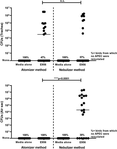
APEC E956 colonization in birds was determined by the number of CFUs of APEC isolated from swabs taken of the trachea and left abdominal air sac. APEC was reisolated from the trachea of 10/19 of the birds after exposure via the atomizer, and 8/19 of the birds after exposure via the nebulizer. There was almost no significant difference in the number of birds which yielded positive cultures of APEC from the trachea using either of the exposure methods. In addition, the median number of APEC reisolated from the trachea in both of these groups was not statistically significant in birds exposed to APEC using either the nebulizer or atomizer method (). In contrast, APEC was not reisolated from the air sacs of any of the birds exposed via the atomizer but was reisolated from the air sacs of 13/19 of the birds exposed using the nebulizer (). The difference in the reisolation rates from the air sacs between the two groups of birds was statistically significant (P < 0.001).
Figure 1. Reisolation of colony-forming units from the trachea and air sacs of birds.
Note: The number of colony-forming units (CFUs) reisolated from swabs taken from either the trachea (top panel) or left abdominal air sac (bottom panel) of each bird exposed to aerosols of APEC E956 or sterile media via the atomizer or nebulizer. Each dot represents the number of CFUs isolated from a single bird and the horizontal line represents the medians of the CFUs for each group. The number of birds from which APEC was not reisolated is indicated by the percentages (%) at the base of the graph. P < 0.001*** significant difference in number of CFUs reisolated, n.s. = non-significant; Mann–Whitney test.
Reduced weight gain in APEC-exposed birds using nebulization method
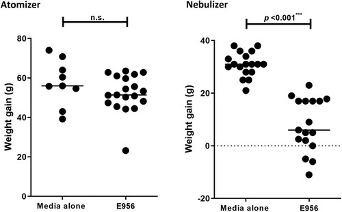
Body weight gains of birds exposed to aerosols were calculated as the difference between the weight of the bird at the beginning of the experiment and at the time of necropsy. The mean body weight gains of birds exposed to sterile media or APEC E956 using the atomizer were not significantly different (). Contrarily, for birds exposed to aerosols produced by the nebulizer, body weight gains of birds exposed to APEC were significantly lower (P < 0.001) than those of birds exposed to sterile media (). Notably, for three birds exposed to APEC via the nebulizer, a loss in body weight was observed rather than weight gain ().
Figure 2. Mean body weight gains of birds.
Note: Body weight gains for birds exposed to aerosols produced by the atomizer (left panel) or the nebulizer (right panel). Each dot represents the weight gain of a single bird from day 1 of the experiment until the time of necropsy, and the horizontal bar represents the mean. P < 0.001*** significant difference in weight gains, n.s. = non–significant; Mann–Whitney test.
Reduced survival of APEC E956 exposed birds using nebulization method compared to the atomizer method
Birds exposed to APEC E956 via the nebulizer experienced significantly lower survival rates than birds exposed to APEC E956 via the atomizer (P < 0.001) (). Birds exposed to APEC E956 via the nebulizer experienced higher mortality, with 3/20 dying (15%) within 3 days after the first exposure. Another seven birds (35%) died or were euthanized due to severe clinical signs of disease within 24 h of the second exposure (day 5). The remaining birds (50%) showed clinical signs of disease and were euthanized due to ethical concerns on day 6 of the experiment. In contrast, all except for one bird exposed to APEC via the atomizer survived throughout the 12 days of the experiment, despite being exposed to APEC at three different time-points. Birds exposed to aerosols of sterile media did not display clinical signs of disease and were euthanized at the conclusion of the experiment.
Figure 3. Survival of birds.
Note: Kaplan-Meier analysis was performed such that birds found dead or exhibiting clinical signs and subsequently euthanized were considered mortalities. The “atomizer media” and “nebulizer media” lines represent the per cent survival of birds for the duration of the experiment following exposure to sterile media. The “atomizer E956” and “nebulizer E956” lines represent the per cent survival of birds over the duration of the experiment following exposure to APEC E956.

Nebulization method successfully produced avian colibacillosis-related clinical signs in APEC-exposed birds
Birds were monitored daily for clinical signs of disease associated with APEC infection, such as severe lethargy, unresponsiveness, dehydration, dull expression of eyes, and dropping of head and neck. Approximately 35% birds (seven birds out of 20 in group) exposed to APEC via the nebulizer were found dead without exhibiting any clinical signs despite frequent monitoring (). Disease progression was studied in the remaining 13 birds exposed to APEC via the nebulizer (). These 13 birds did not show any signs of clinical disease in the 72 h after the first exposure. Early clinical signs, (caking of droppings around the vent and abdominal feathers) were observed in the majority of birds within 24 h of the second exposure. These clinical signs became more pronounced between 24 and 48 h after the second exposure, where birds were observed to be lethargic, dehydrated and occasionally sitting on their hocks, with a dull expression in their eyes. Forty-eight hours following the second exposure, the disease progressed to an advanced stage where birds were observed to be unresponsive when approached, sitting in a hunched posture including a dropping of the head, neck and wings, with eyes closed. These birds were humanely euthanized and a third exposure via the nebulizer was not performed (). In contrast, birds exposed on three occasions to APEC E956 via the atomizer did not show any clinical signs of disease (), and only one bird died prior to the conclusion of the experiment ().
Figure 4. Clinical progression of disease in birds after APEC exposure.
Note: Clinical signs observed in birds throughout the duration of the experiment were defined as mild, moderate or advanced, and were shaded in increasing intensity as illustrated in the key. No detectable clinical signs were observed in birds exposed to APEC E956 via the atomizer, whilst the clinical signs 72 h after exposure to APEC E956 using the nebulizer progressed from mild to advanced.
Table 1. Number of birds displaying clinical signs of disease following exposure to APEC E956.
Nebulization method successfully produced avian colibacillosis-related lesions in APEC-exposed birds
Lesions of airsacculitis, perihepatitis and pericarditis were scored at the time of necropsy and were observed in only three birds exposed to APEC via the atomizer. In the majority of birds exposed to APEC via the atomizer (85–90%), no evidence of pathological lesions was observed in the air sacs, heart or liver during the post-mortem examination (). In contrast, characteristic lesions of avian colibacillosis were observed in birds exposed to APEC via the nebulizer, including airsacculitis, which is a main indicator of the severity of avian colibacillosis. The most predominant and severe lesions were observed in the left abdominal air sacs, and almost all the birds exposed to APEC E956 via the nebulizer presented with airsacculitis. The lesions observed in the right abdominal air sacs were relatively less severe than those observed in the left abdominal air sacs, and were observed in a smaller number of birds. Some birds exposed to APEC E956 via the nebulizer also developed fibrinous pericarditis (10/20) and perihepatitis (13/20); however, the severity of these lesions varied between the two organs and birds mostly displayed a mild form of perihepatitis and a severe form of pericarditis. Overall, lesions seen in the birds exposed to aerosols produced by the nebulizer were significantly higher than those of birds exposed to aerosols produced by the atomizer.
Figure 5. Lesion scores of air sacs, liver and heart following exposure to APEC E956.
Note: At post-mortem examination, lesions typical of airsacculitis, pericarditis and perihepatitis were scored. Each dot represents the score given for the left abdominal air sac, right abdominal air sac, liver and heart for an individual bird and horizontal lines represent the median lesion score. The number of birds without lesions is indicated by the percentage at the base of the graph. P < 0.05*, P < 0.01**, P < 0.001*** significant difference in lesion scores using a Mann–Whitney test.
Discussion
A reproducible infection model that mimics avian colibacillosis is essential for the assessment of APEC gene deletion vaccine candidates. Such a model must be compatible with regulatory requirements for dealing with genetically modified organisms. Infection and immunity studies exposing animals to genetically modified pathogens via aerosols require more stringent experimental settings to prevent the spread of the pathogen outside of the testing environment. This study compared the efficacy of a handheld atomizer and a nebulizer to establish an effective infection model in birds by producing aerosols directly within a negative pressure housing isolator. Despite birds being exposed to the same dose of APEC E956 which had been cultured under the same growth conditions, the nebulizer method to generate aerosols of APEC within an isolator was not only superior in reproducing avian colibacillosis when compared to the method using the atomizer, but also worked better in disease progression than the original method described by Ginns et al. (Citation1998), which provided the baseline for the design of the nebulizer method in this study. Whilst the original method was employed in several studies to assess the virulence of various APEC strains and vaccine candidates (Holden, et al., Citation2012; Citation2014), the disease developed much more slowly than the acute development under field conditions. On the other hand, the use of the nebulizer within the isolator as described in the current study resulted in the robust development of clinical signs, indicating that the nebulizer used in the current study successfully mimicked the disease pathogenesis that would occur in the field.
Differences observed between the exposure methods in establishing an infection in the lower respiratory tract could be attributed to differences in the particle size generated by each method. Particle size is an important physical factor that dictates where aerosolized pathogens deposit within the respiratory tract and can also have a dramatic effect on disease pathogenesis (Gralton et al., Citation2011; Thomas, Citation2013). The particle size delivered by the nebulizer was previously determined to range between 1 and 10 µm, with the majority of the particles under 5 μm (Whithear et al., Citation1996; Wijesurendra et al., Citation2015). In contrast, the particle size generated by the handheld atomizer is known to range between 30 and 100 µm (Wolfe et al., Citation2002). Previous studies have described smaller particles of around 3 µm or less in size to be homogenously distributed throughout the respiratory tract of avian species including chickens and pigeons, whereas larger-sized particles ≥ 5 µm are relatively confined to the upper respiratory tract (Corbanie et al., Citation2006; Tell et al., Citation2006; Citation2012). The size of the particles is also important for effective vaccination with live vaccines against IBV and Newcastle disease viruses (Nolan et al., Citation2013). Similar observations have also been made with murine and macaque models of respiratory tract disease that demonstrated differential deposition of the pathogen in the lower and upper respiratory tract by changing the size of aerosolized particles (Day & Berendt, Citation1972; Thomas et al., Citation2009; Citation2010; Citation2012), further supporting the critical role of the size of aerosolized particles in determining the deposition of pathogens within the respiratory tract. The effects of the physical characteristics of droplets on the development of respiratory tract diseases and their progression have also been extensively highlighted in more recent studies on respiratory viruses including severe acute respiratory syndrome coronavirus 2 (SARS-CoV2/COVID 19 virus) (Jayaweera et al., Citation2020). Viruses enclosed in small aerosol particles (< 5 µm) remain in the environment for a prolonged time and are therefore more effective in disease transmission (Wang et al., Citation2021). One study has reported that aerosols of 1, 5 and 100 µm in size remain in the air for 12.5 h, 33 min and 5 s, respectively (Wang et al., Citation2021). The particle size used in nebulizer method of infection is 1–10 µm which is also similar to the natural APEC infection in field conditions. A previous study described that in poultry houses most of the E. coli (98.8%) are attached to dust particles and can migrate in and out of poultry houses through air movement. Importantly, the aerodynamic diameter of dust particles carrying E. coli is about 2.1–10 µm (Nguyen et al., Citation2022). This combined effect of smaller-sized particles remaining suspended in the air for longer periods, results in deeper penetration of the maximum number of available pathogens during normal respiratory activities (Jayaweera et al., Citation2020; Wang et al., Citation2021). Another important factor that works in conjunction with particle size and can also impact the aerosol performance is the size of the exposure chamber. Birds were exposed to aerosols via the nebulizer in groups of 10 within a confined space of 13,312 cm3 over a 10-min duration. On the other hand, birds were exposed individually to aerosols of APEC via the atomizer (10 sprays over a 1-min period) within the isolator which has a 47 times larger volume than the plastic container used for the nebulizer. Consequently, the physical nature of smaller aerosolized particles (1–10 µm) produced by the nebulizer within a more confined space of exposure chamber potentially enabled the particles to remain suspended in the air for a longer duration, facilitating their inhalation by the birds. Larger-sized droplets (30–100 µm) produced by the atomizer within a larger space will quickly fall to the base of the isolator decreasing the number of APEC droplets available for inhalation. This highlights a possible reason for the poor efficiency of the atomizer to establish an infection even after exposure to APEC on three separate occasions, whilst exposure to APEC on just two occasions via the nebulizer was highly effective in producing clinical disease.
Another reason for improved efficacy of nebulizer method could be deeper inhalation of bacterial inoculum over a longer period of time. In contrast, birds exposed to same number of bacteria using the atomizer within a shorter period did not develop clinical disease, indicating exposure time as another determinant of disease development under experimental conditions. These findings are consistent with previous studies highlighting significance of exposure time in relation to disease development. For instance, the impact of the duration of exposure time has been demonstrated previously in pigeons where the number of aerosolized particles inhaled by each bird was proportional to the exposure periods (Tell et al., Citation2012). Similarly, exposure time is also critical for aerosolized treatments used to combat respiratory tract infections in pigeons, as a short duration of exposure to aerosolized drugs results in limited success due to insufficient penetration of the lower respiratory system by the drug and the corresponding low absorption from the respiratory system into the bloodstream (Beernaert et al., Citation2009). In summary, the combination of particle size, exposure time and the physical space in which the aerosols were generated facilitated the production of clinical disease following exposure to APEC via the nebulizer.
Experiments in adult birds also found that exposure to aerosols of APEC using a nebulizer was an effective method of replicating clinical disease (Landman et al., Citation2013; Kromann et al., Citation2021). In the present study, an avian colibacillosis infection model was developed for young birds as they are more susceptible to develop infection than adults (Rodriguez-Siek et al., Citation2005). Another recent study also found that the aerosol method is the most suitable approach to generate clinical disease in 2-week-old broiler birds, further supporting the idea that, under the right conditions, a nebulizer can be used successfully to reproduce avian colibacillosis in a wide range of age groups (Paudel et al., Citation2021).
The inability of the atomizer method to establish colibacillosis was demonstrated by a lack of APEC-related signs such as weight loss or clinical signs of disease in exposed birds throughout the experiment. In addition, post-mortem examination of birds exposed via the atomizer also revealed an absence of lesions in the air sacs, heart and liver. By contrast, birds exposed to APEC E956 via the nebulizer showed a significant reduction in weight gain, developed severe APEC-related clinical signs and displayed various lesions during post-mortem examination. The smaller weight gains observed in birds exposed to sterile media via the nebulizer as compared to birds exposed to sterile media via the atomizer was due to the shorter duration of the nebulizer experiment.
In conclusion, the current study successfully refined the infection model for avian colibacillosis, using a nebulizer to produce aerosols of APEC within a negative pressure isolator, making it suitable to prevent the accidental spread of GMOs. It also produced a rapidly progressing and clinically relevant avian colibacillosis in young birds making it suitable for studies evaluating genetically modified organisms as potential vaccine candidates against avian colibacillosis.
Supplemental Material
Download MS Word (226 KB)Disclosure statement
No potential conflict of interest was reported by the authors.
References
- Albanese, G.A., Tensa, L.R., Aston, E.J., Hilt, D.A. & Jordan, B.J. (2018). Evaluation of a coccidia vaccine using spray and gel applications. Poultry Science, 97, 1544–1553.
- Antão, E.M., Glodde, S., Li, G., Sharifi, R., Homeier, T., Laturnus, C., Diehl, I., Bethe, A., Philipp, H.C., Preisinger, R., Wieler, L.H. & Ewers, C. (2008). The chicken as a natural model for extraintestinal infections caused by avian pathogenic Escherichia coli (APEC). Microbial Pathogenesis, 45, 361–369.
- Atlas, R.M. (2004). Handbook of Microbiological Media. London: CRC Press.
- Beernaert, L.A., Baert, K., Marin, P., Chiers, K., De Backer, P., Pasmans, F. & Martel, A. (2009). Designing voriconazole treatment for racing pigeons: balancing between hepatic enzyme auto induction and toxicity. Medical Mycology, 47, 276–285.
- Carlson, H.C. & Whenham, G.R. (1968). Coliform bacteria in chicken broiler house dust and their possible relationship to coli-septicemia. Avian Diseases, 12, 297–302.
- Corbanie, E.A., Matthijs, M.G.R., van Eck, J.H.H., Remon, J.P., Landman, W.J.M. & Vervaet, C. (2006). Deposition of differently sized airborne microspheres in the respiratory tract of chickens. Avian Pathology, 35, 475–485.
- Cox, G.J.M., Griffith, B., Reed, M., Sandstrom, J.D., Peterson, M.P., Emery, D. & Straub, D.E. (2021). A vaccine to prevent egg layer peritonitis in chickens. Avian Diseases, 65, 198–204.
- Day, W.C. & Berendt, R.F. (1972). Experimental tularemia in Macaca mulatta: relationship of aerosol particle size to the infectivity of airborne Pasteurella tularensis. Infection and Immunity, 5, 77–82.
- Dho-Moulin, M. & Fairbrother, J.M. (1999). Avian pathogenic Escherichia coli (APEC). Veterinary Research, 30, 299–316.
- Dziva, F. (2010). Deciphering the infection biology of avian pathogenic Escherichia coli: role of experimental infection models. Current Research, Technology and Education Topics in Applied Microbiology and Microbial Biotechnology, 1, 746–753.
- Dwars, R.M., Matthijs, M.G., Daemen, A.J., van Eck, J.H., Vervelde, L. & Landman, W.J. (2009). Progression of lesions in the respiratory tract of broilers after single infection with Escherichia coli compared to superinfection with E. coli after infection with infectious bronchitis virus. Veterinary Immunology and Immunopathology, 127, 65–76.
- Ewers, C., Janssen, T. & Wieler, L.H. (2003). Avian pathogenic Escherichia coli (APEC). Berl Munch Tierarztl Wochenschr, 116, 381–395.
- Filho, F.T., Fávaro, C., Jr., Ingberman, M., Beirão, B.C., Inoue, A., Gomes, L. & Caron, L.F. (2013). Effect of spray Escherichia coli vaccine on the immunity of poultry. Avian Diseases, 57, 671–676.
- Gibbs, P.S., Petermann, S.R. & Wooley, R.E. (2004). Comparison of several challenge models for studies in avian colibacillosis. Avian Diseases, 48, 751–758.
- Ginns, C.A., Benham, M.L., Adams, L.M., Whithear, K.G., Bettelheim, K.A., Crabb, B.S. & Browning, G.F. (2000). Colonization of the respiratory tract by a virulent strain of avian Escherichia coli requires carriage of a conjugative plasmid. Infection and Immunity, 68, 1535–1541.
- Ginns, C.A., Browning, G.F., Benham, M.L. & Whithear, K.G. (1998). Development and application of an aerosol challenge method for reproduction of avian colibacillosis. Avian Pathology, 27, 505–511.
- Goonewardene, K., Ahmed, K.A., Gunawardana, T., Popowich, S., Kurukulasuriya, S., Karunarathna, R., Gupta, A., Ayalew, L.E., Lockerbie, B., Foldvari, M., Tikoo, S., Willson, P. & Gomis, S. (2020). Mucosal delivery of CpG-ODN mimicking bacterial DNA via the intrapulmonary route induces systemic antimicrobial immune responses in neonatal chicks. Scientific Reports, 10, 5343.
- Gralton, J., Tovey, E., McLaws, M.L. & Rawlinson, W.D. (2011). The role of particle size in aerosolised pathogen transmission: a review. The Journal of Infection, 62, 1–13.
- Guabiraba, R. & Schouler, C. (2015). Avian colibacillosis: still many black holes. FEMS Microbiology Letters, 362, 1–8.
- Holden, K.M., Browning, G.F., Noormohammadi, A.H., Markham, P. & Marenda, M.S. (2014). Avian pathogenic Escherichia coli ΔtonB mutants are safe and protective live-attenuated vaccine candidates. Veterinary Microbiology, 173, 289–298.
- Holden, K.M., Browning, G.F., Noormohammadi, A.H., Markham, P.F. & Marenda, M.S. (2012). Tonb is essential for virulence in avian pathogenic Escherichia coli. Comparitive Immunology, Microbiology and Infectious Diseases, 35, 129–138.
- Jayaweera, M., Perera, H., Gunawardana, B. & Manatunge, J. (2020). Transmission of COVID-19 virus by droplets and aerosols: a critical review on the unresolved dichotomy. Environmental Research, 188, 109819–109819.
- Johnson, T.J., Wannemuehler, Y., Doetkott, C., Johnson, S.J., Rosenberger, S.C. & Nolan, L.K. (2008). Identification of minimal predictors of avian pathogenic Escherichia coli virulence for use as a rapid diagnostic tool. Journal of Clinical Microbiology, 46, 3987–3996.
- Kariyawasam, S., Wilkie, B.N. & Gyles, C.L. (2004). Construction, characterization, and evaluation of the vaccine potential of three genetically defined mutants of avian pathogenic Escherichia coli. Avian Diseases, 48, 287–299.
- Kromann, S., Olsen, R.H., Bojesen, A.M., Jensen, H.E. & Thøfner, I. (2021). Development of an aerogenous Escherichia coli infection model in adult broiler breeders. Scientific Reports, 11, 19556.
- Kabir, S.M.L. (2010). Avian colibacillosis and salmonellosis: a closer look at epidemiology, pathogenesis, diagnosis, control and public health concerns. International Journal of Environmental Research and Public Health, 7, 89–114.
- Landman, W.J., Heuvelink, A. & van Eck, J.H. (2013). Reproduction of the Escherichia coli peritonitis syndrome in laying hens. Avian Pathology, 42, 157–162.
- Miles, A.A., Misra, S.S. & Irwin, J.O. (1938). The estimation of the bactericidal power of the blood. The Journal of Hygiene, 38, 732–749.
- Nolan, L.K., Barnes, H.J., Vaillancourt, J.P., Abdul-Aziz, T. & Logue, C.M. (2013). Colibacillosis. In Diseases of Poultry (pp. 751–805).
- Nguyen, X.D., Zhao, Y., Evans, J.D., Lin, J. & Purswell, J.L. (2022). Survival of Escherichia coli in airborne and settled poultry litter particles. Animals, 12, 284.
- Parreira, V.R. & Gyles, C.L. (2003). A novel pathogenicity island integrated adjacent to the thrW tRNA gene of avian pathogenic Escherichia coli encodes a vacuolating autotransporter toxin. Infection and Immunity, 71, 5087–5096.
- Paudel, S., Fink, D., Abdelhamid, M.K., Zöggeler, A., Liebhart, D., Hess, M. & Hess, C. (2021). Aerosol is the optimal route of respiratory tract infection to induce pathological lesions of colibacillosis by a lux-tagged avian pathogenic Escherichia coli in chickens. Avian Pathology, 50, 417–426.
- Peighambari, S.M., Julian, R.J. & Gyles, C.L. (2000). Experimental Escherichia coli respiratory infection in broilers. Avian Diseases, 44, 759–769.
- Rodriguez-Siek, K.E., Giddings, C.W., Doetkott, C., Johnson, T.J. & Nolan, L.K. (2005). Characterizing the APEC pathotype. Veterinary Research, 36, 241–256.
- Tell, L.A., Smiley-Jewell, S., Hinds, D., Stephens, K.E., Teague, S.V., Plopper, C.G. & Pinkerton, K.E. (2006). An aerosolized fluorescent microsphere technique for evaluating particle deposition in the avian respiratory tract. Avian Diseases, 50, 238–244.
- Tell, L.A., Stephens, K., Teague, S.V., Pinkerton, K.E. & Raabe, O.G. (2012). Study of nebulization delivery of aerosolized fluorescent microspheres to the avian respiratory tract. Avian Diseases, 56, 381–386.
- Thomas, R., Davies, C., Nunez, A., Hibbs, S., Eastaugh, L., Harding, S., Jordan, J., Barnes, K., Oyston, P. & Eley, S. (2012). Particle-size dependent effects in the Balb/c murine model of inhalational melioidosis. Frontiers in Cellular and Infection Microbiology, 2, 101.
- Thomas, R., Davies, C., Nunez, A., Hibbs, S., Flick-Smith, H., Eastaugh, L., Smither, S., Gates, A., Oyston, P., Atkins, T. & Eley, S. (2010). Influence of particle size on the pathology and efficacy of vaccination in a murine model of inhalational anthrax. Journal of Medical Microbiology, 59, 1415–1427.
- Thomas, R.J. (2013). Particle size and pathogenicity in the respiratory tract. Virulence, 4, 847–858.
- Thomas, R.J., Webber, D., Collinge, A., Stagg, A.J., Bailey, S.C., Nunez, A., Gates, A., Jayasekera, P.N., Taylor, R.R., Eley, S. & Titball, R.W. (2009). Different pathologies but equal levels of responsiveness to the recombinant F1 and V antigen vaccine and ciprofloxacin in a murine model of plague caused by small- and large-particle aerosols. Infection and Immunity, 77, 1315–1323.
- Tivendale, K.A., Allen, J.L., Ginns, C.A., Crabb, B.S. & Browning, G.F. (2004). Association of iss and iucA, but not tsh, with plasmid-mediated virulence of avian pathogenic Escherichia coli. Infection and Immunity, 72, 6554–6560.
- Tivendale, K.A., Noormohammadi, A.H., Allen, J.L. & Browning, G.F. (2009). The conserved portion of the putative virulence region contributes to virulence of avian pathogenic Escherichia coli. Microbiology, 155, 450–460.
- Wang, C.C., Prather, K.A., Sznitman, J., Jimenez, J.L., Lakdawala, S.S., Tufekci, Z. & Marr, L.C. (2021). Airborne transmission of respiratory viruses. Science, 373, eabd9149.
- Whithear, K.G., Harrigan, K.E. & Kleven, S.H. (1996). Standardized method of aerosol challenge for testing the efficacy of Mycoplasma gallisepticum vaccines. Avian Diseases, 40, 654–660.
- Wijesurendra, D.S., Kanci, A., Tivendale, K.A., Bacci, B., Noormohammadi, A.H., Browning, G.F. & Markham, P.F. (2015). Development of a Mycoplasma gallisepticum infection model in turkeys. Avian Pathology, 44, 35–42.
- Wolfe, T.R., Hillman, T.A. & Bossart, P.J. (2002). The comparative risks of bacterial contamination between a venturi atomizer and a positive displacement atomizer. American Journal of Rhinology and Allergy, 16, 181–186. https://www.teleflex.com/usa/en/product-areas/anesthesia/atomization/madomizer-device/.