
Article title: “Evaluation of Tissue-Clearing Techniques for Intraorgan Imaging of Distribution of Polymeric Nanoparticles as Drug Carriers”
Authors: Ishizawa, K., Togami, K., Tada, H., Chono, S.
Journal: Drug Development and Industrial Pharmacy
DOI: 10.1080/03639045.2020.1843476
When the above article was first published online, figures 1 and 2 were interchanged. It should appear as below:
Figure 2. Fluorescent images of DiR-loaded nanoparticles in mice. DiR-loaded nanoparticles (33 mmol/kg DiR) were intravenously administered in mice. Images were obtained by multi-functional in vivo imager (A). Ex vivo fluorescent images were obtained 24 h after administration (B).
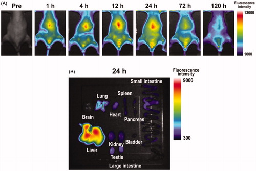
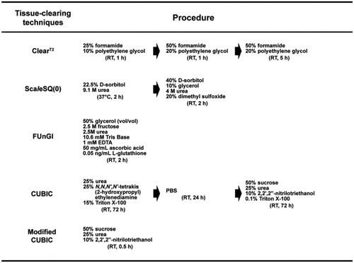
Figure 1. Composition of clearing reagents and protocols for five tissue-clearing techniques (ClearT2, ScaleSQ(0), FUnGI, CUBIC, and modified CUBIC).
The online version of this article has been corrected.