
Abstract
Functional near-infrared spectroscopy (fNIRS) is a noninvasive brain function detection technique based on the principle of neuro-vascular coupling. Based on a bibliometric approach, the present study visualizes and analyses the number of publications, countries, institutions, authors, co-citations and keywords of fNIRS with the help of Web of Science core collection database platform, CiteSpace and VOS-viewer software, and provides a narrative review of the literature in the past decade to comprehensively analyze the application and future development trend of fNIRS in clinical practice. The findings reveal that fNIRS is a clinically valuable tool with numerous advantages. It is therefore widely utilized to capture brain cortical activity data in both resting and task-related states. It enables the analysis of brain functional states from multiple dimensions and can be combined with other brain imaging techniques, improving the identification and detection of various neurological disorders, psychiatric conditions, pediatric medicine, and sports medicine. In the future, fNIRS technology is expected to achieve higher spatiotemporal resolution, increased data collection capacity, reduced interference and errors, and expanded application scope, thereby supporting clinical and research endeavors.
Introduction
Research on brain science and brain-like technology has become a hot spot in the current international science and technology frontier and is regarded in many countries as a new economic growth point and an engine of change leading to the creation of new technologies. Brain science research has grown worldwide, such as the Brain Research Through Advancing Innovative Neurotechnologies (BRAIN) Initiative in the United States and the European Brain Research Area (EBRA) project.[Citation1] The implementation of China Science and Technology Innovation 2030—“Brain Science and Brain-Like Research” major project—has also made great progress.[Citation2] Brain science is of great interest because the human brain is a complex system, with different brain regions showing corresponding activation states for different tasks, and also relies on various functional regions to interact and coordinate with each other. Therefore, studying the human brain system from a network perspective may help understand the brain information processing and expression mechanisms.[Citation3] Understanding the mechanisms of the effects of different brain functional areas is important for the pathological study of neuropsychiatric diseases.[Citation4] Cortical activation and neural remodeling are the paths to recovery for most brain diseases, and accurate assessment is a prerequisite for the continuous optimization of treatment and rehabilitation programs.[Citation5] However, the study of human brain functions such as cognitive, emotional and behavioral changes cannot be completely replaced by animal experiments. From a medical ethics perspective, studying the human brain with invasive techniques is difficult.[Citation6] Therefore, the real-time detection function of functional brain imaging technology for clinical diseases has become an important way to explore the neural mechanism of the human brain and is playing an increasingly important role in brain science research.[Citation7]
Functional brain imaging techniques are one of the most important tools to explore the mechanistic features and functional connections between brain regions. When we think, feel and act, neurons in the brain undergo chemical reactions and energy transformations that can be reflected by changes in blood flow.[Citation8] Thus, changes in blood oxygen levels can be used to assess neuronal activity in different regions of the brain. Most functional brain imaging techniques work based on this principle. Common noninvasive brain functional imaging techniques include functional magnetic resonance imaging (fMRI) and functional near-infrared spectroscopy (fNIRS), of which fNIRS is a new functional brain imaging technique that is safe and radiation-free.[Citation9] It measures the amount of blood flow changes in the cerebral cortex by near-infrared light, thus reflecting the activity of neurons in the brain, which is noninvasive, sensitive, with high resolution, well tolerated by the subject, and easy to carry out.[Citation10] It has obvious economic and cost advantages over fMRI and is both easy to fix and motion-tolerant. It is particularly suitable for functional testing of elderly patients, children and patients with poor mental status.[Citation11] Furthermore, its efficacy in capturing functional brain activity during task performance has contributed to a growing adoption in clinical settings. fNIRS is expected to grow in its application in the auxiliary diagnosis and treatment of neuropsychiatric diseases, pediatric diseases, sports medicine and other fields through the extraction and analysis of blood oxygen signals in the brain.
There has been a surge in fNIRS-related clinical studies within the last 10 years, but no up-to-date quantitative analyses have been reported. Bibliometrics is an emerging analytical approach that uses statistics and visual mapping to rapidly explore the structure and trends of a topic or field.[Citation12] As opposed to descriptive literature reviews (traditional reviews), bibliometrics can quickly assess a specific discipline or field,[Citation13] identify key information,[Citation14] and crystallize scientific questions to guide future research. For example, a bibliometric analysis of studies on the topic of "Intranasal Delivery Route" concluded that the United States dominates this field and that research related to the use of nanostructures as drug delivery vehicles has received much attention in recent years.[Citation15] A bibliometric analysis of studies on the topic of "drug repurposing" concluded that >60% of the approximately 35,000 drugs or drug candidates had been tried in more than one disease, with 189 drugs having been tried in each of >300 diseases. The study will encourage researchers to focus on finding new uses for existing drugs.[Citation16] A bibliometric analysis of advanced healthcare materials (AHM) summarizes current trends in AHM-based research, several considerations for clinical translation of biomaterials, and the future of the field.[Citation17]
CiteSpace and VOSviewer are the most commonly used bibliometric software,[Citation18,Citation19] in addition to bibExcel,[Citation20] Science of Science (SCI2)[Citation21] and HistCite.[Citation22] Citespace, as an effective bibliometric tool, is a data analysis software developed by Prof. Chaomei Chen’s team based on java environment running.[Citation23,Citation24] VOSviewer was created by the Center for Scientific and Technological Research at Leiden University to produce visual knowledge graphs for free.[Citation25,Citation26] The synergy of these two approaches enables the creation of a comprehensive map of scientific knowledge. By interpreting this map, one can disseminate existing information, retrace the entire evolution of the discipline, pinpoint the drivers of change, and venture into new realms of research.[Citation27,Citation28]
The present study is based on the fNIRS-related literature obtained from the Web of Science (WoS) core collection database. Visual analysis of the number of publications, countries, institutions, authors, co-citations, and keywords was performed using professional bibliometric tools CiteSpace and VOS-viewer. On this basis, traditional literature review was conducted to comprehensively analyze the principle of action and application of fNIRS, cerebral cortex detection, design of related trial protocols, and clinical applications in multiple fields, to objectively reflect the research status and future development trend of fNIRS, and to help researchers better utilize fNIRS for clinical and scientific research services.
Data and methods
Data source
Relevant articles were searched in the WoS core collection with TS = (“functional near-infrared spectroscopy” OR “functional near-infrared brain imaging” OR “fNIRS”). The language was English, the document type was Article and Review and the period was 2013-2023. The search date was January 5, 2023, and 3153 articles were obtained after screening and cleaning.
Research methodology
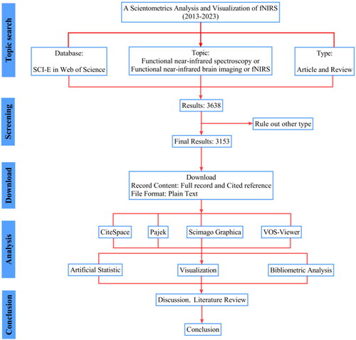
CiteSpace 6.1.R6, VOS-viewer version 1.6.18, Pajek and Scimago Graphica software were used to visualize and analyze the screened data. Some data were manually extracted to produce graphs. A narrative review of the key literature was also conducted. The flow chart of the bibliometric analysis is shown in .
Figure 1. Flow chart of the bibliometric analysis.
Results
Article research hotspots and trends
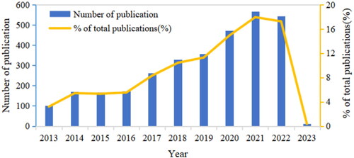
The number of articles published, both annual and cumulative, is a common indicator of research trends in a field.[Citation12] Based on the annual volume of published articles on fNIRS and the percentage of the annual volume (), we refer that the overall development of fNIRS has shown a growing trend in the past decade. The specific analysis has roughly undergone four phases, namely the initial growth phase (2013-2014), the stable development phase (2014-2016), the steady growth phase (2016-2019) and the rapid growth phase (2019-present). The number of articles published in this field reached the peak within a decade in 2021 (566 articles), with a gradually increasing proportion of articles in the experimental category.
Figure 2. Annual publications between 2013 and 2023.
Analysis by country
fNIRS is a tool to monitor functional brain activity in a wide range of applications and populations, with a global research trend involving several countries and relatively close cooperation between countries. The visualization of the number of national publications and the intensity of cooperation between countries in this field was analyzed by VOS-viewer combined with Scimago Graphica software (). The node size indicated the number of national publications, which was positively correlated; the redder the color of the node, the more the total number of collaborative research publications between the country and other countries. The thickness of the connecting line indicated the intensity of cooperation between two countries; the thicker the connecting line, the more the number of cooperation.
Figure 3. Cooperation map of countries/regions.
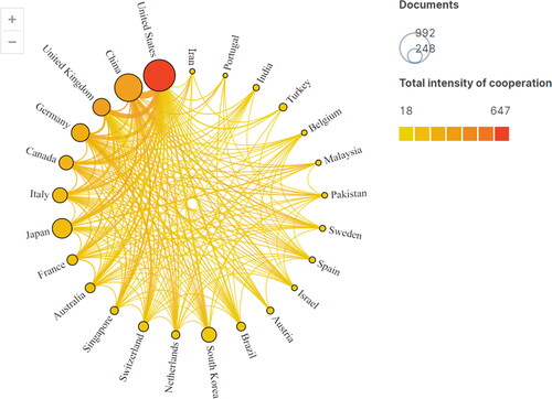
Countries with ≥100 publications included the United States (848), China (720), Japan (358), Germany (305), the United Kingdom (288), South Korea (211), Italy (203), Canada (184), the France (105). Meanwhile, the intermediary centrality of the number of articles issued by these countries was also high, and the top countries were the United States (0.6), the United Kingdom (0.22), Germany (0.15), China (0.13), South Korea (0.13), Canada (0.13) and the France (0.13) ().
Table 1. Top 10 countries with the highest number of publications and their centrality.
Analysis by institution
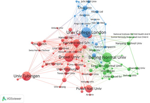
Institutions are the backbone of scientific research. The size of an institution and the degree of cooperation among institutions are crucial to the depth of research. Institutions bring together resources such as research scholars, experimental equipment, research funding and technical support, which are closely related to the quantity and quality of published literature. VOS-viewer was used to map research institution cooperation (). Institutions with the largest number included Univ College London (100 publications), Beijing Normal Univ (93 publications), Drexel Univ (89 publications), Univ Tubingen (85 publications), Pusan Natl Univ (73 publications), Harvard Med Sch (68 articles), Univ Pittsburgh (54 articles), East China Normal Univ (48 articles), Univ Padua (47 articles) and Chuo Univ (46 articles). Institutions with high intermediary included Univ College London (centrality 0.30), Harvard Med Sch (centrality 0.20), Drexel Univ (centrality 0.11), Univ Tubingen (centrality 0.11), Chuo Univ (centrality 0.11) and Beijing Normal Univ (centrality 0.10). They also belonged to the high publication volume institutions, which indicates that these institutions play a key role in the fNIRS research field and have relatively good institutional cooperation.
Figure 4. Cooperation map of institutions.
Analysis by authors
Authorship analysis reflects the key players in a research field and includes two parts: authorship volume analysis and authorship citation analysis. Author publications reflect the contribution of authors to the quantity of research in the field and author citations reflect the contribution of authors to the quality of research in the field. Authorship volume and citation analyses are useful tools to measure the key players in the field.[Citation12] CiteSpace was used to analyze the cooperative relationship among authors. shows the key authors in the fNIRS research field, and shows the top 10 authors in terms of the number of publications and citation frequency. In terms of the number of publications, the main researchers in fNIRS included Ehlis AC, Hong KS, Fallgatter AJ, Balconi M and Ayaz H. Considering the centrality of authors, the high-impact authors were Dan I and Brigadoi S. In terms of citation frequency, the high-impact authors in fNIRS research included Scholkmann F, Cui X, Huppert TJ, Ferrari M and Hoshi Y. However, the centrality of highly cited authors was low.
Figure 5. Cooperation map of authors.
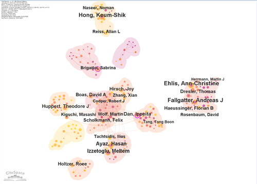
Table 2. Top 10 authors with the highest number of publications and citations.
Co-citation analysis
Co-citation is defined as two publications that are cited together in one article. The higher the number of co-cited literature, the greater the influence of the article in the field.[Citation12] CiteSpace was used to construct the knowledge graph of co-citation relationships and their clustering, and the results are shown in . The top 10 highly co-cited literature is shown in . The main literature included the papers of Scholkmann F in 2014, Ferrari M in 2012, Pinti P in 2020, Brigadoi S in 2014 and Santosa H in 2018. Notably, 5 of the top 10 highly co-cited articles were from the Neuroimage journal, suggesting that this journal had a great influence in the fNIRS field.
Figure 6. Co-citation knowledge map.
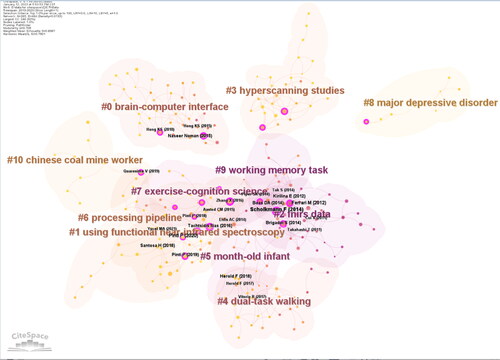
Table 3. Highly co-cited literature on fNIRS research.
Analysis of keywords
In bibliometric analysis, high-frequency keywords often reflect current situations and hot spots in the research field. Keyword emergence analysis was performed using CiteSpace, as shown in . The top 15 high-frequency keywords and their linkage intensity were respectively extracted from the keyword co-occurrence knowledge graph (). Keyword clustering analysis was performed on fNIRS research data using VOS-Viewer combined with Pajek software, and six large clusters were formed, as shown in .
Figure 7. Top 20 keywords with the strongest citation bursts.
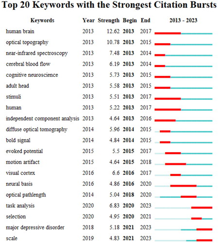
Figure 8. Knowledge map of keyword clustering analysis.
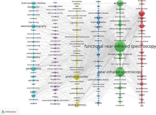
Table 4. Top 10 high-frequency keywords.
Discussion
fNIRS is a noninvasive optical functional brain imaging technology with good penetration power into tissues, which can convert the detected light signal into blood oxygen signal and further reflect the brain functional activity and brain metabolism through the change in hemoglobin concentration in the cerebral cortex.[Citation29] Considering that oxygenated and deoxygenated hemoglobin in human tissues differentially absorb light at different wavelengths, fNIRS measures the absorbed and scattered light by active areas of the cerebral cortex through a “banana-shaped” light path. After the detector receives the light signal, the relative concentration changes of oxyhemoglobin (HbO2) and deoxyhemoglobin (HbR) in the local area are monitored in real-time according to a modified Beer-Lambert law using the corresponding algorithm,[Citation30] which in turn provides a better understanding of the interrelationship between the brain regions.[Citation8]
Analysis of the current research status of fNIRS
The present study visualized the number of articles, countries, institutions, authors, co-citations and keywords of fNIRS. It was found that the number of articles has been increasing in the past 10 years, and the proportion of experimental articles has gradually increased, which shows that research in this field has been transferred from the theoretical analysis stage to the clinical research stage, and has gradually entered the field of brain science mechanism exploration. The visual analysis showed that countries with more fNIRS researches generally have strong economic power, all of which were ranked among the top 10 in 2022 in the world in terms of the Gross Domestic Product (GDP). Simultaneously, the highest-ranked institutions and authors also hail from these nations, underscoring a robust correlation between economic prowess and scientific research proficiency. It is noteworthy that based on the WoS database, China was ranked second in terms of the number of publications and fourth in terms of intermediary centrality, and thus China can continue to strengthen its cooperation with other countries and their institutions to deepen the scientific and clinical research on fNIRS. The analysis of the important literature showed that in the early stage of fNIRS research in the past decade, the co-citation network was relatively dense, and the literature in this period mainly studied topics such as working memory task, exercise-cognition science, month-old infant and fNIRS data. The co-citation network is relatively sparse, and research topics tend to be more diversified, including topics such as major depressive disorder, Chinese coal mine workers and processing pipeline. A study by Scholkmann F, with the highest number of co-citations, is a basic introduction to fNIRS in which the authors review the relevant instruments, detection methods and data analysis of fNIRS, and the authors emphasize that fNIRS can be used as a clinical tool for disease diagnosis of patients in the future.[Citation31] The visualization and analysis of the keyword aspect of fNIRS indicates that fNIRS can complete brain function testing through multiple experimental paradigms, and the interpretation of the indexes has also been expanded from pure blood oxygen signal acquisition to activation analysis, functional network analysis, etc., which can also play an auxiliary role in the diagnosis and treatment of neuropsychiatric and other systemic disorders in the clinic.
Analysis of the content of fNIRS
The current study presents a literature review based on keyword clustering themes, which are divided into four relevant aspects of fNIRS research content, namely fNIRS principles of action and application, cerebral cortex detection, experimental protocol design and multi-disciplinary clinical applications.
Principle of action and application
fNIRS works based on the principle of the neuro-vascular coupling mechanism. It is well established that the brain does not store energy, and all the energy required for neuronal activity comes from the real-time supply of the blood supply system. When the brain is externally stimulated, brain neurons start to move, thus increasing the brain tissue metabolism, which consumes the oxygen carried by hemoglobin and leads to changes in the concentration of HbO2 and HbR in the region.[Citation9] To compensate for the oxygen consumed by neuronal activity, local blood vessels dilate and cerebral blood flow and volume increase accordingly to meet the oxygen requirements of neuronal activity. In this process, cerebral blood flow increases far beyond the actual oxygen consumption required by the neurons. These series of changes eventually lead to excess oxygenated hemoglobin in the blood of local brain regions, which is manifested as an increase in HbO2 and a decrease in HbR.[Citation5] fNIRS monitors the functional status of the corresponding brain regions precisely by the altered blood flow-oxygen levels in different brain regions.
The fNIRS technique is based on traditional near-infrared spectroscopy, which was originally used for food quality monitoring and drug analysis. fNIRS became an effective brain imaging tool in 1977, during which it was used to assess changes in blood oxygenation in the human brain due to hyperventilation, demonstrating the feasibility of fNIRS technology in monitoring brain function.[Citation32] Later, Chance et al. used fNIRS to monitor prefrontal blood oxygen changes induced by cognitive activity during the execution of the classical paradigm, completing one of the first fNIRS studies.[Citation33] As the research progressed, the fNIRS device was upgraded from a single-channel mode to a multi-channel mode, enabling simultaneous monitoring of different brain regions and even the whole head, to fully demonstrate the spatial distribution of brain activation patterns.[Citation34] Since the twenty first century, fNIRS technology has become increasingly mature and has been used for scientific research in several disease systems and different interventions, especially in the field of neurobrain science.[Citation35] The fNIRS technique has been widely used in neuroscience, mainly for the observation of central effect mechanisms in psychoneurological disorders, such as insomnia, depression, anxiety, stroke, cognitive disorders and child development, involving tactile, language, emotional-cognitive, motor-sensory and writing functional areas.[Citation36] It is also used to study the effect of interventions on the function of central brain areas and can be combined with noninvasive neuromodulation techniques, such as transcranial magnetism, to localize and evaluate the efficacy in transcranial magnetic brain areas.[Citation37–39] In recent years, with the development of Chinese medicine, several studies on the changes in the brain area status and functional connectivity have been conducted in patients receiving acupuncture, Chinese medicine and moxibustion interventions based on fNIRS technology.[Citation40–42]
Cerebral cortex detection
fNIRS is a neuroimaging technique used to measure the distribution of blood oxygenation levels within the cerebral cortex, which reflects changes in cerebral blood flow in cortical regions. The cerebral cortex is considered to be the center of cognitive processes, action control, emotional processing and perceptual experience, and is the higher center of the body’s vital activities.[Citation43] The impairment of cortical function disrupts normal biological activities. Speech, movement and other body functions are processed by a collaborative cortical network with multiple brain regions as nodes.[Citation44] It has been found that damage to the cortical center does not result in permanent loss of the cortical center function, the function can be restored to a certain extent through subsequent rehabilitation and compensatory work by undamaged cortical brain areas.[Citation45] The fNIRS device can be used to detect the cortical brain area. With the fNIRS device, an area 3 cm below the cerebral cortex can be detected.[Citation46] The response patterns of the cerebral cortex in different brain regions have been extensively studied using this technique.
The fNIRS technique can be used to assess the risk and prognosis of different diseases and procedures by examining the cerebral cortex and can also be used to study neurodevelopmental and learning processes. In recent years, the fNIRS technology was used to explore the posterior occipital cortical activity in the brain of normal and Parkinson’s disease (PD) patients. The results showed that posterior occipital cortical activity was lower in PD patients than in the normal population under resting conditions, suggesting that the technique could be used as a tool for diagnosing PD.[Citation47] Another study explored the use of fNIRS in surgical oncology.[Citation48] Researchers used the fNIRS technique to assess cortical activity in patients before and after brain tumor surgery and found that cortical activity was suppressed before tumor removal and restored after tumor removal. This indicates that the fNIRS technique can be used to assess the risk of tumor surgery and predict recovery after surgery.
In addition, fNIRS can be used to study cortical neurodevelopment and brain area function. Lawrence et al.[Citation49] used fNIRS to study cortical activation patterns in children with normal hearing for different speech intelligibility—a pattern that could help find new solutions for individuals with delayed speech development or fine hearing through hearing aids (e.g., cochlear implants)—and demonstrated that the fNIRS technique may become an auxiliary tool for clinical studies of listening and comprehension abilities. Several studies have also shown that changes in HbO2 concentrations in subjects’ prefrontal cortical and parietal regions were more pronounced during the working memory load.[Citation50] In addition, cortical activation under different conditions is considered to be a useful predictor of cognitive function. Blum et al.[Citation51] used the Trail Making Test (TMT) and fNIRS to examine the effect of age on behavioral performance and changes in cortical blood oxygen levels in healthy older adults performing a task and showed a positive correlation between age and changes in cortical blood oxygen levels in the TMT, suggesting that cognitive function declines and cortical activity increases compensatorily with an increase in age in older adults. These findings highlight the potential of fNIRS as a tool for investigating the functional state of the cerebral cortex across various modalities, making it a valuable asset in the study of the brain and potentially in the treatment of brain disorders. The fNIRS technique can be used to study the function and structure of the cerebral cortex, providing detailed information that helps to understand neural activity patterns in specific brain regions. The activation status of the damaged brain regions in different states, the compensatory patterns of healthy brain regions and the differences in functional connectivity between brain regions can be observed to assist in the diagnosis and prognosis of diseases.
Experimental protocol design
The main experimental paradigms for fNIRS are block design, event-related and hybrid design in the task state, and resting state design. The resting-state model is often used to evaluate the state of functional brain connectivity, but also to perform functional analysis of specific brain regions by setting the Region of Interest (ROI).[Citation52] The resting-state design proves especially useful for assessing poorly cooperating patients, including children and the elderly. It is commonly employed in evaluating conditions such as ischemic-hypoxic encephalopathy, severe traumatic brain injury, and coma.[Citation53] Task state mode is one of its unique advantages over fMRI, especially the open application of fNIRS portable devices, which can observe the functional state of the brain area of the subjects in different scenarios, such as brisk walking, bicycling, playing chess, piloting an airplane, teamwork, and couple interactions, to obtain more objective and realistic monitoring data.[Citation54–57]
Concerning the indexes for the calculation of fNIRS, the main clinical indicators are the degree of activation and functional connectivity of brain regions based on changes in the concentration of oxygenated hemoglobin and deoxyhemoglobin. Functional connectivity can be analyzed using undirected functional connectivity analysis methods such as Pearson’s correlation coefficient, coherence, and phase synchronization, directed functional connectivity methods such as Granger’s causation, and task-state functional connectivity analysis methods such as wavelet coherence. In addition, the data analysis of fNIRS involves various methods such as graph theory analysis, efficiency analysis, correlation analysis, and complex network analysis.[Citation58–60]
The application parameters of fNIRS are not completely uniform. The wavelength range of near-infrared light is 600-900 nm.[Citation61] The clinical wavelengths are mostly two-segment wavelengths, such as 690, 830, 760 and 850 nm. Three wavelength devices, 730, 808 and 850 nm, have also been developed to better eliminate interference and provide accurate and corrected changes in blood oxygen concentration. Different values of wavelength penetration exist in different ranges, and it is generally believed that the shorter the wavelength, the stronger the penetration, and the greater the interval between the two wavelengths, the less interference and the more accurate the quality of the detection signal. The common clinical sampling rate has different parameter values such as 10, 11, 20, 50, and 100 Hz. The higher the sampling rate, the more comprehensive the data collected. The fNIRS device is compatible and can be used in combination with various brain stimulation and testing devices, such as fNIRS-transcranial magnetic stimulation (TMS), fNIRS-electroencephalography (EEG), fNIRS-motor evoked potential (MEP), fNIRS-transcranial direct-current stimulation (tDCS), etc., to comprehensively assist in the diagnosis, treatment and prognosis of brain diseases.[Citation62]
The differences in clinical equipment lead to inconsistencies in test parameters, which to some extent affects the scientific validity and accuracy of test results. Moreover, the environment in which the patient is tested and the patient’s state also have different degrees of influence on the test results; therefore, it is recommended to analyze the test results objectively and scientifically by continuously recording the test process. The relevant information should be recorded in detail during the experiment, including the subject, instrumentation, stimulation parameters, testing steps, test environment, testing personnel, etc., to ensure the credibility and accuracy of the test results.
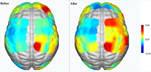
In accordance with the experimental paradigm, we present a case of fNIRS-related data collected by our team. The subject was a 63-year-old male patient in the postictal phase of cerebral infarction in the right basal ganglia region. In this experiment, NirScan-6000B (Danyang Huichuang Medical Equipment Co., Ltd., China) was used to continuously measure and record the functional connectivity status of brain regions in the resting state and the changes in the concentration of cerebral oxyhemoglobin during the task. The device contains 24 light sources and 24 detectors, forming a total of 63 acquisition channels, and the distribution of the channels is shown in , where “S” represents the light sources, “D”represents the detectors, and the connecting line between S and D represents the channels. The sampling rate was 11 HZ and the wavelengths were 730, 808 and 850 nm. During the resting state, the subject remained sedentary for 8 min. For the task paradigm, the subject was instructed to move the upper limb on the affected side. The subjects rested for 10 s in a quiet state, followed by a 15-s movement of the upper limb on the affected side. Afterward, they rested for 20 s. This task paradigm was repeated for a total of 5 groups. The task paradigm is illustrated in . The NirSpark software (Danyang Huichuang Medical Equipment Co., Ltd, China) package was used to preprocess fNIRS signals, it has been used in previous experiments.[Citation63] shows a graph of the change in Hbo concentration after averaging of the subjects’ task-state blocks, which shows that after the start of the task, the patient’s Hbo began to rise, peaked near 15 s, and gradually returned to the baseline level at the end of the 15-s task. shows the resting-state brain functional connectivity atlas and matrix of the subjects, where asymmetry in the functional connectivity of the bilateral cerebral hemispheres can be observed, with a significantly reduced degree of connectivity in the affected brain region. shows the activation changes in the task state brain regions of the subjects during the baseline period and after 10 sessions of TMS treatment, and it can be observed that after 10 sessions of TMS intervention, the activation area and the degree of the brain regions on the affected side during moving the affected upper limb increased significantly compared with the baseline period.
Figure 9. Distribution of light sources, detectors and channels.
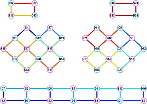
Figure 10. fNIRS task-state design paradigm.
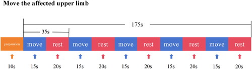
Figure 11. Plot of Hbo concentration change after task-state block averaging.
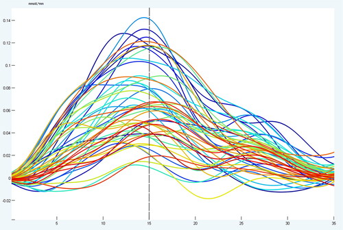
Figure 12. Brain functional connectivity mapping and matrices in the resting state.
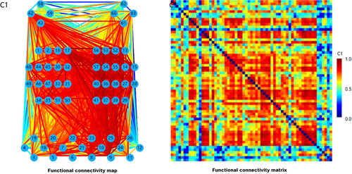
Figure 13. Changes in activation of task-state brain regions before and after treatment.
Multi-disciplinary clinical applications
Neuroscience field
In the field of neuroscience, fNIRS is often used to explore the relationship between neuronal activity and cognition, behavioral and emotional tasks. In recent years, fNIRS has been widely used to explore brain responses to various stimuli (e.g., visual, auditory, tactile, olfactory, etc.). A previous study investigated individual differences in head hemodynamic responses to long-term stimulation by colored light and cognitive tasks based on systemic physiology augmented (SPA)-fNIRS.[Citation64] fNIRS has also been used to study brain activity in learning tasks, through machine learning to predict student academic performance based on fNIRS data.[Citation65]
Furthermore, fNIRS is often used in the study of clinical disorders such as dementia, stroke, PD, and epilepsy to help physicians understand the physiological mechanisms of diseases and guide the treatment and evaluation of brain disorders. Yeung and colleagues[Citation66] systematically reviewed the use of fNIRS in patients with mild cognitive impairment (MCI) and AD. Both amnestic MCI and AD were found to be associated with decreased frontal lobe function in the resting state and decreased concentration of oxygenated hemoglobin in the task state. This phenomenon was more pronounced in AD patients. It was concluded that fNIRS can explain brain function changes in MCI and AD with higher accuracy and ease of use compared with other neuroimaging techniques. Li[Citation67] examined the functional status of brain regions in patients with a cerebral ischemic stroke under the resting mode and of intermittent sequential pneumatic compression (ISPC) intervention by fNIRS and concluded that ISPC has a positive effect on the motor-related network and can improve the recovery effect of patients’ motor function, which demonstrated that fNIRS may assist in the diagnosis and prognosis of the disease and provide objective indicators for the evaluation of the effects of clinical therapies. Another study explored the effects of aerobic exercise combined with tDCS on cortical activity, gait and cognitive performance in PD patients based on fNIRS technology. The results showed that the addition of anodic tDCS at the prefrontal cortex during aerobic exercise had a direct positive effect on gait variability, processing speed and walk execution control in PD patients,[Citation37] which shows that fNIRS can be used as an assessment tool to optimize clinical treatment protocols. Moreover, fNIRS can be used for clinical screening and diagnosis of diseases. Epilepsy is a seizure disorder and the search for an effective detection tool is a constant goal of medical practitioners. fNIRS can assist in the diagnosis of the disease by comparing and analyzing the difference in the activation status of diseased and healthy brain regions. A study of 32 participants showed that fNIRS had better diagnostic ability in monitoring seizures,[Citation68] suggesting that the local cerebral blood volume on the side of the lesion is strongly increased during a seizure, and the localization of the seizure lesion can be observed by the fNIRS system; however, single photon emission computed tomography (SPECT) and fNIRS performed poorly in the localization of the seizure lesion. This study demonstrates that fNIRS can detect the onset of episodic or chronic neurological disorders in advance, promoting early diagnosis and early treatment of the disease.
Spiritual science field
Neurofunctional imaging has revolutionized the field of psychiatric research by offering precise and unbiased biomarkers for diagnosing and treating psychiatric disorders. fNIRS is widely applied in psychiatric disorders such as Major Depressive Disorder, Schizophrenia, Bipolar Affective Disorder and other psychiatric disorders, and the following is an example of its application in the field of psychiatric disorders with depression.
fNIRS is extensively used in the detection of brain function in patients with depression to explore the functional connectivity and activation status of the cortex, which helps to identify and diagnose as well as treat depression. Wong[Citation58] conducted a resting-state fNIRS study to evaluate the efficacy of acupuncture combination therapy in patients with depression. Participants were asked to perform resting-state recordings in the supine position for 3-5 min before the first and last acupuncture sessions to monitor functional connectivity (FC) in the DLPFC. It was observed that compared to patients treated with antidepressants alone, those treated with acupuncture combined antidepressants for 3 wk had stronger brain functional connectivity in the DLPFC and exhibited significant improvement in clinical symptoms. Wang et al.[Citation69] constructed an AlexNet model based on the fNIRS technique, with a recognition accuracy of 0.90 and precision of 0.91, both of which are higher compared to those of the ResNet18 model as well as other machine learning algorithms. A deep neural network-based depression recognition method has been established to improve the clinical diagnosis and treatment of depression. Wei[Citation70] used the frontal lobe integral values of oxyhemoglobin and temporal lobe centroid values to distinguish patients with mental disorders from healthy individuals, and reported that these indicators were highly sensitive.
The representative task state paradigm of the NIRS is the Verbal Fluency Test (VFT).[Citation71] The VFT task reflects complex cognitive activity and serves as a reliable measure of frontal lobe activation.[Citation72] Studies have found significantly lower activation of the prefrontal and superior temporal gyrus in depressed patients during the VFT task.[Citation73] Combined with VFT, fNIRS has become a convenient and cost-effective approach for investigating psychiatric disorders. The fNIRS-VFT paradigm can reveal the diagnosis of psychiatric disorders and is expected to be a suitable brain imaging biomarker of various psychiatric disorders.[Citation74] The studies mentioned above demonstrate that fNIRS is a robust biomarker for elucidating the pathogenesis of depression, diagnosing psychiatric disorders such as depression, and promoting the development of individualized treatment.
Pediatric medicine field
The fNIRS has a wide range of applications in pediatrics, and is often used to examine brain function in children, emotional and cognitive development, and psychiatric disorders. It can also be used to assess the neurodevelopmental status of children. A previous study showed that children exhibit relatively symmetrical network efficiency in the bilateral brain, whereas adults show significant leftward asymmetry and functional separation in the left hemisphere, which may underlie language development from childhood to adulthood.[Citation75] The fNIRS can be used to explore the development of emotional and cognitive functions in children, such as, attention, memory, and decision making. The study by Liu which assessed cognitive function in children with new-onset epilepsy based on fNIRS suggested that fNIRS can reveal the effects of epilepsy on cognitive function in children.[Citation76] Other studies have demonstrated that the fNIRS can be used to study psychiatric disorders in children, such as autism and attention deficit hyperactivity disorder, and explore their neurophysiological basis. Another study[Citation77] in which NIRS was used to explore brain activity in children with autism spectrum disorders showed that brain functional connectivity measured using NIRS could predict social communication skills in children with autism spectrum disorders.
In contrast to fMRI, fNIRS presents a more favorable option for assessing children’s brain function due to its noise-free environment. Researchers provide instructions and reassurance, ensuring that children perform the task accurately.[Citation78] For these reasons, fNIRS is suitable for detecting brain function in children. Due to the limited task cooperation of pediatric patients, fNIRS is predominantly employed in resting-state mode to minimize bias. This enables the assessment of brain functional connectivity, global efficiency, local efficiency, as well as graph theory-based analyses such as modularity, normalized clustering coefficients, and other brain network metrics. Overall, the application of fNIRS in the field of child neurological research is rapidly evolving. This noninvasive technique allows researchers to study brain function in children in more detail relative to other brain functional imaging techniques. As the technology continues to evolve, fNIRS is expected to play an even more important role in pediatric neuroscience and pediatric disease research.
Sports medicine field
Muscle oxygen supply is a crucial process in various sports and physical activities. fNIRS can be used to monitor muscle oxygen supply and help athletes better control exercise intensity, prevent sports injuries, and diagnose complex conditions on time. For example, given that exercise-induced Flow Limitations in the Iliac Arteries (FLIA) is a fairly rare condition,[Citation79] timely and accurate diagnosis should be performed to ensure responsive treatment. fNIRS is more sensitive and specific in the diagnosis of FLIA athletes, and inclusion of fNIRS in the clinical diagnostic pathway can improve the accuracy of diagnosis according to Bender’s study.[Citation80] In addition, NIRS can be potentially applied to assess the physiological state of the brain in athletes at different training stages. Chen et al[Citation81] used the fNIRS resting-state model to study HbO2 changes in the Prefrontal Cortex (PFC), Motor Cortex (MC) and Occipital Cortex (OC) of Taichi athletes over five frequency intervals. Wavelet phase coherence analysis was employed to examine the transient phase interactions between two signals, enabling precise measurement of brain functional connectivity. The study revealed that Taichi players exhibited significantly elevated functional brain connectivity in the PFC, MC, and OC compared to Taichi novices. This suggests that fNIRS can be used to assess physiological changes in the brain during exercise. In the meantime, for patients who need long-term rehabilitation, exercise techniques such as Taichi and Baduanjin are green and effective means of intervention.
Conclusion and future perspectives
As global brain science research programs make significant strides, there has been extensive progress in understanding the neurological foundations of brain science. This includes advancements in diagnosing and treating brain-related disorders. Furthermore, research in brain-inspired artificial intelligence has also seen noteworthy development. fNIRS, as a noninvasive and efficient brain functional neuroimaging technology, possesses unique advantages. It can be applied across various disease types and in diverse scenarios. Its portability, extended detection time, robust resistance to interference, high flexibility, and broad applicability to different demographic groups have all contributed to the growing clinical utilization of fNIRS. This technology is increasingly employed in the diagnosis of functional brain disorders and in assessing treatment efficacy. In addition to the clinical application areas covered in the above text, fNIRS has also been widely used in pain,[Citation82] insomnia,[Citation83,Citation84] sensory disorders,[Citation85–87] and Chinese medicine interventions.[Citation88,Citation89] It is also used to guide and optimize the design of clinical diagnosis and treatment and rehabilitation protocols by evaluating the functional state of the brain under pathological states or during external interventions.
Given its technical constraints, fNIRS has some limitations. ① Currently, fNIRS can only detect superficial cortical functions of the brain, but can be used in conjunction with neurophysiological techniques such as MEP and electromyographic evoked potentials and structural imaging techniques such as DTI, which in turn can be used for the detection of cortical-subcortical whole neural network pathways.[Citation90] ② The interference caused by hair may hinder the effective penetration of near-infrared light, potentially impacting data acquisition quality for certain individuals. Although tools like hair dialers currently exist to enhance signal quality, we foresee future technological advancements that will provide even more efficient methods for improving data acquisition, thus minimizing interference and errors. This progress is expected to broaden the scope of applications for this technology.[Citation31]
The fNIRS is an optical device that operates independently from electromagnetic-based devices and demonstrates high resistance to motion artifacts. Leveraging these characteristics, fNIRS holds the potential to open up new avenues for future research, building upon previous studies. ① Multi-technique fusion of fNIRS with fMRI, ERPs, and EEG for multimodal functional brain imaging to reveal the mechanisms of neural loop and network reconstruction during rehabilitation. ② Combine fNIRS with noninvasive neuromodulation devices such as TMS and tDCS for immediate evaluation after stimulation intervention, provide timely feedback on the treatment effect, and accomplish precise judgment on the treatment and rehabilitation efficacy, which will help to adjust the clinical diagnosis and treatment plan. ③ Promote the application development of portable fNIRS equipment and hyperscan equipment to improve the possibility of scientific research in natural scenes. In conclusion, we demonstrate that fNIRS is a promising technique for functional brain imaging, diagnosis and efficacy assessment of functional brain disorders.
This paper provides a thorough examination, combining bibliometric insights with narrative reviews, to explore the essential features and associated experimental protocols of fNIRS technology. In addition, it systematically explains the importance and relevance of using fNIRS in clinical contexts, particularly within the realm of neurological disorders. psychiatric disorders, pediatric disorders, and sports medicine from the perspective of clinical application. This acts as an indispensable resource for clinical practitioners, offering guidance on research design and the uniform implementation of fNIRS. It empowers them to unlock the complete capabilities of fNIRS technology for improved diagnosis and treatment of clinical conditions.
| Abbreviations | ||
| fNIRS | = | functional Near-infrared Spectroscopy |
| BRAIN | = | Brain Research Through Advancing Innovative Neurotechnologies |
| EBRA | = | European Brain Research Area |
| fMRI | = | functional Magnetic Resonance Imaging |
| AHM | = | advanced healthcare materials |
| SCI2 | = | Science of Science |
| WoS | = | Web of Science |
| HbO2 | = | oxyhemoglobin |
| HbR | = | deoxyhemoglobin |
| GDP | = | Gross Domestic Product |
| PD | = | Parkinson’s disease |
| TMT | = | Trail Making Test |
| AD | = | Alzheimer’s disease |
| TMS | = | Transcranial Magnetic Stimulation |
| EEG | = | electroencephalography |
| MEP | = | motor evoked potential |
| tDCS | = | transcranial direct-current stimulation |
| SPA | = | systemic physiology augmented |
| MCI | = | mild cognitive impairment |
| ISPC | = | intermittent sequential pneumatic compression |
| SPECT | = | single photon emission computed tomography |
| FC | = | functional connectivity |
| VFT | = | Verbal Fluency Test |
| FLIA | = | Flow Limitations in the Iliac Arteries |
| PFC | = | Prefrontal Cortex |
| MC | = | Motor Cortex |
| OC | = | Occipital Cortex |
Authors’ contributions
All authors made a significant contribution to the work reported, whether that is in the conception, study design, execution, acquisition of data, creation of images and tables, analysis and interpretation, or in all these areas; took part in drafting, revising or critically reviewing the article; gave final approval of the version to be published; have agreed on the journal to which the article has been submitted; and agree to be accountable for all aspects of the work.
Disclosure statement
No potential conflict of interest was reported by the authors.
Additional information
Funding
References
- Zhang, X. B.; Yuan, T. W.; Zhang, L. W.; et al. Brain Science and Brain-like Intelligence Development Trends in 2022. Life Sci. 2023, 35, 9–17.
- Ministry of Science and Technology of the People’s Republic of China. Science and Technology Innovation 2030. Brain Science and Brain-like Research. Major Project Guidelines for 2021 [R/OL]. (2021-09-16) [2022-06-01]. https://service. most. gov. cn/kjjh_tztg_all/20210916/4583. html.
- Zhang, H. Q. Looking at the Future Path of Brain Science Development. Dongguan Daily, Jul 7, 2022.
- Chen, Z. H.; Wang, R.; Li, Y. X.; et al. Functional Magnetic Resonance Imaging Dynamic Brain Functional Connectivity Network Analysis Method and Its Application in Brain Diseases. Chin. Clin. Neurosci. 2020, 28, 571–578.
- Expert Consensus on the Clinical Application of near-Infrared Functional Brain Imaging. China Geriatric Health Med. 2021, 19, 3–9.
- Song, Q. Reverse Thinking for Brain Science. Physician’s J. 2023, 2, 1–2.
- Wang, X. L.; Huang, X. Q.; Gong, Q. Y. Advances in Functional Magnetic Resonance Imaging of Neuropsychiatric Disorders. Magn. Reson. Imaging 2012, 3, 61–68.
- Koike, S.; Satomura, Y.; Kawasaki, S.; Nishimura, Y.; Kinoshita, A.; Sakurada, H.; Yamagishi, M.; Ichikawa, E.; Matsuoka, J.; Okada, N.; et al. Application of Functional near Infrared Spectroscopy as Supplementary Examination for Diagnosis of Clinical Stages of Psychosis Spectrum. Psychiatry Clin. Neurosci. 2017, 71, 794–806. DOI: 10.1111/pcn.12551
- Wang, Z.; Zhang, J.; Xia, Y.; Chen, P.; Wang, B. A General and Scalable Vision Framework for Functional near-Infrared Spectroscopy Classification. IEEE Trans. Neural Syst. Rehabil. Eng. 2022, 30, 1982–1991. DOI: 10.1109/TNSRE.2022.3190431
- Naseer, N.; Hong, K. S. fNIRS-Based Brain-Computer Interfaces: A Review. Front. Hum. Neurosci. 2015, 9, 3. DOI: 10.3389/fnhum.2015.00003
- Chen, W.-L.; Wagner, J.; Heugel, N.; Sugar, J.; Lee, Y.-W.; Conant, L.; Malloy, M.; Heffernan, J.; Quirk, B.; Zinos, A.; et al. Functional near-Infrared Spectroscopy and Its Clinical Application in the Field of Neuroscience: Advances and Future Directions. Front. Neurosci. 2020, 14, 724. DOI: 10.3389/fnins.2020.00724
- Qiu, J. P. Bibliometrics. 2nd ed. Beijing: Science Press, 2019; pp 1–315.
- Bordons, M.; Zulueta, M. A. Evaluation of the Scientific Activity through Bibliometric Indices. Rev. Esp. Cardiol. 1999, 52, 790–800. DOI: 10.1016/s0300-8932(99)75008-6
- Jiang, M.; Qi, Y.; Liu, H.; Chen, Y. The Role of Nanomaterials and Nanotechnologies in Wastewater Treatment: A Bibliometric Analysis. Nanoscale Res. Lett. 2018, 13, 233. DOI: 10.1186/s11671-018-2649-4
- Wu, H.; Zhou, Y.; Wang, Y.; Tong, L.; Wang, F.; Song, S.; Xu, L.; Liu, B.; Yan, H.; Sun, Z.; et al. Current State and Future Directions of Intranasal Delivery Route for Central Nervous System Disorders: A Scientometric and Visualization Analysis. Front. Pharmacol. 2021, 12, 717192. DOI: 10.3389/fphar.2021.717192
- Baker, N. C.; Ekins, S.; Williams, A. J.; Tropsha, A. A Bibliometric Review of Drug Repurposing. Drug Discovery Today 2018, 23, 661–672. DOI: 10.1016/j.drudis.2018.01.018
- Zhu, S.; Liu, Y.; Gu, Z.; Zhao, Y. A Bibliometric Analysis of Advanced Healthcare Materials: Research Trends of Biomaterials in Healthcare Application. Adv. Healthcare Mater. 2021, 10, e2002222. DOI: 10.1002/adhm.202002222
- Wang, W. H.; Lu, C. Visualization Analysis of Big Data Research Based on Citespace. Soft Comput. 2020, 24, 8173–8186. DOI: 10.1007/s00500-019-04384-7
- Xie, L.; Chen, Z.; Wang, H.; Zheng, C.; Jiang, J. Bibliometric and Visualized Analysis of Scientific Publications on Atlantoaxial Spine Surgery Based on Web of Science and VOSviewer. World Neurosurg. 2020, 137, 435–442.e4. DOI: 10.1016/j.wneu.2020.01.171
- Dai, S. L.; Duan, X.; Zhang, W. Knowledge Map of Environmental Crisis Management Based on Keywords Network and co-Word Analysis, 2005-2018. J. Cleaner Prod. 2020, 262, 121168. DOI: 10.1016/j.jclepro.2020.121168
- Wang, M. H.; Ho, Y. S.; Fu, H. Z. Global Performance and Development on Sustainable City Based on Natural Science and Social Science Research: A Bibliometric Analysis. Sci. Total Environ. 2019, 666, 1245–1254. DOI: 10.1016/j.scitotenv.2019.02.139
- Shah, S. H. H.; Lei, S.; Ali, M.; Doronin, D.; Hussain, S. T. Prosumption: Bibliometric Analysis Using HistCite and VOSviewer. Kybernetes 2019, 49, 1020–1045. DOI: 10.1108/K-12-2018-0696
- Chen, Y.; Chen, C. M.; Liu, Z. Y.; et al. Methodological Functions of the CiteSpace Knowledge Graph. Stud. Sci. Sci. 2015, 33, 242–253.
- Chen, C. M. CiteSpace II: Detecting and Visualizing Emerging Trends and Transient Patterns in Scientific Literature. J. Am. Soc. Inf. Sci. 2006, 57, 359–377. DOI: 10.1002/asi.20317
- van Eck, N. J.; Waltman, L. Software Survey: VOSviewer, a Computer Program for Bibliometric Mapping. Scientometrics 2010, 84, 523–538. DOI: 10.1007/s11192-009-0146-3
- Xu, D.; Wang, Y.-L.; Wang, K.-T.; Wang, Y.; Dong, X.-R.; Tang, J.; Cui, Y.-L. A Scientometrics Analysis and Visualization of Depressive Disorder. Curr. Neuropharmacol. 2021, 19, 766–786. DOI: 10.2174/1570159X18666200905151333
- Chen, C.; Dubin, R.; Kim, M. C. Emerging Trends and New Developments in Regenerative Medicine: A Scientometric Update (2000 - 2014). Expert Opin. Biol. Ther. 2014, 14, 1295–1317. DOI: 10.1517/14712598.2014.920813
- Ma, W.; Xu, D.; Zhao, L.; Yuan, M.; Cui, Y.-L.; Li, Y. Therapeutic Role of Curcumin in Adult Neurogenesis for Management of Psychiatric and Neurological Disorders: A Scientometric Study to an in-Depth Review. Crit. Rev. Food Sci. Nutr. 2022, 28, 1–13. DOI: 10.1080/10408398.2022.2067827
- Fan, C. Y.; Xie, H. Y.; Wu, Y. Progress of Functional near-Infrared Spectroscopy in Patients with Mild Cognitive Impairment. Chin. J. Rehabil. Med. 2022, 37, 830–833.
- Murkin, J. M.; Arango, M. Near-Infrared Spectroscopy as an Index of Brain and Tissue Oxygenation. Br J Anaesth 2009, 103, i3–13. DOI: 10.1093/bja/aep299
- Scholkmann, F.; Kleiser, S.; Metz, A. J.; Zimmermann, R.; Mata Pavia, J.; Wolf, U.; Wolf, M. A Review on Continuous Wave Functional near-Infrared Spectroscopy and Imaging Instrumentation and Methodology. Neuroimage 2014, 85, 6–27. DOI: 10.1016/j.neuroimage.2013.05.004
- Jöbsis, F. F. Noninvasive, Infrared Monitoring of Cerebral and Myocardial Oxygen Sufficiency and Circulatory Parameters. Science 1977, 198, 1264–1267. DOI: 10.1126/science.929199
- Almajidy, R. K.; Mankodiya, K.; Abtahi, M.; Hofmann, U. G. A Newcomer’s Guide to Functional near Infrared Spectroscopy Experiments. IEEE Rev. Biomed. Eng. 2020, 13, 292–308. DOI: 10.1109/RBME.2019.2944351
- Yücel, M. A.; Lühmann, A. V.; Scholkmann, F.; Gervain, J.; Dan, I.; Ayaz, H.; Boas, D.; Cooper, R. J.; Culver, J.; Elwell, C. E.; et al. Best Practices for fNIRS Publications. Neurophotonics 2021, 8, 012101. DOI: 10.1117/1.NPh.8.1.012101
- Obrig, H. NIRS in Clinical Neurology - A 'Promising’ Tool? Neuroimage 2014, 85, 535–546. DOI: 10.1016/j.neuroimage.2013.03.045
- Li, R.; Mayseless, N.; Balters, S.; Reiss, A. L. Dynamic Inter-Brain Synchrony in Real-Life Inter-Personal Cooperation: A Functional near-Infrared Spectroscopy Hyperscanning Study. Neuroimage 2021, 238, 118263. DOI: 10.1016/j.neuroimage.2021.118263
- Conceição, N. R.; Gobbi, L. T. B.; Nóbrega-Sousa, P.; Orcioli-Silva, D.; Beretta, V. S.; Lirani-Silva, E.; Okano, A. H.; Vitório, R. Aerobic Exercise Combined with Transcranial Direct Current Stimulation over the Prefrontal Cortex in Parkinson Disease: Effects on Cortical Activity, Gait, and Cognition. Neurorehabil. Neural Repair 2021, 35, 717–728. DOI: 10.1177/15459683211019344
- Curtin, A.; Tong, S.; Sun, J.; Wang, J.; Onaral, B.; Ayaz, H. A Systematic Review of Integrated Functional near-Infrared Spectroscopy (fNIRS) and Transcranial Magnetic Stimulation (TMS) Studies. Front. Neurosci. 2019, 13, 84. DOI: 10.3389/fnins.2019.00084
- Huang, J.; Zhang, J.; Zhang, T.; Wang, P.; Zheng, Z. Increased Prefrontal Activation during Verbal Fluency Task after Repetitive Transcranial Magnetic Stimulation Treatment in Depression: A Functional near-Infrared Spectroscopy Study. Front. Psychiatry 2022, 13, 876136. DOI: 10.3389/fpsyt.2022.876136
- Chen, L.; Qu, Y.; Cao, J.; Liu, T.; Gong, Y.; Tian, Z.; Xiong, J.; Lin, Z.; Yang, X.; Yin, T.; et al. The Increased Inter-Brain Neural Synchronization in Prefrontal Cortex between Simulated Patient and Acupuncturist during Acupuncture Stimulation: Evidence from Functional near-Infrared Spectroscopy Hyperscanning. Hum. Brain Mapp. 2023, 44, 980–988. DOI: 10.1002/hbm.26120
- Li, H.; Hou, C. W.; Bai, Y. L.; et al. Near-Infrared Light Imaging Technique to Study the Cortical Effects of Electronic Moxibustion. Chin. Acupuncture 2010, 30, 925–927.
- Guo, J. L. Clinical Study on the Treatment of Chronic Insomnia by Adding and Subtracting Sour Jujube Soup Combined with Cognitive Behavioral Therapy. Beijing: Chin. Acad. Trad. Chin. Med. 2020, 1, 32–47.
- Yang, N.-N.; Lin, L.-L.; Li, Y.-J.; Li, H.-P.; Cao, Y.; Tan, C.-X.; Hao, X.-W.; Ma, S.-M.; Wang, L.; Liu, C.-Z.; et al. Potential Mechanisms and Clinical Effectiveness of Acupuncture in Depression. Curr. Neuropharmacol. 2022, 20, 738–750. DOI: 10.2174/1570159X19666210609162809
- Si, X.; Zhou, W.; Hong, B. Cooperative Cortical Network for Categorical Processing of Chinese Lexical Tone. Proc. Natl. Acad. Sci. USA 2017, 114, 12303–12308. DOI: 10.1073/pnas.1710752114
- Liu, X.; Cheng, F.; Hu, S.; Wang, B.; Hu, C.; Zhu, Z.; Zhuang, W.; Mei, X.; Li, X.; Zhou, Q.; et al. Cortical Activation and Functional Connectivity during the Verbal Fluency Task for Adolescent-Onset Depression: A Multi-Channel NIRS Study. J. Psychiatr. Res. 2022, 147, 254–261. DOI: 10.1016/j.jpsychires.2022.01.040
- Agbangla, N. F.; Maillot, P.; Vitiello, D. Mini-Review of Studies Testing the Cardiorespiratory Hypothesis with near-Infrared Spectroscopy (NIRS): Overview and Perspectives. Front. Neurosci. 2021, 15, 699948. DOI: 10.3389/fnins.2021.699948
- Maidan, I.; Rosenberg-Katz, K.; Jacob, Y.; Giladi, N.; Deutsch, J. E.; Hausdorff, J. M.; Mirelman, A. Altered Brain Activation in Complex Walking Conditions in Patients with Parkinson’s Disease. Parkinsonism Relat. Disord. 2016, 25, 91–96. DOI: 10.1016/j.parkreldis.2016.01.025
- Muto, J.; Mine, Y.; Nakagawa, Y.; Joko, M.; Kagami, H.; Inaba, M.; Hasegawa, M.; Lee, J. Y. K.; Hirose, Y. Intraoperative Real-Time near-Infrared Optical Imaging for the Identification of Metastatic Brain Tumors via Microscope and Exoscope. Neurosurg. Focus 2021, 50, E11. DOI: 10.3171/2020.10.FOCUS20767
- Lawrence, R. J.; Wiggins, I. M.; Hodgson, J. C.; Hartley, D. E. H. Evaluating Cortical Responses to Speech in Children: A Functional near-Infrared Spectroscopy (fNIRS) Study. Hear. Res. 2021, 401, 108155. DOI: 10.1016/j.heares.2020.108155
- Meidenbauer, K. L.; Choe, K. W.; Cardenas-Iniguez, C.; Huppert, T. J.; Berman, M. G. Load-Dependent Relationships between Frontal fNIRS Activity and Performance: A Data-Driven PLS Approach. Neuroimage 2021, 230, 117795. DOI: 10.1016/j.neuroimage.2021.117795
- Blum, L.; Rosenbaum, D.; Röben, B.; Dehnen, K.; Maetzler, W.; Suenkel, U.; Fallgatter, A. J.; Ehlis, A.-C.; Metzger, F. G. Age-Related Deterioration of Performance and Increase of Cortex Activity Comparing Time- versus Item-Controlled fNIRS Measurement. Sci. Rep. 2021, 11, 6766. DOI: 10.1038/s41598-021-85762-w
- Rahman, M. A.; Siddik, A. B.; Ghosh, T. K.; Khanam, F.; Ahmad, M. A Narrative Review on Clinical Applications of fNIRS. J. Digital Imaging 2020, 33, 1167–1184. DOI: 10.1007/s10278-020-00387-1
- Kazazian, K.; Norton, L.; Laforge, G.; Abdalmalak, A.; Gofton, T. E.; Debicki, D.; Slessarev, M.; Hollywood, S.; Lawrence, K. S.; Owen, A. M.; et al. Improving Diagnosis and Prognosis in Acute Severe Brain Injury: A Multimodal Imaging Protocol. Front. Neurol. 2021, 12, 757219. DOI: 10.3389/fneur.2021.757219
- Pan, Y.; Cheng, X.; Zhang, Z.; Li, X.; Hu, Y. Cooperation in Lovers: An fNIRS-Based Hyperscanning Study. Hum. Brain Mapp. 2017, 38, 831–841. DOI: 10.1002/hbm.23421
- Astolfi, L.; Toppi, J.; Borghini, G.; Vecchiato, G.; He, E. J.; Roy, A.; Cincotti, F.; Salinari, S.; Mattia, D.; He, B.; et al. Cortical Activity and Functional Hyperconnectivity by Simultaneous EEG Recordings from Interacting Couples of Professional Pilots. Annu. Int. Conf. IEEE Eng. Med. Biol. Soc. 2012, 2012, 4752–4755. DOI: 10.1109/EMBC.2012.6347029
- Liu, T.; Duan, L.; Dai, R.; Pelowski, M.; Zhu, C. Team-Work, Team-Brain: Exploring Synchrony and Team Interdependence in a Nine-Person Drumming Task via Multiparticipant Hyperscanning and Inter-Brain Network Topology with fNIRS. Neuroimage 2021, 237, 118147. DOI: 10.1016/j.neuroimage.2021.118147
- Liu, N. Cognitive Advantage Performance and Brain Basis of GO Experts. Shanghai: East China Normal University, 2020, 2, 46–80. DOI: 10.27149/d.cnki.ghdsu.2019.000024
- Wong, Y. K.; Wu, J. M.; Zhou, G.; Zhu, F.; Zhang, Q.; Yang, X. J.; Qin, Z.; Zhao, N.; Chen, H.; Zhang, Z.-J.; et al. Antidepressant Monotherapy and Combination Therapy with Acupuncture in Depressed Patients: A Resting-State Functional near-Infrared Spectroscopy (fNIRS) Study. Neurotherapeutics 2021, 18, 2651–2663. DOI: 10.1007/s13311-021-01098-3
- Mihara, M.; Fujimoto, H.; Hattori, N.; Otomune, H.; Kajiyama, Y.; Konaka, K.; Watanabe, Y.; Hiramatsu, Y.; Sunada, Y.; Miyai, I.; et al. Effect of Neurofeedback Facilitation on Poststroke Gait and Balance Recovery: A Randomized Controlled Trial. Neurology 2021, 96, e2587–e2598. DOI: 10.1212/WNL.0000000000011989
- Liao, Y.-Y.; Chen, I.-H.; Hsu, W.-C.; Tseng, H.-Y.; Wang, R.-Y. Effect of Exergaming versus Combined Exercise on Cognitive Function and Brain Activation in Frail Older Adults: A Randomised Controlled Trial. Ann. Phys. Rehabil. Med. 2021, 64, 101492. DOI: 10.1016/j.rehab.2021.101492
- Fantini, S.; Sassaroli, A. Frequency-Domain Techniques for Cerebral and Functional near-Infrared Spectroscopy. Front. Neurosci. 2020, 14, 300. DOI: 10.3389/fnins.2020.00300
- Yang, D.; Hong, K.-S.; Yoo, S.-H.; Kim, C.-S. Evaluation of Neural Degeneration Biomarkers in the Prefrontal Cortex for Early Identification of Patients with Mild Cognitive Impairment: An fNIRS Study. Front. Hum. Neurosci. 2019, 13, 317. DOI: 10.3389/fnhum.2019.00317
- Huang, W.; Li, X.; Xie, H.; Qiao, T.; Zheng, Y.; Su, L.; Tang, Z.-M.; Dou, Z. Different Cortex Activation and Functional Connectivity in Executive Function between Young and Elder People during Stroop Test: An fNIRS Study. Front. Aging Neurosci. 2022, 14, 864662. DOI: 10.3389/fnagi.2022.864662
- Zohdi, H.; Scholkmann, F.; Wolf, U. Individual Differences in Hemodynamic Responses Measured on the Head Due to a Long-Term Stimulation Involving Colored Light Exposure and a Cognitive Task: A SPA-fNIRS Study. Brain Sci. 2021, 11, 54.
- Oku, A.; Sato, J. R. Predicting Student Performance Using Machine Learning in fNIRS Data. Front. Hum. Neurosci. 2021, 15, 622224. DOI: 10.3389/fnhum.2021.622224
- Yeung, M. K.; Chan, A. S. Functional near-Infrared Spectroscopy Reveals Decreased Resting Oxygenation Levels and Task-Related Oxygenation Changes in Mild Cognitive Impairment and Dementia: A Systematic Review. J. Psychiatr. Res. 2020, 124, 58–76. DOI: 10.1016/j.jpsychires.2020.02.017
- Li, W.; Xu, G.; Huo, C.; Xie, H.; Lv, Z.; Zhao, H.; Li, Z. Intermittent Sequential Pneumatic Compression Improves Coupling between Cerebral Oxyhaemoglobin and Arterial Blood Pressure in Patients with Cerebral Infarction. Biology (Basel) 2021, 10, 869. DOI: 10.3390/biology10090869
- Watanabe, E.; Nagahori, Y.; Mayanagi, Y. Focus Diagnosis of Epilepsy Using near-Infrared Spectroscopy. Epilepsia 2002, 43, 50–55. DOI: 10.1046/j.1528-1157.43.s.9.12.x
- Wang, R.; Hao, Y.; Yu, Q.; Chen, M.; Humar, I.; Fortino, G. Depression Analysis and Recognition Based on Functional near-Infrared Spectroscopy. IEEE J. Biomed. Health Inf. 2021, 25, 4289–4299. DOI: 10.1109/JBHI.2021.3076762
- Wei, Y.; Chen, Q.; Curtin, A.; Tu, L.; Tang, X.; Tang, Y.; Xu, L.; Qian, Z.; Zhou, J.; Zhu, C.; et al. Functional near-Infrared Spectroscopy (fNIRS) as a Tool to Assist the Diagnosis of Major Psychiatric Disorders in a Chinese Population. Eur. Arch. Psychiatry Clin. Neurosci. 2021, 271, 745–757. DOI: 10.1007/s00406-020-01125-y
- Satomura, Y.; Sakakibara, E.; Takizawa, R.; Koike, S.; Nishimura, Y.; Sakurada, H.; Yamagishi, M.; Shimojo, C.; Kawasaki, S.; Okada, N.; et al. Severity-Dependent and -Independent Brain Regions of Major Depressive Disorder: A Long-Term Longitudinal near-Infrared Spectroscopy Study. J. Affect. Disord. 2019, 243, 249–254. DOI: 10.1016/j.jad.2018.09.029
- Husain, S. F.; Yu, R.; Tang, T.-B.; Tam, W. W.; Tran, B.; Quek, T. T.; Hwang, S.-H.; Chang, C. W.; Ho, C. S.; Ho, R. C.; et al. Validating a Functional near-Infrared Spectroscopy Diagnostic Paradigm for Major Depressive Disorder. Sci. Rep. 2020, 10, 9740. DOI: 10.1038/s41598-020-66784-2
- Noda, T.; Yoshida, S.; Matsuda, T.; Okamoto, N.; Sakamoto, K.; Koseki, S.; Numachi, Y.; Matsushima, E.; Kunugi, H.; Higuchi, T.; et al. Frontal and Right Temporal Activations Correlate Negatively with Depression Severity during Verbal Fluency Task: A Multi-Channel near-Infrared Spectroscopy Study. J. Psychiatr. Res. 2012, 46, 905–912. DOI: 10.1016/j.jpsychires.2012.04.001
- Yeung, M. K.; Lin, J. Probing Depression, Schizophrenia, and Other Psychiatric Disorders Using fNIRS and the Verbal Fluency Test: A Systematic Review and Meta-Analysis. J. Psychiatr. Res. 2021, 140, 416–435. DOI: 10.1016/j.jpsychires.2021.06.015
- Cai, L.; Dong, Q.; Wang, M.; Niu, H. Functional near-Infrared Spectroscopy Evidence for the Development of Topological Asymmetry between Hemispheric Brain Networks from Childhood to Adulthood. Neurophotonics 2019, 6, 025005. DOI: 10.1117/1.NPh.6.2.025005
- Liu, Z.; Han, J. X.; Zhu, X. H.; et al. Application of Functional near-Infrared Brain Imaging to Mathematical Computational Cognitive Function in Children with Benign Epilepsy with Central Temporal Area Spikes. Beijing Med. 2019, 41, 980–983.
- Wu, X.; Lin, F.; Zhang, T.; Sun, H.; Li, J. Acquisition Time for Functional near-Infrared Spectroscopy Resting-State Functional Connectivity in Assessing Autism. Neurophotonics 2022, 9, 045007. DOI: 10.1117/1.NPh.9.4.045007
- Vannasing, P.; Cornaggia, I.; Vanasse, C.; Tremblay, J.; Diadori, P.; Perreault, S.; Lassonde, M.; Gallagher, A. Potential Brain Language Reorganization in a Boy with Refractory Epilepsy; an fNIRS-EEG and fMRI Comparison. Epilepsy Behav. Case Rep. 2016, 5, 34–37. DOI: 10.1016/j.ebcr.2016.01.006
- Bender, M. H. M.; Schep, G.; de Vries, W. R.; Hoogeveen, A. R.; Wijn, P. F. F. Sports-Related Flow Limitations in the Iliac Arteries in Endurance Athletes: Aetiology, Diagnosis, Treatment and Future Developments. Sports Med. 2004, 34, 427–442. DOI: 10.2165/00007256-200434070-00002
- van Hooff, M.; Arnold, J.; Meijer, E.; Schreuder, P.; Regis, M.; Xu, L.; Scheltinga, M.; Savelberg, H.; Schep, G. Diagnosing Sport-Related Flow Limitations in the Iliac Arteries Using near-Infrared Spectroscopy. J. Clin. Med. 2022, 11, 7462. DOI: 10.3390/jcm11247462
- Chen, W.; Zhang, X.; Xie, H.; et al. Brain Functional Connectivity in Middle-Aged Hong Chuan Tai Chi Players in Resting State. Int. J. Environ. Res. Public Health 2022, 19, 12232.
- Karunakaran, K. D.; Peng, K.; Berry, D.; Green, S.; Labadie, R.; Kussman, B.; Borsook, D. NIRS Measures in Pain and Analgesia: Fundamentals, Features, and Function. Neurosci. Biobehav. Rev. 2021, 120, 335–353. DOI: 10.1016/j.neubiorev.2020.10.023
- Sun, J.-J.; Liu, X.-M.; Shen, C.-Y.; Zhang, X.-Q.; Sun, G.-X.; Feng, K.; Xu, B.; Ren, X.-J.; Ma, X.-Y.; Liu, P.-Z.; et al. Reduced Prefrontal Activation during Verbal Fluency Task in Chronic Insomnia Disorder: A Multichannel near-Infrared Spectroscopy Study. Neuropsychiatr. Dis. Treat. 2017, 13, 1723–1731. DOI: 10.2147/NDT.S136774
- Chao, J.; Zheng, S.; Wu, H.; Wang, D.; Zhang, X.; Peng, H.; Hu, B. fNIRS Evidence for Distinguishing Patients with Major Depression and Healthy Controls. IEEE Trans. Neural Syst. Rehabil. Eng. 2021, 29, 2211–2221. DOI: 10.1109/TNSRE.2021.3115266
- Liu, J. Y.; Huang, F. B.; Zhang, T. Functional near Infrared Spectroscopy in Post Cerebral Infarction Patients in Somatosensory Assessment. Chin. J. Rehabil. 2022, 37, 355–358.
- Mateus, V.; Osório, A.; Miguel, H. O.; Cruz, S.; Sampaio, A. Maternal Sensitivity and Infant Neural Response to Touch: An fNIRS Study. Soc. Cogn. Affect. Neurosci. 2021, 16, 1256–1263. DOI: 10.1093/scan/nsab069
- Tian, X. X. Cortical Processing Related to Sound Source Localization- a Functional near-Infrared Spectroscopy Study. Guangzhou: Southern Medical University, 2023, 1, 1–11.
- Fernandez Rojas, R.; Liao, M.; Romero, J.; Huang, X.; Ou, K.-L. Cortical Network Response to Acupuncture and the Effect of the Hegu Point: An fNIRS Study. Sensors (Basel) 2019, 19, 394. DOI: 10.3390/s19020394
- Gao, C.; Jia, L.; Ma, M.; Zhang, X.; Li, T. Hemodynamic Alterations Response to Chinese Acupuncture Therapy Monitored by a Custom near-Infrared Spectroscopy Probe with an Open Hole. J. Biophotonics 2023, 16, e202300124. DOI: 10.1002/jbio.202300124
- Chen, S.; Zhang, X.; Chen, X.; Zhou, Z.; Cong, W.; Chong, K.; Xu, Q.; Wu, J.; Li, Z.; Lin, W.; et al. The Assessment of Interhemispheric Imbalance Using Functional near-Infrared Spectroscopic and Transcranial Magnetic Stimulation for Predicting Motor Outcome after Stroke. Front. Neurosci. 2023, 17, 1231693. DOI: 10.3389/fnins.2023.1231693