Abstract
A new blight disease on fruit stems of kiwifruit was observed in Fengxin County, Jiangxi Province, China in 2011 and 2012. Twenty isolates from diseased fruit stems were obtained and their pathogenicity was confirmed. The pathogen was identified as Glomerella septospora based on morphological characters and molecular methods. This is the first report of fruit stem blight disease of kiwifruit caused by G. septospora.
Résumé
En 2011 et 2012, une nouvelle brûlure qui s'attaque aux tiges des plants de kiwi a été observée dans le comté de Fengxin, dans la province du Jiangxien, en Chine. Vingt isolats provenant de tiges infectées ont permis de confirmer la pathogénicité de l'agent. Ce dernier, Glomerella septospora, a été identifié grâce à ses caractères morphologiques et par analyse moléculaire. Il s'agit de la première observation rapportée de brûlure de la tige chez le kiwi, causée par G. septospora.
Introduction
Kiwifruit (Actinidia spp.) is an economically important fruit crop grown worldwide in more than 30 countries and regions (Everett et al., Citation2011). One of the largest kiwifruit producing regions in South China is Fengxin County in Jiangxi Province. As a dominant agricultural industry in Fengxin, kiwifruit production plays an important role in local economic development and enhancing farmer's income (Li et al., Citation2012). With rapid increases in kiwifruit production, various diseases are becoming problems for the local kiwifruit orchardists. During 2011–2012, a new disease infecting fruit stems of kiwifruit with blight symptoms occurred during the blossom and fruit-set periods, causing considerable early drop of blossom and fruitlets. To date, no similar disease on kiwifruit has been reported. In this study, the fungal pathogen was identified and characterized using morphological and molecular techniques.
Materials and methods
Pathogen isolation and morphological identification
In June 2011, sixty-two diseased fruit stem samples were collected from Shan Kou Kiwifruit Orchard in Fengxin County during the blossom period and the fruit-set period, and brought back to the laboratory for pathogen isolation. Symptomatic fruit stem tissues at the junction of diseased and healthy tissues were cut into 3 mm-long segments. The segments were surface-sterilized in 70% ethanol for 10 s and then in 0.1% mercuric chloride solution for 1–3 min. After being rinsed in sterile distilled water three times, the segments were transferred onto potato dextrose agar (PDA) plates and incubated at 25 °C with a 12 h light–dark cycle. Mycelial fragments emerging from stem tissues on the agar were then transferred to new PDA plates to obtain pure cultures. The cultures were grown under the same conditions to examine the characteristics of colony morphology and colour changes in the medium. Colony diameters were measured after five days, and the mean linear growth rate was calculated from sets of three plates. After incubation on PDA for 10 days, morphological characteristics of the isolates were examined by microscopy, and compared with previous descriptions (Sivanesan & Hsieh, Citation1993; Lu, Citation2004; Hyde et al., Citation2009).
Pathogenicity assays
Twenty representative isolates, obtained from infected fruit stem tissues, were selected for pathogenicity tests on detached young fruit stems of kiwifruit ‘JinKui’. Each isolate was inoculated onto three healthy fruit stems. Prior to the treatment, the healthy fruit stems were surface-sterilized with 70% ethanol, rinsed in sterile distilled water and air-dried on clean benches. Each stem was slightly wounded with a sterile scalpel and inoculated with a 5 mm diameter colonized PDA plug, while control stems were treated with the agar disk not containing mycelium. The inoculated fruit stems were incubated at 25 °C with a 12 h light–dark cycle, and a relative humidity of 60–70% in a growth chamber. After four days of incubation, the pathogenicity of the isolates was assessed by presence and absence of symptoms.
Molecular identification of pathogen
Six isolates of the fungus were selected for molecular identification. Each isolate was cultured in potato dextrose broth for three days at 25 °C on a shaker (150 rpm). The mycelium was collected by vacuum filtration and ground in a previously cooled mortar with addition of liquid nitrogen. Genomic DNA was extracted by using the CTAB method (Murray & Thompson, Citation1980), and the rDNA-ITS region was amplified by using primers ITS1 (5′-TCCGTAGGTGAACCTGCGG-3′) and ITS4 (5′-TCCTCCGCTTATTGATATGC-3′) (White et al., Citation1990). Primers were synthesized by Sangon Biotech (Shanghai) Co., Ltd. rDNA-ITS amplification was performed in a 50 μL reaction mixture containing 1 μL ITS 1 (10 pmol L−1), 1 μL ITS 4 (10 pmol L−1), 2 μL DNA template, 25 μL 2×Taq PCR MasterMix, 21 μL ddH2O. The PCR reaction was carried out in a Gen Amp PCR system 9700 thermal cycler (Applied Biosystems, Foster City, CA), starting with a denaturation step at 94 °C for 5 min, followed by 30 cycles each consisting of denaturation at 94 °C for 1 min, annealing at 60 °C for 30 s and extension at 72 °C for 1 min and final extension step at 72 °C for 10 min. The amplified products were confirmed by 1% agarose gel electrophoresis and then purified by using TIANgel Midi Purification Kit [TIANGEN Biotech (Beijing) Co., Ltd.] and DNA was sequenced by Sangon Biotech (Shanghai) Co., Ltd. Sequences were analysed by using DNAStar software (Madison Wisconsin, USA) and compared with sequences in the GenBank database by using BLAST program (Perrone et al., Citation2004).
Results and discussion
All 62 isolates had very similar colony morphology on PDA plates with initial white and later dark green colour (). They grew at an average rate of 13.75 mm/day. After 25 days of incubation, black and subglobose perithecia appeared in the centre of the colonies (). Through microscopic examination of perithecia at 400× magnification, club-shaped asci, usually containing 8 ascospores, were found (). Ascospores were cylindrical to narrowly fusiform, straight or slightly curved, mostly three-septate, and 54.5 to 107.5 × 5.0 to 7.5 μm in size. All of these cultural and morphological characteristics indicated that the pathogen belonged to a Glomerella species.
Fig. 1. a–b, Colony morphology of pathogen on PDA (a: 3 days; b: 25 days). c, Morphology of ascospores. d–f, Symptoms of inoculated fruit stems with pathogen in laboratory. g, h, Symptoms in field. i, Negative control.
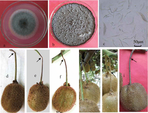
Results of pathogenicity experiments showed that all isolates were highly pathogenic to detached young fruit stems of kiwifruit. The initial symptoms on fruit stems were small, light brown lesions that surrounded the colonized PDA plugs. After four days, extensive necrosis and tissue constriction occurred (–f). The symptoms were similar to those observed on infected fruit stems of kiwifruit in the field (, h). No symptom was observed on control fruit stems treated with the sterile PDA plugs (). Furthermore, Glomerella species was successfully re-isolated onto PDA from the symptomatic fruit stems of kiwifruit, fulfilling Koch's postulates.
Genomic DNA (approximately 50 ng μL−1) was extracted from each of the six fungus isolates by using the standard CTAB method. The internal transcribed space (ITS) region of rDNA from each isolate was amplified by primers ITS1 and ITS4, and then sequenced. The six sequences were all 521 bp long (not including the primer sequence) and were identical (GenBank Accession No. JX885687). Comparison with the sequences in the NCBI–GenBank database revealed that the ITS sequence shared 100% homology with the sequence of Glomerella septospora (GenBank Accession No. GU935911). Therefore, the pathogen was identified as Glomerella septospora based on the morphological characters, pathogenicity test and the ITS sequence. G. septospora, classified in Glomerellaceae, Sordariomycetes and Ascomycota, is a teleomorph, and its anamorph is Colletotrichum taiwanense (Sivanesan & Hsieh, Citation1993).
At present, only a few fungal diseases are reported to infect kiwifruit. These include fruit ripe rot caused by either Botryosphaeria dothidea or Phomopsis sp. (Pennycook, Citation1985; Lee et al., Citation2001; Koh et al., Citation2003; Manning et al., Citation2003), shoot blight caused by Botryosphaeria dothidea (Thomidis & Exadaktylou, Citation2010) and gray mould caused by Botrytis cinerea (Karakaya & Bayraktay, Citation2009). In addition, Colletotrichum gloeosporioides (teleomorph Glomerella cingulata) and Colletotrichum acutatum have also been reported as kiwifruit anthracnose pathogens in New Zealand, Japan and Korea (Hawthorne & Otto, Citation1986; Ushiyama et al., Citation1996; Jeong et al., Citation2008). Glomerella septospora, the pathogen found in this study, has not previously been reported causing disease in kiwifruit. This is the first report of fruit stem blight disease of kiwifruit caused by G. septospora. Besides infecting kiwifruit, G. septospora also infects other hosts, including Styrax formosanus, Capsicum annuum and Dendropanax arboreus (Sivanesan & Hsieh, Citation1993; Hyde et al., Citation2009; Toyozo et al., Citation2012).
Acknowledgements
This work was supported by the Scientific Research Foundation of the Education Department of Jiangxi Province, China (No. GJJ13269) and National Natural Science Foundation of China (No. 31160358).
Related Research Data
References
- Everett , K.R. , Taylor , R.K. , Romberg , M.K. , Rees-George , J. , Fullerton , R.A. , Vanneste , J.L. and Manning , M.A. 2011 . First report of Pseudomonas syringae pv. actinidiae causing kiwifruit bacterial canker in New Zealand . Austral. Plant Dis , 6 : 67 – 71 .
- Hawthorne , B.T. and Otto , C. 1986 . Pathogenicity of fungi associated with leaf spots of kiwifruit . N. Z. J. Agric. Res. , 29 : 533 – 538 .
- Hyde , K.D. , Cai , L. , Cannon , P.F. , Crouch , J.A. , Crous , P.W. , Damm , U. , Goodwin , P.H. , Chen , H. , Johnston , P.R. and Jones , E.B.G. 2009 . Colletotrichum – names in current use . Fungal Div. , 39 : 147 – 182 .
- Jeong , I.N. , Lim , M.T. , Kim , G.H. , Han , T.W. , Kim , H.C. , Kim , M.J. , Park , H.S. , Shin , S.H. , Hur , J.S. , Shin , J.S. and Koh , Y.J. 2008 . Incidences of leaf spots and blights on kiwifruit in Korea . Plant Path. J. , 24 : 125 – 130 .
- Karakaya , A. and Bayraktay , H. 2009 . Botrytis disease of kiwifruit in Turkey . Austral. Plant Dis. Notes , 4 : 87 – 88 .
- Koh , Y.J. , Lee , J.G. , Lee , D.H. and Hur , J.S. 2003 . Botryosphaeria dothidea, the causal organism of ripe rot of kiwifruit (Actinidia deliciosa) in Korea . Plant Path. J. , 19 : 227 – 230 .
- Lee , J.G. , Lee , D.H. and Park , S.Y. 2001 . First report of Diaporthe actinideae, the causal organism of stem-end rot of kiwifruit in Korea . Plant Path. J. , 17 : 110 – 113 .
- Li , C. , Jiang , J.X. , Leng , J.H. , Li , B.M. , Tu , G.Q. and Yu , Q. 2012 . Isolation and identification of pathogenic fungi causing fruit rot of kiwifruit in Fengxin County . Axta Agricult. Universit. Jiangxiensis , 34 : 259 – 263 .
- Lu , J.Y. 2004 . Plant Pathogenic Mycology , Beijing : China Agriculture Press . in Chinese
- Manning , M.A. , Meie , X. and Olsen , T.L. 2003 . Fungi associated with fruit rots of Actinidia chinensis “Hort16A” in New Zealand . N.Z. J. Crop Horticul. Sci. , 31 : 315 – 324 .
- Murray , M.G. and Thompson , W.F. 1980 . Rapid isolation of high molecular weight plant DNA . Nucleic Acids Res. , 8 : 4321 – 4325 .
- Pennycook , S.R. 1985 . Fungal fruit rots of Actinidia deliciosa (kiwifruit) . N.Z. J. Expt. Agric. , 13 : 289 – 299 .
- Perrone , G. , Susca , A. , Stea , G. and Mule , G. 2004 . PCR assay for identification of Aspergillus carbonarius and Aspergillus japonicus . Eur. J. Plant Pathol , 110 : 641 – 649 .
- Sivanesan , A. and Hsieh , W.H. 1993 . A new ascomycete, Glomerella septospora sp. nov. and its coelomycete anamorph, Colletotrichum taiwanenes sp. nov. from Taiwan . Mycol. Res. , 97 : 1523 – 1529 .
- Thomidi , T. and Exadaktylou , E. 2010 . First report of Botryosphaeria dothidea causing shoot blight of kiwifruit (Actinidia deliciosa) in Greece . Plant Dis. , 94 : 1503
- Toyozo , S. , Jouji , M. , Shihomi , U. , Yousuke , D. , Tsuyoshi , O. and Kazuko , N. 2012 . Molecular phylogenetic analyses and morphological re-examination of strains belonging to three rare Colletotrichum species in Japan . Microbiol. Culture Collect. , 28 : 121 – 134 .
- Ushiyama , K. , Aono , N. , Kita , N. and Ogawa , J. 1996 . First report of Pestalotia disease, anthracnose and angular leaf spot of kiwifruit and their pathogens in Japan . Ann. Phytopathol. Soc. Jpn , 62 : 61 – 68 .
- White , T.J. , Bruns , T. , Lee , S. and Taylor , J.W. 1990 . Amplification and direct sequencing of fungal ribosomal RNA genes for phylogenetics . PCR Protocols Guide Methods Applic. , 18 : 315 – 322 .