
Abstract
Young olive trees showing wilting and trunk cankers were observed in an orchard in Antalya province, Turkey. The aerial symptoms associated with reddish brown cankers expanding from the root collar to the stem were leaf discolouration, defoliation, and twig dieback. A Phytophthora sp. was consistently isolated from necrotic root and crown tissues of symptomatic trees. The causal agent of the disease was identified as Phytophthora inundata by morphological characteristics and comparison of sequences of the Internal Transcribed Spacer (ITS) region. Pathogenicity of the isolates was tested by stem inoculation and soil infestation using olive saplings. At the end of the experiments, lesions developed on both stems and roots, and root weight reduction occurred. This is the first report of P. inundata causing disease of olive trees in Turkey.
Résumé
On a observé de jeunes oliviers affichant des signes de flétrissement et portant des chancres sur leurs troncs dans la province d’Antalya, en Turquie. Les symptômes aériens associés aux chancres brun-rouge, comme la décoloration des feuilles, la défoliation et le dépérissement des rameaux, se répandaient de la cime à la tige. On a invariablement isolé une espèce de Phytophthora des tissus nécrotiques des racines et des cimes prélevés sur les arbres symptomatiques. Grâce à ses caractéristiques morphologiques et à la comparaison des séquences de la région de l’espaceur transcrit interne (ITS), l’agent causal de la maladie a été identifié en tant que Phytophthora inundata. La pathogénicité des isolats a été testée par inoculation des tiges et infestation du sol avec des gaules d’olivier. À la fin des expériences, des lésions s’étaient développées sur les tiges et les racines, et le poids des racines avait diminué. Il s’agit de la première mention de P. inundata infectant des oliviers en Turquie.
Introduction
Olive (Olea europaea L.) is grown on approximately 820 000 ha in Turkey with an annual production of 1 768 000 tonnes (Anon Citation2014). Root and crown rot is not common in olive trees, but the disease may kill the trees in flooded or poorly drained soils. Several species of Phytophthora affecting olive trees have been reported. Phytophthora citricola Sawada and P. megasperma Drechsler were reported as olive tree pathogens in Greece (Kouyeas & Chitzanidis Citation1968, Citation1978; Erwin & Ribeiro Citation2005). Phytophthora citricola and P. drechsleri Tucker have been associated with root and crown rot of olive trees in California, USA (Teviotdale Citation1994). In Spain, P. inundata Brasier, Sanch. Hern. & S.A. Kirk, P. megasperma and P. palmivora (Butler) Butler caused root rot and death of young olive trees (Sanchez Hernandez et al. Citation1998; Sanchez-Hernandez et al. Citation2001; Brasier et al. Citation2003). In Italy, five Phytophthora species, including P. citricola, P. inundata, P. nicotianae Breda de Haan, P. megasperma and P. palmivora have been associated with root rot and decline of olives in orchards and nurseries (Cacciola et al. Citation2000, Citation2001, Citation2011). Also P. nicotianae and P. palmivora were responsible for olive decline in Argentina (Lucero et al. Citation2006; Vettraino et al. Citation2009).
In May 2014, a severe decline of young olive trees was observed in an orchard in Antalya, Turkey, and a Phytophthora sp. was consistently isolated from necrotic root and crown tissues of symptomatic trees. The aim of this study was to identify the causal agent of this olive disease.
Materials and methods
Field sampling and Phytophthora isolation
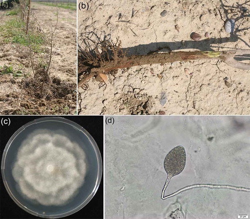
Olive trees showing decline symptoms in recently flooded parts of the orchard next to an irrigation canal in Manavgat district of Antalya province (southwestern Turkey) were observed in May 2014. In this newly planted orchard containing approximately 500 2–3-year-old olive trees ‘Gemlik’, approximately 80 trees showed poor growth, leaf discolouration, and eventual death (). Examination of the lower stem revealed the presence of a reddish brown canker expanding from root collar to stem, and feeder roots were decayed (). Nine symptomatic trees were uprooted and brought to the laboratory for causal agent identification.
Fig. 1 (Colour online) Symptomatic olive tree: a, Canopy wilt symptoms. b, Lower stem cankering visible when the outer bark is removed. c, Seven-day old culture on PDA. d, Sporangium of P. inundata obtained in soil water.
Diseased olive roots were washed in tap water and air-dried. Small sections of 3 to 5 mm diameter were cut from the margin of the lesions on the root collar and 10 sections per agar dish were directly plated on 1.7% corn meal agar (CMA) amended with PARP (per litre: pimaricin, 4 mg; ampicillin, 250 mg; rifampicin, 10 mg and pentachloronitrobenzene [PCNB], 75 mg). Three dishes per tree were incubated at 22°C in the darkness and resulting colonies were examined under the light microscope after 2 days. Emerging colonies were transferred to carrot agar (CA) (200 mL boiled carrot juice, 800 mL distilled water, 20 g agar) containing 20 mg L−1 β-sitosterol to obtain pure cultures for identification.
Morphological and cultural characteristics
Isolates were studied in different growth media including CA, CMA, malt extract agar (MEA) and potato dextrose agar (PDA). The presence of asexual structures including sporangia, hyphal swellings, or chlamydospores was assessed. For this purpose, 5-mm-diameter discs were cut from the growing edge of 4-day-old cultures grown on CA at 24°C in the dark, and placed in 6-cm-diameter Petri dishes previously filled with 7 mL of 1.5% non-sterile soil extract. The dishes were kept under continuous light for 12–48 h at 24°C. Approximately 40 to 50 sporangia per isolate were measured. The ability to grow at 35°C was observed during 7 days on CA and PDA. Identification of Phytophthora sp. was carried out using the identification key of Gallegly & Hong (Citation2008), and by comparison with morphological characteristics described in Brasier et al. (Citation2003).
PCR and sequence analysis
Internal transcribed spacer (ITS) regions of ribosomal DNA (rDNA) of two isolates were amplified using the universal primer pairs ITS-1 (5ʹ TCC GTA GGT GAA CCT GCGG 3ʹ) and ITS-4 (5ʹ TCC TCC GCT TAT TGA TATGC 3ʹ). PCR products were separated in 2% agarose gels, stained with ethidium bromide and visualized under UV light. Sequence analysis was done by GENOKS (Turgut Reis Cad. 36/2 Anıttepe 06570 Çankaya-Ankara/Turkey). The nucleotide sequences were deposited in GenBank.
Pathogenicity tests
Two isolates obtained from the root collar of olive trees in Turkey were used in both stem inoculation and soil infestation tests. For each isolate, five 3-year-old olive saplings ‘Gemlik’ were inoculated. Five non-inoculated plants were used as controls. In the first experiment, stems were inoculated with a 5-mm plug from a 5-day-old culture grown on CA. Sterile agar plugs were used for control plants. Stems of each plant were wounded with a sterile scalpel and agar plugs were placed on the exposed cambium. Then the wounds were covered with moist, autoclaved cotton wool and wrapped with aluminium paper to prevent drying. Plants were kept in a climate-controlled glasshouse at 25 ± 1°C and watered as needed. Tissue under the outer bark was examined 2 months after inoculation. Necrotic tissues excised from cankers were plated onto CMA-PARP to re-isolate the pathogen.
In the second experiment, olive saplings were transplanted to 4 L pots containing a soil mixture (1:1, vol:vol, mixture of orchard soil:sand) mixed with inoculum at a rate of 5% (v/v). The inoculum was prepared by autoclaving wheat grains moistened with distilled water and inoculating them with mycelial agar discs followed by incubation for a month at 22°C in the dark. Plants were grown for 6 months from October 2014 to April 2015 in an uncontrolled glasshouse. They were kept constantly wet by placing them in trays filled with 2 to 3 cm of water 4 days a week. At the end of the experiment, roots were examined and lateral and hairy roots were weighed. Re-isolations were made as described above. Results of root weight were subjected to analysis of variance (ANOVA) according to a completely randomized design replicated five times, and means separated according to the LSD test.
Results and discussion
Six isolates of Phytophthora sp. were obtained from nine symptomatic olive trees. Isolates did not produce sexual structures in single culture. They grew rapidly and formed colonies with a rosette pattern on MEA and PDA (). They produced non-papillate and non-caducous sporangia (), usually ovoid, but sometimes obpyriform or nearly spherical, with rounded and tapered bases. Sporangia were 44.3–74.3 μm long (mean 57.9 μm) and 31.1–56.9 μm wide (mean 41.0 μm), with a 1.4 length-width ratio. Internal proliferation of sporangia and nesting occurred. Cultures also produced rounded hyphal swellings in single or catenulate in non-sterile soil extract. Slow colony growth was observed at 35ºC. Based on these morphological characteristics, the isolates were identified as Phytophthora inundata Brasier, Sanch. Hern. & S.A. Kirk.
Morphological identification was confirmed by ITS sequences of rDNA. Sequences (Acc. No: KP420011, KP420012) showed a 99–100% homology with other P. inundata isolates in GenBank (e.g. Acc. No: AF266791, DQ439963, FJ196751, FJ801484, JF896567). Our isolates also exhibited 99% similarity with some P. humicola isolates present in GenBank (e.g. Acc. No: JQ757060, GU111608). Being heterothallic and growing colonies at 35ºC distinguished our isolates of P. inundata from P. humicola. Phytophthora humicola is a homothallic species and its maximum temperature for growth is 32ºC (Erwin & Ribeiro Citation2005; Gallegly & Hong Citation2008).
Isolates of P. inundata proved to be pathogenic in both experiments. In the stem inoculation test, the isolates caused 1–2 cm long, oval-elliptic cankers with smooth margins, while no cankers developed on control plants. Phytophthora inundata was re-isolated from symptomatic tissues. At the end of the soil infestation tests, poor growth and leaf discolouration on inoculated plants were observed. The pathogen was re-isolated from the necrotic lesions with sizes ranging from 0.5 to 2.5 cm on woody roots. The roots of control plants remained symptomless. Root weights of olive saplings inoculated with the two P. inundata isolates were significantly different from those of controls (P < 0.01) (), while these were not significantly different between the isolates.
Table 1. Root weight of olive saplings inoculated with two isolates of Phytophthora inundata obtained from olive trees in Turkey.
Although Phytophthora root and crown rot has long been known as one of the diseases of olive in some countries (Kouyeas & Chitzanidis Citation1968, Citation1978; Teviotdale Citation1994; Sanchez Hernandez et al. Citation1998; Cacciola et al. Citation2000, Citation2001, Citation2011; Sanchez-Hernandez et al. Citation2001; Brasier et al. Citation2003; Erwin & Ribeiro Citation2005; Lucero et al. Citation2006; Vettraino et al. Citation2009), it has not been detected in olive orchards of Turkey, one of the most important producers of the fruit worldwide. This study showed that P. inundata was associated with decline and mortality of young olive trees and this is the first report of P. inundata causing disease of olive trees in Turkey.
Phytophthora inundata, provisionally referred to as Group B by Sanchez-Hernandez et al. (Citation2001) and then formally named P. inundata by Brasier et al. (Citation2003) was reported to cause root rot, wilt and death of both young and mature olive trees in Italy and Spain, and was shown to be highly aggressive in pathogenicity tests (Cacciola et al. Citation2011; Sanchez-Hernandez et al. Citation2001). The presence of P. inundata in roots after soil flooding events was in accordance with Sanchez-Hernandez et al. (Citation2001) and Brasier et al. (Citation2003) stating these conditions to be favourable for P. inundata root infections.
Control of Phytophthora crown and root rot by chemical methods after foliar symptoms appear is generally ineffective, but preventing flooding or waterlogging, and establishing new olive plantings in well-drained soils may help to avoid the disease caused by P. inundata.
References
- Anon. 2014. Agricultural production statistics of Turkish Statistical Institute [Internet]; [cited 2015 Mar 13]. Available from: http://tuikapp.tuik.gov.tr/bitkiselapp/bitkisel.zul
- Brasier CM, Sanches-Hernandez E, Kirk SA. 2003. Phytophthora inundata sp. nov., a part heterothallic pathogen of trees and shrubs in wet or flooded soils. Mycol Res. 107:477–484.
- Cacciola SO, Agosteo GE, Pane A. 2000. First report of Phytophthora palmivora as a pathogen of olive in Italy. Plant Dis. 84:1153.
- Cacciola SO, Agosteo GE, San Lio GM. 2001. Collar and root rot of olive trees caused by Phytophthora megasperma in Sicily. Plant Dis. 85:96.
- Cacciola SO, Faedda R, Pane A, Scarito G. 2011. Root and crown rot of olive caused by Phytophthora spp. In: Schena L, Agosteo GE, Cacciola SO, editors. Olive diseases and disorders. Trivandrum India: Transworld Research Network; p. 305–327.
- Erwin DC, Ribeiro OK. 2005. Phytophthora diseases worldwide. 2nd ed. St. Paul (MN): APS Press; p. p 562.
- Gallegly ME, Hong C. 2008. Phytophthora, ıdentifying species by morphology and DNA fingerprints. St. Paul (MN): The American Phytopathological Society.
- Kouyeas H, Chitzanidis A. 1968. Notes on Greek species of Phytophthora. Ann Inst Phytopathol Benaki N.S. 8:175–192.
- Kouyeas H, Chitzanidis A. 1978. Host list of Phytophthora spp. in Greece. Phytophthora Newsl. 6:53–54.
- Lucero G, Vettraino AM, Pizzuolo P, Di Stefano C, Vannini A. 2006. First report of Phytophthora palmivora on olive trees in Argentina. New Dis Rep. 14:32.
- Sanchez Hernandez ME, Ruiz Davila A, Perez De Algaba A, Blanco Lopez MA, Trapero Casas A. 1998. Occurrence and etiology of death of young olive trees in southern Spain. Eur J Plant Pathol. 104:347–357.
- Sanchez-Hernandez E, Munoz-Garcia M, Brasier CM, Trapero-Casas A. 2001. Identity and pathogenicity of two Phytophthora taxa associated with a new root disease of olive trees. Plant Dis. 85:411–416.
- Teviotdale BE. 1994. Diseases of olive. In: Ferguson L, Sibbett GS, Martin GC, editors. Olive production manual. Publication 3353. University of California; p. 107–109. Richmond, CA, USA.
- Vettraino AM, Lucero G, Pizzuolo P, Franceschini S, Vannini A. 2009. First report of root rot and twigs wilting of olive trees in Argentina caused by Phytophthora nicotianae. Plant Dis. 93:765.