
Abstract
Objective: Accumulating evidence in both humans and animal models indicates that dietary intake of long-chain polyunsaturated fatty acids (PUFAs) can improve response to chemotherapy. The intent of this study was to determine the mechanisms by which PUFAs affect the response to anticancer chemotherapy.
Methods: Human colorectal cancer cell line Caco-2 was used as a model system in this study. Caco-2 cells were treated with different concentrations of three PUFAs: eicosapentaenoic acid (EPA), docosahexaenoic acid (DHA), and arachidonic acid (AA). Real-time polymerase chain reaction was used to determine mdr1 gene (codes for P-glycoprotein [P-gp]) expression. Western blotting and calcein-acetoxymethylester efflux assay were used for P-gp expression and functional evaluation, respectively. Furthermore, apoptosis assay was conducted by adding PUFAs with paclitaxel to confirm the synergetic effect. Finally, gene expression of nuclear receptors CAR and PXR were estimated to evaluate the possible mechanisms.
Results: Both classes of PUFAs, omega-3 (ω-3) and omega-6 (ω-6), can cause a modest but very reproducible reduction of gene expression, protein production, and pump activity of MDR1. Incubation of cells with PUFAs greatly enhanced the cytotoxicity of the anticancer drug paclitaxel, manifested mainly through enhanced paclitaxel-induced apoptosis. Furthermore, PUFAs increased the messenger RNA (mRNA) levels of the nuclear receptors CAR and PXR, thus implicating these two transcription factors as cellular targets of PUFAs in cells but not directly affecting MDR1 regulation.
Conclusions: Our results suggest that inhibition of the multidrug resistance MDR1/P-gp is one mechanism through which dietary polyunsaturated fatty acids exert a synergetic effect on the response of tumor cells to anticancer drugs.
ACKNOWLEDGMENTS
We are indebted to Dr. Zena Indik at the University of Pennsylvania School of Medicine for critical reading of the manuscript. We appreciate the colleagues at Clemson University, Dr. Meredith Morris, Dr. Chih-Chao Yang, Mr. Chun-Huai Cheng, and Dr. Guohui Huang, for their technical assistance in flow cytometer, Western blotting, qPCR, and cell culture, respectively. This research received no specific grant from any funding agency in the public, commercial, or not-for-profit sectors.
Fig. 1 The effect of PUFAs on viability of Caco-2 cells. Cells were incubated for 24, 48, and 72 hours with 25, 50 and 100 μM of the following PUFAs: (A) AA, (B) EPA, and (C) DHA. The viability of treated cells is presented as the means of cell number (± standard deviation) compared with control (untreated) cells. For each experiment, n = 3. *p < 0.05, **p < 0.01, ***p < 0.001 indicate significant differences from control values.

Fig. 2 (A) The effect of PUFAs on MDR1 gene expression. Caco-2 cells were incubated with 100 μM PUFAs for 4, 12, and 24 hours. (B) The effect of EPA on MDR1 gene expression at concentrations of 50 μM and 100 μM. mRNA levels were determined by qPCR (see Materials and Methods), from samples obtained from three independent experiments. *p < 0.05, **p < 0.01, ***p < 0.001 indicate significant differences from control values.
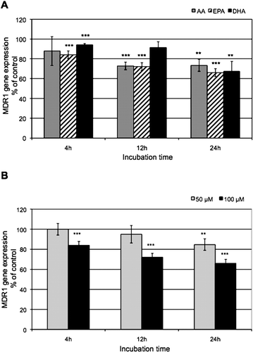
Fig. 3 The effect of PUFAs on cellular levels of the MDR1 gene product P-gp. Western blot analysis of P-gp (top), β-actin (bottom). Caco-2 cells were treated with 100 μM PUFAs for 24 hours. These images are representative of three independent experiments.
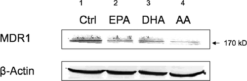
Fig. 4 The effect of PUFAs on calcein-AM efflux in Caco-2 cells: intracellular fluorescence intensity. Caco-2 cells were incubated with 100 μM PUFA for 24 hours followed by the addition of 0.25 μM of calcein-AM. The results are averages of three independent experiments. *p < 0.05, **p < 0.01, ***p < 0.001 indicate significant differences from control values.
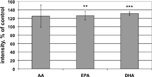
Fig. 5 The effect of PUFAs on apoptosis. Exposure to PUFAs increased apoptosis of Caco-2 cells and enhanced paclitaxel-induced apoptosis in these cells. Images in column 1 are cells incubated with solvent controls; column 2, cells treated with paclitaxel (PTX); column 3, cells treated with PUFAs (from top to bottom: AA, DHA, and EPA, respectively); column 4, cells treated with paclitaxel plus PUFAs (from top to bottom: AA, DHA, and EPA, respectively). One of the apoptotic cells (the lighter spots) is indicated with an arrow. Each photograph was taken with the same magnitude (100×), and the white bar represents 200 μm.
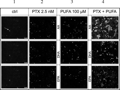
Table 1. Primers and Cycling Information
Table 2. The Effect of PUFAs on Cytotoxicity of Paclitaxel in the Caco-2 Cells
Table 3. The Effect of PUFAs on the Expression of the CAR and PXR Genes
Notes
Conflict of interest: All authors declared that they had no conflict of interest.