

Abstract
Methods for sequence-specific microRNA (miRNA) analysis are crucial for miRNA research and guiding nursing strategies. We have devised a colorimetric technique for detecting miRNA using a dumbbell probe-based polymerase/endonuclease assisted chain displacement, along with silver ions (Ag+) aptamer assisted color reaction. The suggested approach enables precise measurement of miRNA-21 within the concentration range of 100 fM–5 nM, with a low detection limit of 45.32 fM. Additionally, it exhibits exceptional capability in distinguishing variations at the level of individual nucleotides. Furthermore, the detection technique may be utilized to precisely measure the amount of miRNA-21 in serum samples, demonstrating a high level of concordance with the findings obtained from a commercially available miRNA detection kit.
METHOD SUMMARY
This method utilizes the hairpin structure in the dumbbell probe to significantly enhance the accuracy of target recognition in the polymerase/endonuclease assisted chain displacement strategy. As a result, the method gains a superior capability to discriminate between target miRNA and interfering miRNAs. The combination of target recycling and the polymerase/endonuclease assisted chain displacement strategy allows for highly sensitive analysis of miRNA, which is either superior or comparable to previous colorimetric methods for miRNA detection. This approach is simple, economical and extremely specific for detecting miRNA. It holds great potential for clinical use, particularly in guiding the adaptation of nursing techniques.
Background
The microRNA (miRNA) have emerged as diagnostic, prognostic and treatment-response biomarkers for various diseases.
The existing miRNA detection methods are commonly criticized for the drawbacks of low sensitivity and accuracy.
Materials & methods
Construction of the dumbbell probe.
Fluorescence experiments verified the recovery of target miRNA.
Target miRNA detection.
Results & discussion
Optimization of experimental parameters.
The suggested approach enables precise measurement of miRNA-21 within the concentration range of 100 fM–5 nM, with a low detection limit of 45.32 fM.
Conclusion
This research offers a sensitive and reliable approach for miRNA detection.
1. Background
Recent findings indicate that microRNA (miRNA) signatures have significant potential as diagnostic, prognostic and treatment-response biomarkers for various diseases [Citation1,Citation2]. miRNAs, acting as key regulators, have the ability to influence the expression of numerous genes in various tissues [Citation3]. This regulation extends to almost all cellular processes at the transcriptional level [Citation4], encompassing cellular development, proliferation, migration, survival, metabolism, homeostasis and regeneration. Prior studies have shown that a miRNA-based biomarker can be used to effectively guide the selection of nursing techniques for patients undergoing surgery [Citation5,Citation6]. Therefore, a precise analysis of the expression level of miRNAs can offer vital insights into their regulation and their use as biomarkers for the early diagnosis and treatment of malignancies. Hence, there is a significant need to create simple and reliable techniques for miRNA detection.
The majority of current techniques, such as northern blotting, microarray [Citation7,Citation8] and polymerase chain reaction (PCR)-based methods [Citation9,Citation10], used signal amplification, target conversion or enrichment strategy to achieve a relatively high sensitivity and selectivity because of the low abundance, short sequence and high sequence homology of miRNAs. PCR-based techniques have gained significant popularity due to their excellent performance and broad applicability. However, the use of multiple enzymes, particularly in reverse transcription PCR-based techniques, leads to increased experimental costs and design complexities, as well as the requirement for a thermal cycler to accurately regulate the reaction temperature [Citation11]. These approaches cannot be implemented in point-of-care settings or locations with limited resources, in addition to the aforementioned disadvantages. Enzyme-assisted isothermal amplification approaches have demonstrated significant potential for “on the spot” miRNA testing [Citation12,Citation13,Citation14]. The polymerase/endonuclease assisted chain displacement technique has gained significant attention because of its excellent amplification efficiency and reproducibility [Citation13,Citation14]. However, the polymerase/endonuclease assisted chain displacement approach often necessitates integration with fluorescence transducers and suffers from insufficient selectivity. The colorimetric approach with naked-eye recognition offers significant advantages over the fluorescence transducer for miRNA detection [Citation15,Citation16]. Due to their robust distance-dependent optical characteristics and the ability to change color when aggregated, gold nanoparticles (AuNPs) have been extensively applied in colorimetric assays [Citation17,Citation18]. This single-stranded DNA (ssDNA) regulated state alteration of AuNPs is typically achieved through DNA hybridization, which is a one-to-one signal transduction process; its sensitivity is inadequate for the detection of miRNA, which has stringent sensitivity requirements, despite its considerable achievements. Hence, increased selectivity and simplicity are necessary for a viable alternative.
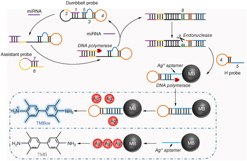
Therefore, we have devised a novel and efficient colorimetric method that ingeniously employs a dumbbell probe to enhance the specificity of polymerase/endonuclease assisted chain displacement, together with silver ions (Ag+) to facilitate the color reaction (). The sensing technique involves the designing of a dumbbell probe that consists of five distinct functional portions according to related references [Citation19,Citation20]. Within this probe, the “1” segment contains sites that bind to the target miRNA, the “2” fragment aids in the recycling of the target, the “3” fragment is capable of transcribing sites that are recognized by the endonuclease and the “5” fragment serves as a primer to initiate the color reaction. When the target miRNA is present, it hybridizes with the “1” fragment and slowly unravels the “stem” region in the hairpin structure, revealing the “2” fragment to serves as a recognition site for the “6” fragment in the assistance probe. The “6” fragment substitutes for the target miRNA from the dumbbell probe by binding to the “2” fragment. The target miRNA that was released forms a signal cycle by binding with a next dumbbell probe. The “6” fragment serves as a primer to commence the chain extension with the assistance of DNA polymerase, encoding the “8” fragment using “3” as the template. The endonuclease identifies the “8” segment and creates a nicking site. Through the collaboration of DNA polymerase and endonuclease, a substantial quantity of ssDNA sequences, consisting of “9” and “10” fragments are generated. The H probe is formed when the ssDNA products assemble into a hairpin structure; this structure is responsible for inducing the Ag+ aptamer based color response. The aptamer was utilized to bind silver ions (Ag+) through the interaction between Ag+ and the N3 of cytosine (C), which connects two cytosine residues to create a strong and stable “C–Ag+-C” hairpin structure. Specifically, the “10” fragment binds with the Ag+ aptamer that is fixed on the surface of magnetic bead (Ag+ aptamer@MB), and works as a primer to encode a complementary sequence under the assistance of the DNA polymerase. The Ag+ aptamer's capacity to chelate Ag+ is hindered by the creation of a double-stranded DNA structure (dsDNA). Therefore, Ag+ was unable to chelate with the aptamer and instead underwent a reaction with TMB, resulting in the color reaction. Under these circumstances, the solution containing oxidized TMB (TMBox) had a blue color and a distinctive UV–vis absorption peak at 652 nm. If miRNA is not present, the formation of dsDNA product will not take place, and the aptamer will remain intact and can be extracted using a magnetic rack. Through this procedure, Ag+ ions can attach to the aptamer that has been magnetically enhanced by interacting with cytosine to create a hairpin structure known as “C–Ag+-C”. As a result, the solution loses its color in the absence of TMBox.
Figure 1. The working principle of the polymerase/endonuclease assisted colorimetric microRNA detection method.
2. Experimental section
2.1. Materials & instrumentation
The enzymes, including the DNA polymerase and Nicking endonuclease Nb.BbvCI, were purchased from Sigma-Aldrich (Shanghai, China). SYBR Green I for characterization of dsDNA section was purchased from New England Biolabs (NEB, Beijing, China). TaKaRa PCR Amplification Kit was bought from Tsingke Biological Technology Company (Beijing, China). Deoxyribonucleoside 5′-triphosphate mixture (dNTPs) was provided by Tiangen Biotech. Co. Ltd (Beijing, China). All oligonucleotides used in this experiment (Supplementary Table S1) were purchased from Sangon Biotech. Co. Ltd (Shanghai, China).
2.2. Construction of the dumbbell probe
The 5 μl dumbbell probe (500 nM) was mixed with 15 μl PBS buffer and heated to 90°C, then slowly lowered to room temperature. The assembly of the dumbbell probe was verified by fluorescence experiment following the following steps. First, the dumbbell probe with 5 μl, SYBR Green I with 2 μl and PBS buffer with 13 μl were mixed and heated to 90°C, and then slowly lowered to room temperature. The fluorescence intensity of SYBR Green I before and after assembly was compared.
2.3. Fluorescence experiments verified the recovery of target miRNA
5 μl dumbbell probe (500 nM), 2 μl target miRNA and 5 μl assistance probe (500 nM) were added to tubes containing 28 μl PBS buffer. The mixture was cultured at room temperature for 30 min, and the fluorescence intensity of the solution was detected.
2.4. Target miRNA detection
5 μl dumbbell probe (500 nM), 2 μl target miRNA and 5 μl assistance probe (500 nM), 2 μl DNA polymerase, 2 μl nicking endonuclease Nb.BbvCI, 2 μl dNTPs and 25 μl Ag+ aptamer@MB were added to tubes containing 22 μl PBS buffer. Afterward, 35 μl of TMB (5 mM) was added to the mixture for 20 min in a light-avoidance manner. Finally, absorbance at 652 nm was measured while coloration can be easily observed by the naked-eye.
3. Results & discussion
3.1. The feasibility of the polymerase/endonuclease assisted chain displacement & Ag+ based color reaction
Fluorescence assays were employed to validate the construction of the dumbbell probe and its capacity to identify the target miRNA. SYBR Green I, a highly sensitive fluorescent dye with the ability to uniquely identify dsDNA structures, was employed to analyze the “stem” structures that arise during the assembly of the dumbbell probe. A demonstrates that the SYBR-Green I signal exhibited a gradual enhancement after construction, ultimately reaching equilibrium within a 20 min timeframe. This indicates the successful completion of the assembly process for the linear dumbbell probe. In order to confirm the construction of the dumbbell probe, the “1” and “5” segments were tagged with Cy5 and BHQ, respectively. The results demonstrated, as depicted in B, that the fluorescence intensity of the assembled dumbbell probe dramatically decreased compared with that of the linear dumbbell probe. This suggests that the Cy5 fluorescence signal was suppressed by BHQ, and the formation of the dumbbell structure. Furthermore, the target miRNA's recognition potential was confirmed. The findings demonstrated that the fluorescence signal of Cy5 was amplified in the presence of miRNA. The observed outcome could be attributed to the hybridization of miRNA with the “1” segment, which diminishes the quenching impact of BHQ on Cy5. The addition of the assistance probe to the sensing system resulted in a further enhancement of the fluorescence signal, providing evidence for the target recycling based on the assistance probe. The assembly of the assistance probe and the assistance probe's role in assisting the target cycle, were further confirmed using fluorescence assays, as depicted in C.
Figure 2. The feasibility of target recycling and colorimetric approach for miRNA detection. (A) SYBR Green I signals of the DP with different incubation time. (B) Fluorescence intensities of the dumbbell probe during the target recycling process. (C) Fluorescence intensities of the FAM labeled assistance probe during the target recycling process. 1, AP before assembly to hairpin structure; 2, AP; 3, AP + DP; 4, AP + DB + miRNA. (D) Fluorescence intensities of the FAM (carboxyfluorescein) labeled aptamer before and after construction of the aptamer@MB. (E) Absorbance of the approach when several experimental parameters existed or not.
AP: Assistance probe; DP: Dumbbell probe.
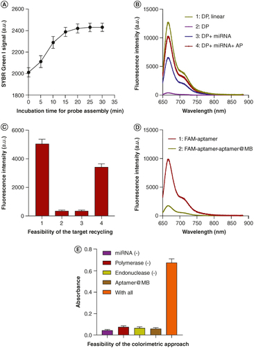
The terminus of the aptamer is labeled with fluorescent dye to verify the assembly of the Ag+ aptamer@MB. Before assembly, the fluorescence intensity of the Cy5-labeled aptamer was 4545 a.u., which decreased by about 86.12% after assembly, indicating that a large number of Cy5-labeled aptamers were loaded on the surface of MB (D). Color reaction was used to verify the feasibility of the method. The results are shown in E. When miRNA, DNA polymerase, endonuclease and Ag+ aptamer @MB are present at the same time, the color of the solution turns significantly blue, indicating the important value of these experimental components in the method.
3.2. Optimization of experimental parameters
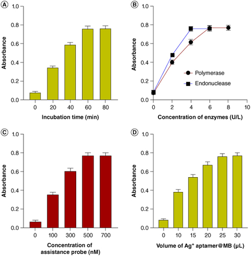
Several critical parameters, including incubation duration, concentration of DNA polymerase and endonuclease, amount of assistance probe and volume of Ag+ aptamer@MB, were investigated in order to attain the intended detection outcomes. The duration of incubation is crucial for achieving optimal analytical results. As the duration increased, the chain extension mechanism became saturated, no longer preventing the aptamer from binding to Ag+. According to the experimental findings, the desired analytical performance was achieved within 60 min (A). The quantity of DNA polymerase and endonuclease is crucial in the signaling cycle and the Ag+ aptamer based color reaction. Hence, the absorbance was measured to assess the efficiency of the method in identifying 1 pM target miRNA under varying concentrations of DNA polymerase and endonuclease, as shown in B. The findings demonstrated a substantial increase in absorbance when the polymerase concentration varied from 0 to 6 U/l, followed by a stabilization of absorbance beyond a polymerase concentration of 6 U/l. Hence, the ideal concentration of polymerase is 6 U/l. Similarly, the concentration of endonuclease is discovered to be 4 U/l. It is important to mention that in this approach; the concentration of the polymerase enzyme is substantially greater than that of the endonuclease. This is because the polymerase plays a critical role in both the DNA polymerase/endonuclease assisted chain displacement and Ag+ aptamer transcription. Conversely, the most efficient quantity of assistance probe was determined to be 500 nM, as shown in C. Furthermore, the utilization of 25 μl of aptamer@MB complex resulted in both the most favorable optical response and the minimal consumption of reagents, as demonstrated in the future studies (D).
Figure 3. Optimization of experimental parameters. Absorbance of the approach when detecting miRNA with different incubation time (A), different enzyme concentrations (B), different amounts of the assistance probe (C) and the volume of the aptamer@MB complex (D).
3.3. Sensitivity & selectivity of the colorimetric approach for miRNA-21 detection
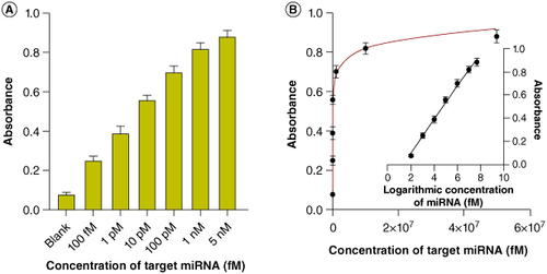
The polymerase/endonuclease assisted chain displacement was activated using various doses of miRNA-21 under optimal conditions. As expected, there was a noticeable and progressive rise in absorbance as the concentration of miRNA-21 increased from 100 fM to 5 nM. This increase in concentration also resulted in a visible shift in color from colorless to blue, which could be noticed without the need for any additional equipment (A). The color change indicates the production of H probes by the polymerase/endonuclease assisted chain displacement, which triggers the Ag+ aptamer based color reaction and results in an increase in absorption intensity. Thus, the absorbance of the method increased proportionally with the quantity of miRNA-21, ranging from 100 fM to 5 nM. The correlation equation between the absorbance and the logarithmic concentration of miRNA-21 was calculated as Y = 0.1428*lgC - 0.1804. This equation shows a strong correlation between the two variables, with a high correlation coefficient (R2) of 0.9943 (B). The estimated detection limit was 45.32 fM, determined using a 3σ/S method, where σ represents the standard deviation of the blank solution.
Figure 4. Sensitivity of the approach. (A) Absorbance of the approach when detecting different concentrations of target miRNA. (B) Correlation between the absorbance anf the concentration of target miRNA.
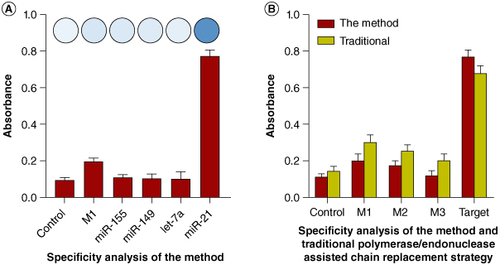
3.4. Selectivity of the colorimetric approach for miRNA-21 detection
In order to assess the potential of the proposed assay to distinguish between different sequences, target miRNA-21, a sequence with single base mismatched with miRNA-21 (M1), as well as three other miRNAs (miRNA-155, miRNA-149 and let-7a) that crucial in postoperative nursing recovery of endoscopic resection for stomach cancer. The synthetic miRNAs were individually evaluated at a concentration of 500 pM. A (inserted) demonstrates that only the target miRNA-21 elicits noticeable color changes, resulting in a distinct blue color in the tube, whereas the other miRNAs do not cause any color change. Similarly, the absorbance value of miRNA-21 is greater than that of the non-target sequences (A). Upon applying the approach to detect a sequence with a single-base mismatch (M1), a faint blue hue was observed in the tube, suggesting the possibility of a nonspecific color reaction. The absorbance for detecting M1 was approximately 21.2% of the absorbance for detecting miRNA-21. The reduced rates of recovery were comparable to or superior to previous recognized methods. In order to determine the efficacy of the dumbbell structure probe in enhancing the accuracy of traditional polymerase/endonuclease assisted chain replacement strategy, we employed the developed method to identify miRNA-21, as well as sequences with one-base mismatch (M1), dual-base mismatch (M2) and triple-base mismatch (M3), both with and without a dumbbell probe. The findings indicated that the suggested method yielded recovery rates of 21.21% (M1), 17.65% (M2) and 12.12% (M3), respectively (B). These recovery rates were notably lower compared with those achieved by the conventional polymerase/endonuclease assisted chain replacement technology (p < 0.05). The aforementioned results demonstrate that the suggested method has superior capability in identifying mismatches compared with conventional methods (Supplementary Table S2).
Figure 5. Selectivity of the approach. (A) Absorbance of the approach when detecting different miRNAs. (B) Absorbance of the approach and traditional method when detecting mismatched sequences.
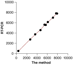
3.5. Biological sample application
The application of the proposed method for the quantitative detection of miRNA-21 in a complex biological matrix was investigated. To determine the quantity of miRNA-21 diluted in commercial serum samples, both the proposed method and a commercial RT-PCR-based miRNA detection kit were employed. The quantities of miRNA-21 detected using the proposed technique were highly consistent with those obtained using the commercially available kit, as depicted in .
Figure 6. Biological sample application of the approach.
4. Conclusion
In summary, a simple and reliable colorimetric technique for sequence-specific miRNA detection has been devised by using Ag+ aptamer for color reaction and dumbbell probe-based polymerase/endonuclease assisted chain displacement. The advantages of the established approach could be concluded as colorimetric method with naked-eye recognition extended the applicable sceneries for the approach; target recycling enhanced the sensitivity of polymerase/endonuclease assisted chain displacement strategy; hairpin structure in the dumbbell probe improved the target recognition capability of the traditional polymerase/endonuclease assisted chain displacement strategy. As a result, the method exhibited a high sensitivity and a low limit of detection of 45.32 fM. Importantly, the established approach showed a greatly improved selectivity to the interfering sequences that even contains only one base mismatched with target miRNA. Given the unique characteristics, we are confident that the established approach holds significant promise as a standard tool for expeditious miRNA analysis in typical laboratory investigations, clinical diagnosis and guiding nursing approaches.
5. Future perspective
Specific and sensitive miRNA analysis is crucial for guiding nursing strategies and early-diagnosis of diseases. We devised here a colorimetric technique for sensitive and accurate miRNA detection by integrating dumbbell probe-based polymerase/endonuclease assisted chain displacement, along with silver ions aptamer assisted color reaction. By utilizing the hairpin structure in the dumbbell probe to significantly enhance the accuracy of target recognition in the polymerase/endonuclease assisted chain displacement strategy, the suggested approach enables precise measurement of miRNA-21 within the concentration range of 100 fM–5 nM, with a low detection limit of 45.32 fM. Considering that the proposed approach is simple, economical and extremely specific for detecting miRNA, it could be a robust tool for clinical use, particularly in guiding the adaptation of nursing techniques. In the future, we will extend the method to various biomarkers detection and establish a versatile platform for disease diagnosis.
Author contributions
S Tao is the supervisor of the team in all research steps including designing, data analysis and manuscript writing. X Xu and P Zhang, as the co-first authors, shared the main role for experimental data collection, data gathering, preparation of results and data analysis.
Financial disclosure
The authors have no financial involvement with any organization or entity with a financial interest in or financial conflict with the subject matter or materials discussed in the manuscript. This includes employment, consultancies, honoraria, stock ownership or options, expert testimony, grants or patents received or pending, or royalties.
Writing disclosure
No writing assistance was utilized in the production of this manuscript.
Supplementary Tables S1 and S2
Download MS Word (23 KB)Acknowledgments
The authors thank the support from Central Laboratory of the People’s Hospital Of Chongqing Liang Jiang New Area.
Supplemental material
Supplementary data for this article can be accessed at https://doi.org/10.1080/07366205.2024.2368394
Competing interests disclosure
The authors have no competing interests or relevant affiliations with any organization or entity with the subject matter or materials discussed in the manuscript. This includes employment, consultancies, stock ownership or options and expert testimony.
References
- Chen L, Heikkinen L, Wang C, et al. Trends in the development of miRNA bioinformatics tools. Brief Bioinform. 2019;20:1836–1852. doi:10.1093/bib/bby054
- Hill M, Tran N. miRNA interplay: mechanisms and consequences in cancer. Dis Model Mech. 2021;14:dmm047662. doi:10.1242/dmm.047662
- Mishra S, Yadav T, Rani V. Exploring miRNA based approaches in cancer diagnostics and therapeutics. Crit Rev Oncol Hematol. 2016;98:12–23. doi:10.1016/j.critrevonc.2015.10.003
- Saliminejad K, Khorram Khorshid HR, Soleymani Fard S, et al. An overview of microRNAs: biology, functions, therapeutics, and analysis methods. J Cell Physiol. 2019;234:5451–5465. doi:10.1002/jcp.27486
- Zloto K, Tirosh-Wagner T, Bolkier Y, et al. Preoperative miRNA-208a as a predictor of postoperative complications in children with congenital heart disease undergoing heart surgery. J Cardiovasc Transl Res. 2020;13:245–252. doi:10.1007/s12265-019-09921-1
- Selvaraj V, Sekaran S, Rajamani Sekar SK. Advancing postoperative pain management in humans with miRNA-based therapeutic strategies: lacunae that need to be addressed. Int J Surg. 2023;109:2849–2850. doi:10.1097/JS9.0000000000000514
- Uso M, Jantus-Lewintre E, Sirera R, et al. miRNA detection methods and clinical implications in lung cancer. Future Oncol. 2014;10:2279–2292. doi:10.2217/fon.14.93
- Li W, Ruan KJA, Chemistry B. MicroRNA detection by microarray. 2009;394:1117–1124. doi:10.1007/s00216-008-2570-2
- Cirillo PDR, Margiotti K, Mesoraca A, et al. Quantification of circulating microRNAs by droplet digital PCR for cancer detection. BMC Res Notes. 2020;13:351. doi:10.1186/s13104-020-05190-3
- Chen C, Tan R, Wong L, et al. Quantitation of microRNAs by real-time RT-qPCR. Methods Mol Biol. 2011;687:113–134. doi:10.1007/978-1-60761-944-4_8
- Deng R, Zhang K, Li J. Isothermal amplification for microRNA detection: from the test tube to the cell. Acc Chem Res. 2017;50:1059–1068. doi:10.1021/acs.accounts.7b00040
- Zhang M, Wang H, Wang H, et al. CRISPR/Cas12a-assisted ligation-initiated loop-mediated isothermal amplification (CAL-LAMP) for highly specific detection of microRNAs. Anal Chem. 2021;93:7942–7948. doi:10.1021/acs.analchem.1c00686
- Kamal Masud M, Islam MN, Haque MH, et al. Gold-loaded nanoporous superparamagnetic nanocubes for catalytic signal amplification in detecting miRNA. Chem Commun (Camb). 2017;53:8231–8234. doi:10.1039/C7CC04789D
- Masud MK, Umer M, Hossain MSA, et al. Nanoarchitecture frameworks for electrochemical miRNA detection. Trends Biochem Sci. 2019;44:433–452. doi:10.1016/j.tibs.2018.11.012
- Kang Y, Zhang J, Zhao L, et al. Colorimetric miRNA detection based on self-primer-initiated CRISPR-Cas12a-assisted amplification. BioTechniques. 2023;74:172–178. doi:10.2144/btn-2023-0008
- Xu Z, Zheng K, Du Z, et al. Colorimetric identification of miRNA-195 sequence for diagnosing osteosarcoma. Biotechnol Appl Biochem. 2022;69:974–980. doi:10.1002/bab.2169
- Aamri ME, Mohammadi H, Amine A. Paper-based colorimetric detection of miRNA-21 using pre-activated nylon membrane and peroxidase-mimetic Activity of cysteamine-capped gold nanoparticles. Biosensors (Basel). 2023;13:74. doi:10.3390/bios13010074
- You Z, Yang Z, Chen Y, et al. Visual detection of heart failure associated MiRNA with DSN enzyme-based recycling amplification strategy. RSC Adv. 2021;11:18068–18073. doi:10.1039/D1RA01500A
- Miao P, Chai H, Tang Y. DNA hairpins and dumbbell-wheel transitions amplified walking nanomachine for ultrasensitive nucleic acid detection. ACS Nano. 2022;16:4726–4733. doi:10.1021/acsnano.1c11582
- Miao P, Tang Y. Dumbbell hybridization chain reaction based electrochemical biosensor for ultrasensitive detection of exosomal miRNA. Anal. Chem. 2020;92:12026–12032. doi:10.1021/acs.analchem.0c02654

