
Abstract
Introduction: The use of oral appliances to enhance sports performance has been advocated by some authors, however, studies addressing the effectiveness of these strategies are inconclusive.
Methods: Here we investigate the effects of dental occlusions on shoulder strength. Fourteen healthy male subjects (age = 21.67 ± 0.86 years) without temporomandibular joint (TMJ) disorder participated in this study. Isokinetic strength was evaluated in shoulder abduction/adduction and arm external/internal rotation tests. Three randomised conditions were assessed: (1) occlusal splint (OS), which repositioned the TMJ in centric relation; (2) placebo splint (PS); and (3) no-splint (N). The strength tests were performed at a speed of 60°/sec in concentric mode. Muscle activity was measured by surface electromyography (EMG) in the main muscles engaged in the movements.
Results: Significant differences in peak torque between OS and both of the other experimental conditions were found in some of the analyzed variables. Moreover, there was significantly higher muscular EMG activation in the OS condition when compared to the other conditions for some of the tested muscles. These data suggest that splints may have a positive ergogenic effect on shoulder muscular strength in healthy male subjects.
Conclusion: OS may provide an advantage for healthy subjects engaged in sports whereby shoulder and arm strength are important for performance.
Occlusal splints in centric relation position have an ergogenic effect, by increasing strength and muscle activation on shoulder in healthy subjects.
These results could have implications for sports or other physical activities were arm and shoulder strength are important.
Key messages
Introduction
Muscular performance and motor skills in sports are determined by a wide array of factors [Citation1]. An extensive number of studies in sports performance research has focussed on motor performance enhancement [Citation1–4]. Sports dentistry is an emerging field with strong potential for preventing sports injury and enhancing performance [Citation5]. As up to 18% of sports-related injuries occur in the maxillofacial area [Citation6], dental medicine is essential for the athletes’ well-being and health, and it has been suggested that sports teams should have a dentist in their staff [Citation7].
Occlusal splints (OS) are removable appliances for treating temporomandibular joint (TMJ) disorders that partially or totally cover the occlusal surfaces of the teeth [Citation8]. In recent years, OS have also been used to enhance sports performance [Citation9–11] based on research suggesting that dental occlusion (i.e. dynamic relationship between the maxillary and mandibular teeth when they approach each other) may affect muscle strength elsewhere in the body [Citation12–14]. For instance, changes in vertical dimension of occlusion (VDO) caused by OS influence the upper limb muscles (e.g. trapezius muscle), which play an important role in fixating the scapula and clavicle [Citation15,Citation16]. Moreover, the proprioceptive feedback from the TMJ also seems to be affected by the VDO [Citation17]. Importantly, these proprioceptive changes are projected via afferent fibres to the accessory nerve nucleus that controls the sternocleidomastoid and trapezius muscles [Citation17]. This modification in afferent information is also likely to influence scapular muscles, which stabilize bones supporting upper limb muscles, including the deltoid, pectoralis major and rotator cuff muscles [Citation18].
Over the past decades, numerous studies have examined the potential of using occlusal appliances for improving muscular strength in athletes [Citation9,Citation13,Citation19–22]. Studies addressing the effects of OS on lower body muscle strength through isokinetic evaluation of knee extension and flexion were unanimous in reporting that OS have no impact on lower limb strength [Citation13,Citation20,Citation22,Citation23]. In contrast, research assessing isometric strength in the upper limbs revealed that OS increased peak strength in a significant manner [Citation24–26], including handgrip tests [Citation11,Citation27] and also in lower limbs [Citation28].
A study measuring electromyography (EMG) activity showed an increase in upper body muscle activity in athletes using OS [Citation10]. Another study found changes in muscle activation on neck muscles [Citation29].
However, others have found opposite results. Greenberg et al. [Citation22] showed that an OS that increased VOD produced no effects on the peak torque (PT) of shoulder abduction/adduction movements, and these findings were corroborated independently by others [Citation13,Citation23]. Finally, results from an alternative upper limb isokinetic task (upright rowing movement) on American football athletes further confirmed that OS increasing VOD caused no effects on PT [Citation21]. Another study with college football players [Citation30] also revealed that OS did not cause any changes in physical performance.
Although some studies suggest a physiological connection between OS and muscle strength, the literature remains controversial and unambiguous evidence supporting this hypothesis is currently lacking. Here, we investigate the effects of OS on shoulder strength in healthy male subjects.
Material and methods
Subjects
Fourteen male subjects were recruited (age = 21.67 ± 0.86 years; body mass = 76.33 ± 7 kg; height = 1.76 ± 0.61 m) for this experiment. A dental examination confirmed that none of the participants had any type of temporomandibular joint (TMJ) disorders. An occlusal splint (OS) that repositioned the TMJ in centric relation (CR) was fabricated for each of the subjects. The CR is considered the most stable position for the mandible, and it is achieved when the TMJ condyles are in their most anterior-superior position in the articular fossae. This position encourages the condyles to seat stably in a congruent skeletal arrangement [Citation31]. The OS were custom-made, using a vacuum former machine (Easy Vac, Baekseokdong, South Korea). A thermoforming foil (Erkodent, Pfalzgrafenweiler, Germany) was adapted over the maxillary casts, trimmed and adjusted in the articulator to the requisites of a stabilization splint in CR position. The thermoforming foil had a thickness of 1 mm and lost about 20%–30% in the manufacturing process. Hence, the interocclusal distance was ∼7 mm. A placebo splint was also fabricated for each subject, using the same manufacturing process, adapted over maxillary casts and trimmed down on the occlusal surface to ensure that they would not interfere with the subjects normal maximum intercuspation This research was approved by the ethics committee of the Faculdade de Motricidade Humana for use of human research (6/2016) and all subjects signed an informed consent.
Experimental protocol
All measurements were performed on a Biodex Medical System Isokinetic dynamometer (Biodex System, Shirley, NY), with a sample rate of 100 Hz. The dominant arm was defined as that used for writing. The subjects started with a warm-up of approximately five minutes of specific light exercises. Subsequently, two tests were conducted: shoulder abduction/adduction and external/internal rotation of the arm. These tests were applied in two different days separated by 72 hours, to avoid fatigue. Each subject performed the tests in a randomized order and was unaware of the differences between the occlusal or placebo splints. Test conditions were: occlusal splint (OS), placebo splint (PS) and normal condition (N, no splint or and no contact between teeth) ().
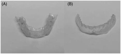
Figure 1. Occlusal splints used (A) Occlusal splint (OS); (B) Placebo splint (PS).
EMG signals were collected using bipolar surface electrodes (Plux, Lisbon, Portugal) and the skin preparation and placement of the electrodes followed guidelines previously described [Citation32,Citation33]. Amplification of the EMG signal was done with a bandpass filter (25–500 Hz), common-mode rejection ratio (CMRR) of 110 dB and a gain of 1000. The signals were sampled at 1000 Hz. The position of the subjects was identical in all tests: seated in a chair in a comfortable position, with the seatback at an angle of 70° relative to the seat. The axis of the dynamometer was aligned with the axis of the movement. Straps were placed around chest, waist and wrist. The arm was statically weighted to provide gravity correction. Five maximal repetitions were performed at a velocity of 60°/sec [Citation34,Citation35] with a rest period of three minutes between test conditions.
For shoulder abduction and adduction movements, measurements were made in the scapular plane (angle of the scapula in its resting position, normally 30°–45° forward from the frontal plane toward the sagittal plane) with the elbow placed at a 45° flexion and a range of motion from 30° (start) abduction to 135° (end) abduction. EMG measurements were done on anterior and middle portions of the deltoid, lower trapezius and pectoralis major muscles. When testing external and internal rotation of the arm, measurements were made with the arm positioned at 45° abduction in the scapular plane, and elbow flexed to 90°. The range of motion was from 0° (start) to 90° (external rotation – end). EMG measurements were collected from the deltoid (anterior and posterior portions) and infraspinatus muscles. Peak EMG and PT were assessed for both isokinetic tests.
Signal processing and analysis
EMG and PT signals were collected for all tests and conditions. To ensure the EMG signals were of good quality, experienced researchers visually examined the EMG patterns before processing. EMG signals were digitally filtered (25–500 Hz), rectified, smoothed through low-pass filter (12 Hz, fourth-order Butterworth digital filter) and the amplitude was normalized using a 100 ms window on peak EMG in the normal condition. In both tests, the peak EMG was selected and measured during the part of the movement when the muscles act as agonists. The peak EMG was determined for every muscle, repetition, and condition by using the average EMG of a 200 ms window on the normalized EMG peak. The mean value of each repetition was retrieved and transformed to the percentage for better comparison. The EMG data were processed with Matlab software VR2013a (The Mathworks Inc., Natick Massachusetts, USA).
Statistical analyses
Statistical analysis was performed with IBM SPSS Statistics 22.0 (IBM Corporation, NY). The data were tested for normality by using the Shapiro–Wilk test. For testing differences between conditions (OS, PS, N), a repeated measures ANOVA test was used. Mauchly’s test was used for sphericity. When the assumption of normality was not supported, the Friedman’s test was applied instead. For the variables that demonstrated significant results, a multiple comparison test with Bonferroni adjustment was applied. For estimation of effect size, Cohen’s D for matched pairs was calculated in GPower 3.1.9.2. (Universitat Dusseldorf, Germany). The magnitudes of the effects were interpreted using thresholds of 0.2, 0.6 and 1 for small, moderate and large, respectively. An effect size lower than 0.2 was considered trivial [Citation36]. The significance level was set at 5%.
Results
The results for the shoulder abduction/adduction test are shown in . The PT for abduction and adduction movements was significantly increased (p < .05) in the OS condition when compared to N condition. The effect size for abduction was 0.72 and for adduction was 1.50. However, no significant differences in mean PT values were found between the OS and PS conditions. Muscular EMG activity was significantly higher in the OS condition when compared to the N and PS conditions in the anterior deltoid (p < .01) and pectoralis major (p < .01), but no changes in muscle activity were detected in the middle deltoid in any condition. The OS condition also had a significant increase in peak EMG when compared to the N condition in the lower trapezius (p < .05), but no differences in EMG activity were found between the OS and PS conditions in this muscle. No significant changes in muscle activity were detected between the N and PS conditions in the abduction/adduction tests.
Table 1. Results of the shoulder abduction/adduction tests.
The results of the arm external/internal rotation tests () revealed that the PT was increased in the OS condition when compared with the N (p < .05) and PS (p < .05) conditions, with large effect sizes (>1). Muscle peak EMG was increased in every muscle in the OS condition when compared to the N condition (p < .01), with effect sizes ranging from 1.5 to 2.9. However, no differences in EMG activity were detected between the OS and the PS conditions in any muscle. Consistent with the results of the abduction/adduction tests, no significant changes in muscle activity were detected between the N and PS conditions in the external/internal rotation tests.
Table 2. Results of the shoulder external/internal rotation tests.
Discussion
The purpose of this study was to determine whether OS improve upper body strength in healthy male subjects by measuring surface EMG, a non-invasive method to assess neuromuscular patterns of activation. Previous research addressing the effects of OS on strength using isokinetic methods focussed exclusively on output performance (peak torque or peak strength). To date, only one report addressing isometric strength has used EMG to measure how shoulder muscle activity is affected by OS [Citation18]. Thus, to our knowledge, our study is the first to use EMG activity measurements to determine the influence of OS on muscular activation during isokinetic movements. In contrast to previous studies [Citation21–23], our results suggest that OS have an ergogenic effect (i.e. enhancing physical performance) and promote the ability of the shoulder to produce movements with higher PT. This hypothesis is supported by the following findings: (1) when compared with both the N and PS conditions, the OS condition had significantly higher PT values in the external/rotation test, and significantly increased peak EMG in the anterior deltoid and pectoralis major in the abduction/adduction test; (2) the OS condition showed consistently significantly higher PT and peak EMG in every muscle and movement when compared to the N condition, except for the middle deltoid muscle; (3) there was no placebo effect in these experiments, since no significant changes in muscle EMG activity were detected between the PS and N conditions in any of the movements tested.
Our results showing that participants using OS had significantly increased EMG activity in the main muscles involved in shoulder movements (except for the middle deltoid) when compared to the control condition (not wearing OS) strongly suggest that OS have an ergogenic effect. Notably, significant increased EMG activation was also observed in the anterior deltoid (during abduction) and pectoralis major (during adduction) when the OS condition was compared with the PS condition. Together these results demonstrate that OS promote robust muscle activation in the anterior deltoid and pectoralis major during abduction/adduction movements, and suggest that splints may also facilitate the activation of other muscles involved in these movements, except for the middle deltoid. This difference in muscle activation in the middle deltoid could be explained by the arm position and range of motion of the abduction/adduction movements. In this study, the participants conducted the abduction/adduction test with the arm in the scapular plane, which may have affected the relative contribution of the different parts of the deltoid muscle [Citation37]. Moreover, a high abduction angle (135°) probably increased the engagement of other muscles (i.e. rotator cuff) in the movement [Citation38].
The ergogenic effect induced by the OS is likely caused by a combination of several factors. The OS manufactured in this study reposition the TMJ in a CR position, which increases the occlusal stability of the jaw through bilateral simultaneous and symmetrical contacts of the teeth in both TMJs. This increased stability leads to an upsurge in EMG activity in the masseter muscles during strength tests, due to an involuntary contraction of the mandible [Citation39]. Moreover, changes in proprioceptive feedback may occur, which project information via afferent fibres from the masticatory system to the accessory nerve nucleus that controls the sternocleidomastoid and trapezius muscles [Citation17]. It was previously shown by measuring H-reflex amplitude that higher EMG activities are associated with increments in spinal [Citation40]. In addition, some authors have suggested that stimuli increasing acute H-reflex activity (e.g. clenching) may potentially increase chronic H-reflex activity and ultimately improve strength in other parts of the body [Citation40–42]. Thus, it is possible that the OS produced an effect in untrained subjects but not in athletes [Citation21–23] because the latter have larger H-reflex amplitudes [Citation43–45], and therefore have less scope for enhancements in excitability and motor activity than untrained subjects. Finally, the cervical spine, cranium, scapula, and clavicle are thought to interact via muscles and ligaments, and variations in VDO may influence several cervical muscles, such as the sternocleidomastoid and trapezius [Citation15,Citation16]. These muscles play an important role in fixating the scapula and the clavicle, which in turn support the muscles analyzed in this study (deltoid, pectoralis major and infraspinatus muscles). Since the stabilizing role of the bones is necessary to potentiate the activation of the muscles attached to them, it is reasonable to assume that OS promote an increase in EMG activation in the muscles attached to the scapula and clavicle, such as the lower trapezius, anterior deltoid, pectoralis major and infraspinatus muscles, as observed in our study.
We can then extrapolate that for healthy, non-athlete’s male subjects, which appear to have a lower H-reflex amplitude, the use of OS in their daily activity could be beneficial. In particular, for subjects that are involved in manual labour tasks, the use of OS could prove to be a good ergogenic aid for their professional routines.
Some of our results contradict previous reports, however, this is likely due to differences in methodological approaches. For instance, Greenberg [Citation22] and Yates [Citation21] used OS that increased VDO without repositioning the TMJ. In addition, in our study, we screened for and excluded subjects with TMJ disorders, whereas some studies tested subjects with a TMJ disorder or malocclusion [Citation13,Citation23]. Finally, studies reporting that OS have no ergogenic effects were conducted on athletes [Citation21–23], whereas in our study and in a previous report showing that OS increase PT, the subjects were not athletes [Citation14].
Recently, it was shown that clenching produces a positive effect on strength [Citation42,Citation45]. In our study, the subjects were not asked to clench during the strength tests, however, it has been previously established that clenching is a natural reaction that occurs involuntarily during strength performance [Citation39,Citation46]. More importantly, clenching seems to increase EMG activity of masseters muscles in healthy subjects using equilibrated splints such as those used in our study [Citation47,Citation48]. Correspondingly, in subjects using an unbalanced splint, clenching caused a decrease in EMG activity of masseters muscles [Citation48]. Thus, we cannot rule out that clenching may have influenced our results, and this is an important limitation of this study. Future research is needed to address this issue. For instance, it will be necessary to measure EMG activity in the masseters and quantify bite force/clenching during the strength exercises. Moreover, future studies should also investigate how the H-reflex is affected by OS, comparing athletes and non-athletes, to assess whether splints have an impact on the arm and shoulder movements required for optimal performance.
Conclusion
Our results from isokinetic strength and EMG activation assays suggest that OS have a positive ergogenic effect on shoulder and arm strength in healthy subjects, when compared to a no-splint condition. Thus, this study may have important implications for healthy subjects engaged in sports or other physical activities, such as manual labour, where upper body strength is relevant for performance.
Disclosure statement
No potential conflict of interest was reported by the authors.
References
- Clarys JP, Cabri J. Electromyography and the study of sports movements: a review. J Sports Sci. 1993;11:379–448.
- Shrier I. Does stretching improve performance? A systematic and critical review of the literature. Clin J Sport Med. 2004;14:267–273.
- Rehn B, Lidström J, Skoglund J, et al. Effects on leg muscular performance from whole-body vibration exercise: a systematic review. Scand J Med Sci Sport. 2006;17:2–11.
- Zech A, Hübscher M, Vogt L, et al. Balance training for neuromuscular control and performance enhancement: a systematic review. J Athl Train. 2010;45:392–403.
- Ramagoni NK, Singamaneni VK, Rao SR, et al. Sports dentistry: a review. J Int Soc Prev Community Dent. 2014;4:S139–S146.
- Sane J. Maxillofacial and dental injuries in contact team sports. Proc Finn Dent Soc. 1988;84 Suppl 6–7:1–45.
- Winters JE. Sports dentistry - The profession’s role in athletics. J Am Dent Assoc. 1996;127:810–811.
- Yunus N. Occulusal splints therapy: a review. Indian J Dent Educ. 2009;2:33–37.
- Arent SM, McKenna J, Golem DL. Effects of a neuromuscular dentistry-designed mouthguard on muscular endurance and anaerobic power. Comp Exerc Physiol. 2010;7:73–79.
- Lee S-Y, Hong M-H, Park M-C, et al. Effect of the mandibular orthopedic repositioning appliance on trunk and upper limb muscle activation during maximum isometric contraction. J Phys Ther Sci. 2013;25:1387–1389.
- Lee S-Y, Park Y-J, Park H-M, et al. Effect of the mandibular orthopedic repositioning appliance (MORA) on forearm muscle activation and grasping power during pinch and hook grip. J Phys Ther Sci. 2014;26:195–197.
- Klineberg I, Jagger RG. Occlusion and clinical practice: an evidence-based approach. London: Wright publishers; 2004.
- Williams M, Chaconas S, Bader P. The effect of mandibular position on appendage muscle strength. J Prosthet Dent. 1983;49:560–567.
- Verban E, Groppel J, Pfautsch E, et al. The effects of a mandibular orthopedic repositioning applianceon shoulder strength. J Craniomandib Pract. 1984;2:232–237.
- Ferrario VF, Sforza C, Dellavia C, et al. Evidence of an influence of asymmetrical occlusal interferences on the activity of the sternocleidomastoid muscle. J Oral Rehabil. 2003;30:34–40.
- Miralles R, Dodds C, Manns A, et al. Vertical dimension. Part 2: the changes in electrical activity of the cervical muscles upon varying the vertical dimension. Cranio. 2002;20:39–47.
- Gangloff P, Louis J, Perrin PP. Dental occlusion modifies gaze and posture stabilization in human subjects. Neurosci Lett. 2000;293:203–206.
- Wang K, Ueno T, Taniguchi H, Ohyama T. Influence on isometric muscle contraction during shoulder abduction by changing occlusal situation. Bull Tokyo Med Dent Univ. 1996;43:1–12.
- Grosdent S, O’Thanh R, Domken O, et al. Dental occlusion influences knee muscular performances in asymptomatic females. J Strength Cond Res. 2014;28:492–498.
- Jung J-K, Chae W-S, Lee K-B. Analysis of the characteristics of mouthguards that affect isokinetic muscular ability and anaerobic power. J Adv Prosthodont. 2013;5:388–395.
- Yates J, Koen T, Semenick D, et al. Effect of a mandibular orthopedic repositioning appliance on muscular strength. J Am Dent Assoc. 1984;108:331–333.
- Greenberg M, Cohen S, Springer P, et al. Mandibular position and upper body strength: a controlled clinical trial. J Am Dent Assoc. 1981;103:576–579.
- Schubert M, Guttu R, Hunter L, et al. Changes in shoulder and leg strength in athletes wearing mandibular orthopedic repositioning appliances. J Am Dent Assoc. 1984;108:334–337.
- Chakfa AM, Mehta NR, Forgione AG, et al. The effect of stepwise increases in vertical dimension of occlusion on isometric strength of cervical flexors and deltoid muscles in nonsymptomatic females. Cranio. 2002;20:264–273.
- Forgione AG, Mehta NR, McQuade CF, et al. Strength and bite, Part 2: testing isometric strength using a MORA set to a functional criterion. Cranio. 1992;10:13–20.
- Alexander CF. A study on the effectiveness of a self-fit mandibular repositioning appliance on increasing human strength and endurance capabilities. Univ Tennessee. 1999.
- Battaglia G, Messina G, Giustino V, et al. Influence of vertical dimension of occlusion on peak force during handgrip tests in athletes. Asian J Sports Med. 2018;9(4):e68274. Available from: https://iris.unipa.it/retrieve/handle/10447/329424/642203/asjsm-9-4-68274-1.pdf
- Maurer C, Heller S, Sure J-J, et al. Strength improvements through occlusal splints? The effects of different lower jaw positions on maximal isometric force production and performance in different jumping types. PLoS One. 2018;13:1–17.
- Gage C, Bliven K, Bay R, et al. Effects of mouthguards on vertical dimension, muscle activation, and athlete preference: a prospective cross-sectional study. Gen Dent. 2015;63:48–55.
- Drum S, Swisher A, Buchanan C, et al. Effects of a custom bite-aligning mouthguard on performance in college football players. J Strength Cond Res. 2016;30:1409–1415.
- Okeson J. Management of temporomandibular disorders and occlusion. 6th ed. Dojan J, editor. St. Louis (MO): Elsevier; 2008.
- Criswell E. Cram’s introduction to surface electromyography. 2nd ed. Criswell E, editor. Mississauga: Jones and Bartlett; 2011.
- Perotto A. Anatomical guide for the electromyographer - the limbs and trund. 5th ed. Vol. 186. Charles C. Thomas, editor. Springfield, Illinois, USA: Journal of Anatomy; 2011.
- Adsuar JC, Olivares PR, Parraca J, et al. Applicability and test-retest reliability of isokinetic shoulder abduction and adduction in women fibromyalgia patients. Arch Phys Med Rehabil. 2013;94:444–450.
- Ruivo R, Pezarat-Correia P, Carita I. Elbow and shoulder muscles strength profile in judo athletes. Ies. 2012;20:41–45.
- Hopkins WG, Marshall SW, Batterham AM, et al. Progressive statistics for studies in sports medicine and exercise science. Med Sci Sports Exerc. 2009;41:3–12.
- Politti JC, Felice CJ, Valentinuzzi ME. Arm EMG during abduction and adduction: hysteresis cycle. Med Eng Phys. 2003;25:317–320.
- Michiels I, Bodem F. The deltoid muscle: an electromyographical analysis of its activity in arm abduction in various body postures. Int Orthop. 1992;16:268–271.
- Yokoyama Y. Involuntary teeth clenching during physical exercise. J Japan Prosthodont Soc. 1998;42:90–101.
- Miyahara T, Hagiya N, Ohyama T, et al. Modulation of human soleus H reflex in association with voluntary clenching of the teeth. J Neurophysiol. 1996;76:2033–2041.
- Ebben WP. A brief review of concurrent activation potentiation: theoretical and practical constructs. J Strength Cond Res. 2006;20:985–991.
- Buscà B, Morales J, Solana-Tramunt M, et al. Effects of jaw clenching while wearing a customized bite-aligning mouthpiece on strength in healthy young men. J Strength Cond Res. 2016;30:1102–1110.
- Nielsen J, Crone C, Hultborn H. H-reflexes are smaller in dancers from The Royal Danish Ballet than in well-trained athletes. Europ J Appl Physiol. 1993;66:116–121.
- Aagaard P, Simonsen EB, Andersen JL, et al. Neural adaptation to resistance training: changes in evoked V-wave and H-reflex responses. J Appl Physiol. 2002;92:2309–2318.
- Garceau L, Petushek E, McKenzie F, et al. Effect of remote voluntary contractions on isometric prime mover torque and electromyography. J Exerc Physiol. 2012;15:40–46.
- Dunn-Lewis C, Hydren JR, Kraemer WJ, et al. The effects of a customized over-the-counter mouth guard on neuromuscular force and power production in trained men and women. J Strength Cond Res. 2012;26:1085–1093.
- Manns A, Miralles R, Pena MC. The effect of different occlusal splints on the electromyographic activity of elevator muscles: a comparative study. J Gnathol. 1988;7:61–73.
- Wood WW, Tobias DL. EMG response to alteration of tooth contacts on occlusal splints during maximal clenching. J Prosthet Dent. 1984;51:394–396.