Abstract
Noninvasive imaging of cardiovascular end organ injury has now been applied to adolescents and young adults to better understand the early natural history of chronic heart disease. Echocardiography, assessment of endothelial function, and subclinical atherosclerosis imaging using carotid intima-media thickness measures and coronary calcium identified on computed tomography scanning have all been applied at young ages to demonstrate adverse effects of cardiovascular risk factors. Intervention trials using these techniques as end points may improve understanding of the natural history of acquired cardiovascular disease.
Noninvasive cardiac imaging in the prediction of cardiovascular morbidity and mortality has attracted considerable interest. Observations in the Framingham study of the importance, independent of traditional risk factors, of electrocardiography and M-mode echocardiographic assessment of left ventricular (LV) hypertrophy in risk algorithms naturally led to similar investigations of newer imaging modalities including computed tomography (CT) scanning, measurement of carotid intima-media thickness (CIMT), and measurements of endothelial function by vascular ultrasound Citation1. Two basic principles were consistently applied to these studies: the measures themselves reflected some form of cardiovascular end organ injury, and abnormalities of the measures predicted cardiovascular morbidity and mortality independently of conventional risk factors Citation2. Currently LV mass, LV geometry, and left atrial diameter from echocardiography, endothelial function assessed by brachial artery reactivity, CIMT, and quantification of coronary calcium are all being evaluated according to these principles. Magnetic resonance (MR) imaging of LV mass and large and medium-sized vessels is on the horizon Citation3.
For younger individuals, a different role for noninvasive imaging has evolved with assessment of LV hypertrophy again providing the model Citation4. Presence of LV hypertrophy is now considered in hypertension treatment algorithms for children as its presence presumably reflects early cardiovascular injury and higher risk for early morbidity Citation5. Studies of other imaging modalities have been used to demonstrate the relationship of cardiovascular risk factors to subclinical atherosclerosis, that is, early vascular injury without morbidity Citation6, Citation7. In epidemiologic studies, the critical observation that cardiovascular risk factors measured in youth or young adulthood, 15 years or more before imaging, are better predictors of subclinical atherosclerosis measured at the time of the study has heightened awareness that cardiovascular risk reduction must begin in youth to be effective Citation8–10. Importantly, there are now studies showing that either pharmacologic interventions to lower cholesterol or behavioral improvements in risk improve markers of subclinical atherosclerosis Citation7, Citation11.
What is the future of this new role for noninvasive assessment of cardiovascular end organ injury? This paper will briefly review the status of echocardiography, endothelial dysfunction assessment, and subclinical atherosclerosis imaging by CIMT and CT scanning. Future use in clinical care, epidemiologic research, and clinical trials will be considered.
Echocardiography
Three different echocardiographic measurements have been used in adults in event prediction: LV mass, LV geometry, and left atrial diameter. LV mass is fully established as an independent risk factor; increased LV mass is associated with cardiovascular morbidity and mortality independently of conventional risk factors, and it has recently been demonstrated that improvement in LV mass independently of risk reduction lowers event rates Citation12, Citation13.
LV mass is directly related to body size and blood pressure. Additional contributors to LV mass found in at least some studies are measures of fitness, tobacco use, race, and gender Citation14. Longitudinal studies have shown that the major determinants of change in LV mass are both initial measures and change in body size and blood pressure Citation15, Citation16. LV mass is conventionally measured by 2D directed M-mode echocardiography using a formula validated from autopsy measures of LV size with a coefficient of variation of about 10% in most studies. Since height and weight are directly related to LV mass, indexing to body size is necessary. There is controversy over whether height alone (as a measure of lean body mass independent of fat mass) or body surface area is the best method for indexing. This issue is critical for growing children. In outcomes prediction, height2.7 appears to be better Citation17.
LV hypertrophy, defined in adults as an LV mass ≥51 g/m2.7, has increased prevalence in hypertensive and morbidly obese adolescents and young adults; this value is above the 98th percentile in population-based samples of children and young adults Citation18, Citation19. Median values in children with hypertension and morbid obesity are at or above the 90th percentile for age and gender. The strong relationship of LV hypertrophy to cardiovascular morbidity has prompted the inclusion of LV hypertrophy as a criterion for more aggressive treatment of hypertension Citation5. LV hypertrophy has not been incorporated into obesity treatment algorithms.
The relationship between adverse LV geometry (increased LV wall thickness relative to cavity diameter or concentric geometry) and events is weaker than for LV mass and not consistently demonstrated in all studies Citation20. There are few evaluations of this parameter in children and young adults; hypertension is associated with concentric geometry and hypertrophy Citation18. Noninvasive calculation of LV wall stress has been used to demonstrate risk for future congestive heart failure in patients with cardiac injury secondary to cancer chemotherapy; for these children LV wall thickness is diminished relative to LV diameter Citation21.
Key messages
Noninvasive imaging studies of youths and young adults confirm that atherosclerosis begins early in life.
Left atrial (LA) diameter has been associated with atrial fibrillation, congestive heart failure, and cardiovascular mortality. There are few studies in children and young adults. The most consistent relationship is with overweight, though blood pressure also contributes to LA size Citation22.
Assessment of LV mass is now a well accepted measure of cardiac end organ injury both in clinical practice and research. The best method for indexing in adolescents and young adults is likely to be dividing mass by height2.7, but consensus on this has not been achieved. A limitation is standardization of the measure across clinical laboratories, though with appropriate training clinical trials have achieved acceptable measurement reliability. Because of the coefficient of variation of 10%, a change of at least this magnitude must be achieved to demonstrate clinical benefit or worsening. Further research is needed before measures of LV wall stress (comparison with LV mass alone in event prediction), left atrial diameter, and LV geometry can be used clinically.
Endothelial dysfunction
The discovery of the important role of nitric oxide in regulating vascular function has sparked interest in endothelial dysfunction assessment as a marker of ongoing vascular injury. Abnormal vascular function assessed noninvasively by brachial artery reactivity and invasively during cardiac catheterization has been related in older adults to events independent of conventional risk factors Citation23, Citation24
In children and young adults, two techniques have been employed. In both, a transient ischemic stress is applied to the arm, and change in blood flow after the stress is measured. Sometimes the effect of an inhibitor of nitric oxide release is also assessed (these latter studies cannot truly be considered noninvasive as they require intravenous infusions). Strain gauge plethysmography measuring forearm blood flow, or ultrasound of the brachial artery to measure change in artery diameter and brachial artery flow, are used as outcome measures Citation23, Citation25
Most pediatric and young adult studies of endothelial dysfunction are performed in smaller cohorts with a defined morbidity. Statistically significant differences between patients and controls have been identified for obesity, hypertension, diabetes mellitus, hypercholesterolemia, passive and active smoke exposure, and Kawasaki disease Citation24–29. Acute infection also alters endothelial function Citation30. However, across studies these relationships are inconsistent. Epidemiologic population-based studies have shown inconsistent relationships with cardiovascular risk factors Citation31, Citation32. Inconsistent effects are seen with gender relationships Citation33. Unfortunately, intrapatient variability is high, and tracking of endothelial function over time in the one study where this could be accomplished showed weak correlation, with a tracking coefficient of only 0.27 over 3 years Citation32, Citation34.
Intervention studies have shown improvements in vascular reactivity after lipid-lowering therapy for familial hypercholesterolemia Citation35. More interesting, there are now several pediatric studies that consistently show improvement in endothelial function after an exercise intervention Citation36–38.
The STRIP study has analyzed the impact of lifetime cholesterol exposure and parental smoking on endothelial function in late childhood/early adolescence. In boys, cholesterol level in infancy and, for both genders, early exposure to tobacco smoke have a small effect on endothelial function later in life Citation33, Citation39.
Whereas assessment of forearm vascular reactivity has provided convincing evidence of endothelial dysfunction secondary to many different exposures in young individuals, there are a number of limitations to its clinical usefulness Citation40. Normal values have not been defined. It is not a marker for chronic exposure and vascular injury, that is differences between patients and controls are resolved during short- to medium-term follow-up. Poor tracking, rapid changes in response to a number of environmental stimuli, and high intrapatient variability further limit its usefulness as a chronic measure.
Subclinical atherosclerosis
Two techniques have emerged in adults that provide direct imaging of atherosclerosis. Ultrasound imaging of CIMT and coronary calcium identified by CT scan have both been shown to relate to the presence of cardiovascular risk factors and independently predict future cardiac events, though it is unclear if these modalities add significantly to risk prediction from conventional risk score algorithms Citation2, Citation41. CIMT has been used in prevention trials to demonstrate regression of atherosclerosis after statin treatment Citation7. Both have been evaluated in adolescents and young adults in epidemiologic and high-risk settings.
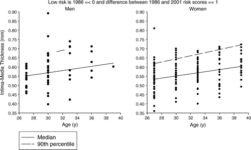
CIMT thickness measures both the intima and media thickness. Discrete plaque can also be seen, but this finding is unusual before the fourth decade of life Citation10. Though the absolute value of these measures is small, advances in ultrasound allow these measurements to be performed reliably. There are two clinical limitations to its use. There is a strong age independence to measured values independent of cardiovascular risk, with age-related increases demonstrated in individuals without cardiovascular risk. This makes determination of normal values difficult (). Also, it is difficult to separate the media from the intima when making measurements; increases in the former may more accurately reflect pathologic smooth muscle cell hypertrophy whereas increases in the latter may reflect atherosclerosis Citation40.
Figure 1. The relationship of age to carotid intima media thickness (CIMT) thickness by gender in young adults with a pathobiological determinants of atherosclerosis in youth study (PDAY) risk score of 0 (age excluded) in 1986 and no increase over time; Cardiovascular Risk in Young Finns cohort.

Increased CIMT has been demonstrated in children with familial hypercholesterolemia, and the presence of familial hypercholesterolemia is associated with a significant increase in rate of change in CIMT measures over time compared to unaffected individuals Citation42. Familial hypercholesterolemia genotype also has an impact on CIMT measures, with more severe mutations associated with higher levels of total cholesterol and CIMT Citation43. In a landmark randomized trial conducted for 2 years, Wiegman et al. have shown that pravastatin treatment can reduce CIMT in adolescents and young adults Citation7. In patients with hypertension, there is a strong relationship between CIMT and LV mass Citation44.
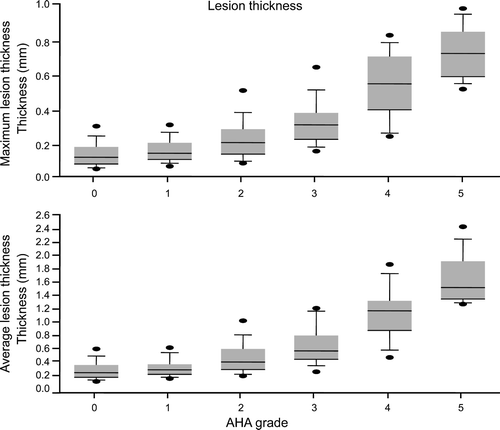
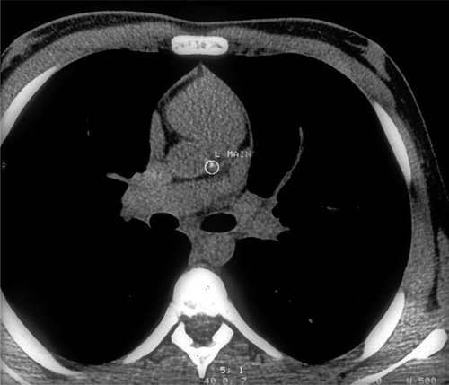
After performance of a chest CT scan, a coronary calcium score is calculated from identified opacities in the distribution of the coronary arteries Citation2. The major limitation of coronary calcium measurement in youth relates to the fact that calcium deposition in atherosclerotic lesions occurs late in the pathologic process unless calcium homeostasis is abnormal, such as in end stage renal disease. Thus, significant atherosclerosis can be present in the absence of calcium, and changes in atheroma volume may not be accurately assessed by changes in coronary calcium Citation45. Nonetheless small amounts of coronary calcium have been identified in 25%–30% of adolescents with familial hypercholesterolemia; higher body mass index makes the presence of calcium more likely () Citation6.
Figure 2. Maximum and mean right coronary artery wall thickness in pathobiological determinants of atherosclerosis in youth study (PDAY) specimens is shown in relation to American Heart Association (AHA) atherosclerosis grade (minimum, median, maximum, 25th and 75th percentiles are shown).

Figure 3. Coronary calcium in the left main (L Main) coronary artery of a 14-year-old boy with familial hypercholesterolemia is shown.

A critical finding related to subclinical atherosclerosis measurement and the pediatric and young adult prevention of atherosclerosis has come from studies where risk factors have been measured in youth or young adulthood, and measures of atherosclerosis have been performed in young adults after a substantial interval (15–20 years) has passed. In four such studies (Muscatine, Bogalusa, Cardiovascular Risk in Young Finns, and CARDIA), some risk factors measured in youth are better predictors of future atherosclerosis, both CIMT and coronary calcium, than are risk factors measured concurrently with the radiologic examination Citation8–11.
Using findings from the Pathobiologic Determinants of Atherosclerosis in Youth study (PDAY), a study of atherosclerosis in late adolescents and young adults dying of accidental causes and with risk factors measured post mortem, a PDAY risk score was developed to provide a global estimate of coronary atherosclerosis risk in a given individual (). Using this score both baseline risk and change in risk over time could be calculated. In both the Young Finns (CIMT) and CARDIA (coronary calcium) studies, change in risk status between initial measurement and measurement at the time of the study impacted outcome Citation11, Citation46. The presence of initial high risk with risk worsening predicts a 9-fold increase in likelihood of the presence of coronary calcium at age 33–45 years compared to sustained low risk over a 15-year interval; lowering of risk in high-risk individuals does not return markers to values seen in low-risk individuals.
Table I. PDAY risk score.
Though both CIMT and coronary calcium have been critical to the understanding of early atherogenesis, each has important limitations. CIMT is the technique of choice for the assessment of the effect of clinical interventions on subclinical markers but, because of the absence of agreed-upon normal values, cannot be used for prevalence estimates. Coronary calcium is ideal for estimates of prevalence but should not be performed before 20–25 years of age, and its role in intervention studies is limited.
Summary
Noninvasive measures of cardiac end organ injury have contributed significantly to our understanding of the evolution of cardiovascular disease from childhood to young adulthood. Current status of these measures has been summarized (). Advances in cardiac imaging, including higher-resolution CT scanning and magnetic resonance imaging (not discussed here), may advance the field further. The PDAY risk score provides a tool whereby high global risk individuals can be identified to have a high likelihood of positive imaging studies as young individuals. Clinical trials of risk reduction can be initiated in these persons as well as those with familial hypercholesterolemia and diabetes mellitus to enhance understanding of the role of more aggressive treatment in those with accelerated vascular disease.
Table II. Current status of noninvasive measures of cardiovascular end organ injury.
Acknowledgements
Figures 1 and 2 were provided by C. Alex McMahan, PhD, of the PDAY Study; Cardiovascular Risk in Young Finns data were provided by Olli Raitakari MD, PhD.
References
- Benjamin EJ, Levy D. Why is left ventricular hypertrophy so predictive of morbidity and mortality?. Am J Med Sci. 1999; 317: 168–75
- Greenland P, Bonow RO, Brundage BH, Budoff MJ, Eisenberg MJ, Grundy SM, et al. ACCF/AHA 2007 clinical expert consensus document on coronary artery calcium scoring by computed tomography in global cardiovascular risk assessment and in evaluation of patients with chest pain: a report of the American College of Cardiology Foundation Clinical Expert Consensus Task Force (ACCF/AHA Writing Committee to Update the 2000 Expert Consensus Document on Electron Beam Computed Tomography). Circulation. 2007; 115: 402–26
- Fuster V, Fayad ZA, Moreno PR, Poon M, Corti R, Badimon JJ. Atherothrombosis and high-risk plaque: Part II: approaches by noninvasive computed tomographic/magnetic resonance imaging. J Am Coll Cardiol. 2005; 46: 1209–18
- Gidding SS. The aging of the cardiovascular system: when should children be treated like adults?. J Pediatr. 2002; 141: 159–61
- National High Blood Pressure Education Program Working Group on High Blood Pressure in Children and Adolescents. The fourth report on the diagnosis, evaluation, and treatment of high blood pressure in children and adolescents. Pediatrics. 2004; 114 Suppl 4th Report:555–76.
- Gidding SS, Bookstein LC, Chomka EV. Usefulness of electron beam tomography in adolescents and young adults with heterozygous familial hypercholesterolemia. Circulation. 1998; 98: 2580–3
- Wiegman A, Hutten BA, de Groot E, Rodenburg J, Bakker HD, Buller HR, et al. Efficacy and safety of statin therapy in children with familial hypercholesterolemia: a randomized controlled trial. JAMA. 2004; 292: 331–7
- Mahoney LT, Burns TL, Stanford W, Thompson BH, Witt JD, Rost CA, et al. Coronary risk factors measured in childhood and young adult life are associated with coronary artery calcification in young adults: the Muscatine Study. J Am Coll Cardiol. 1996; 27: 277–84
- Li S, Chen W, Srinivasan SR, Bond MG, Tang R, Urbina EM, et al. Childhood cardiovascular risk factors and carotid vascular changes in adulthood: the Bogalusa Heart Study. JAMA. 2003; 290: 2271–6
- Raitakari OT, Juonala M, Kahonen M, Taittonen L, Laitinen T, Maki-Torkko N, et al. Cardiovascular risk factors in childhood and carotid artery intima-media thickness in adulthood: the Cardiovascular Risk in Young Finns Study. JAMA. 2003; 290: 2277–83
- Gidding SS, McMahan CA, McGill HC, Colangelo LA, Schreiner PJ, Williams OD, et al. Prediction of Coronary Artery Calcium in Young Adults Using the Pathobiological Determinants of Atherosclerosis in Youth (PDAY) Risk Score: The CARDIA Study. Arch Intern Med. 2006; 166: 2341–7
- Gardin JM, Lauer MS. Left ventricular hypertrophy: the next treatable, silent killer?. JAMA. 2004; 292: 2396–8
- Devereux RB, Wachtell K, Gerdts E, Boman K, Nieminen MS, Papademetriou V, et al. Prognostic significance of left ventricular mass change during treatment of hypertension. JAMA. 2004; 292: 2350–6
- Gardin JM, Wagenknecht LE, Anton-Culver H, Flack J, Gidding S, Kurosaki T, et al. Relationship of cardiovascular risk factors to echocardiographic left ventricular mass in healthy young black and white adult men and women. The CARDIA study. Coronary Artery Risk Development in Young Adults. Circulation. 1995; 92: 380–7
- Urbina EM, Gidding SS, Bao W, Pickoff AS, Berdusis K, Berenson GS. Effect of body size, ponderosity, and blood pressure on left ventricular growth in children and young adults in the Bogalusa Heart Study. Circulation. 1995; 91: 2400–6
- Gardin JM, Brunner D, Schreiner PJ, Xie X, Reid CL, Ruth K, et al. Demographics and correlates of five-year change in echocardiographic left ventricular mass in young black and white adult men and women: the Coronary Artery Risk Development in Young Adults (CARDIA) study. J Am Coll Cardiol. 2002; 40: 529–35
- de Simone G, Devereux RB, Maggioni AP, Gorini M, de Divitiis O, Verdecchia P. Different normalizations for body size and population attributable risk of left ventricular hypertrophy: the MAVI study. Am J Hypertens. 2005; 18: 1288–93
- Daniels SR, Loggie JM, Khoury P, Kimball TR. Left ventricular geometry and severe left ventricular hypertrophy in children and adolescents with essential hypertension. Circulation. 1998; 97: 1907–11
- Gidding SS, Nehgme R, Heise C, Muscar C, Linton A, Hassink S. Severe obesity associated with cardiovascular deconditioning, high prevalence of cardiovascular risk factors, diabetes mellitus/hyperinsulinemia, and respiratory compromise. J Pediatr. 2004; 144: 766–9
- Lorber R, Gidding SS, Daviglus ML, Colangelo LA, Liu K, Gardin JM. Influence of systolic blood pressure and body mass index on left ventricular structure in healthy African-American and white young adults: the CARDIA study. J Am Coll Cardiol. 2003; 41: 955–60
- Lipshultz SE, Lipsitz SR, Sallan SE, Dalton VM, Mone SM, Gelber RD, et al. Chronic progressive cardiac dysfunction years after doxorubicin therapy for childhood acute lymphoblastic leukemia. J Clin Oncol. 2005; 23: 2629–36
- Daniels SR, Witt SA, Glascock B, Khoury PR, Kimball TR. Left atrial size in children with hypertension: the influence of obesity, blood pressure, and left ventricular mass. J Pediatr. 2002; 141: 186–90
- Deanfield JE, Halcox JP, Rabelink TJ. Endothelial function and dysfunction: testing and clinical relevance. Circulation. 2007; 115: 1285–95
- Brunner H, Cockcroft JR, Deanfield J, Donald A, Ferrannini E, Halcox J, et al. Endothelial function and dysfunction. Part II: Association with cardiovascular risk factors and diseases. A statement by the Working Group on Endothelins and Endothelial Factors of the European Society of Hypertension. J Hypertens. 2005; 23: 233–46
- Rocchini AP, Moorehead C, Katch V, Key J, Finta KM. Forearm resistance vessel abnormalities and insulin resistance in obese adolescents. Hypertension. 1992; 19: 615–20
- Woo KS, Robinson JT, Chook P, Adams MR, Yip G, Mai ZJ, et al. Differences in the effect of cigarette smoking on endothelial function in chinese and white adults. Ann Intern Med. 1997; 127: 372–5
- Dhillon R, Clarkson P, Donald AE, Powe AJ, Nash M, Novelli V, et al. Endothelial dysfunction late after Kawasaki disease. Circulation. 1996; 94: 2103–6
- Clarkson P, Celermajer DS, Donald AE, Sampson M, Sorensen KE, Adams M, et al. Impaired vascular reactivity in insulin-dependent diabetes mellitus is related to disease duration and low density lipoprotein cholesterol levels. J Am Coll Cardiol. 1996; 28: 573–9
- Celermajer DS, Adams MR, Clarkson P, Robinson J, McCredie R, Donald A, et al. Passive smoking and impaired endothelium-dependent arterial dilatation in healthy young adults. N Engl J Med. 1996; 334: 150–4
- Charakida M, Donald AE, Terese M, Leary S, Halcox JP, Ness A, et al. Endothelial dysfunction in childhood infection. Circulation. 2005; 111: 1660–5
- Leeson CP, Whincup PH, Cook DG, Donald AE, Papacosta O, Lucas A, et al. Flow-mediated dilation in 9- to 11-year-old children: the influence of intrauterine and childhood factors. Circulation. 1997; 96: 2233–8
- Whincup PH, Gilg JA, Donald AE, Katterhorn M, Oliver C, Cook DG, et al. Arterial distensibility in adolescents: the influence of adiposity, the metabolic syndrome, and classic risk factors. Circulation. 2005; 112: 1789–97
- Raitakari OT, Ronnemaa T, Jarvisalo MJ, Kaitosaari T, Volanen I, Kallio K, et al. Endothelial function in healthy 11-year-old children after dietary intervention with onset in infancy: the Special Turku Coronary Risk Factor Intervention Project for children (STRIP). Circulation. 2005; 112: 3786–94
- Jarvisalo MJ, Jartti L, Marniemi J, Ronnemaa T, Viikari JS, Lehtimaki T, et al. Determinants of short-term variation in arterial flow-mediated dilatation in healthy young men. Clin Sci (Lond) 2006; 110: 475–82
- de Jongh S, Lilien MR, op't Roodt J, Stroes ES, Bakker HD, Kastelein JJ. Early statin therapy restores endothelial function in children with familial hypercholesterolemia. J Am Coll Cardiol. 2002; 40: 2117–21
- Watts K, Beye P, Siafarikas A, O'Driscoll G, Jones TW, Davis EA, et al. Effects of exercise training on vascular function in obese children. J Pediatr. 2004; 144: 620–5
- Meyer AA, Kundt G, Lenschow U, Schuff-Werner P, Kienast W. Improvement of early vascular changes and cardiovascular risk factors in obese children after a six-month exercise program. J Am Coll Cardiol. 2006; 48: 1865–70
- Abbott RA, Harkness MA, Davies PS. Correlation of habitual physical activity levels with flow-mediated dilation of the brachial artery in 5–10 year old children. Atherosclerosis. 2002; 160: 233–9
- Kallio K, Jokinen E, Raitakari OT, Hamalainen M, Siltala M, Volanen I, et al. Tobacco smoke exposure is associated with attenuated endothelial function in 11-year-old healthy children. Circulation. 2007; 115: 3205–12
- Roman MJ, Naqvi TZ, Gardin JM, Gerhard-Herman M, Jaff M, Mohler E. American society of echocardiography report. Clinical application of noninvasive vascular ultrasound in cardiovascular risk stratification: a report from the American Society of Echocardiography and the Society for Vascular Medicine and Biology. Vasc Med. 2006; 11: 201–11
- Lorenz MW, Markus HS, Bots ML, Rosvall M, Sitzer M. Prediction of clinical cardiovascular events with carotid intima-media thickness: a systematic review and meta-analysis. Circulation. 2007; 115: 459–67
- Wiegman A, de Groot E, Hutten BA, Rodenburg J, Gort J, Bakker HD, et al. Arterial intima-media thickness in children heterozygous for familial hypercholesterolaemia. Lancet. 2004; 363: 369–70
- Koeijvoets KC, Rodenburg J, Hutten BA, Wiegman A, Kastelein JJ, Sijbrands EJ. Low-density lipoprotein receptor genotype and response to pravastatin in children with familial hypercholesterolemia: substudy of an intima-media thickness trial. Circulation. 2005; 112: 3168–73
- Sorof JM, Alexandrov AV, Cardwell G, Portman RJ. Carotid artery intimal-medial thickness and left ventricular hypertrophy in children with elevated blood pressure. Pediatrics. 2003; 111: 61–6
- Nicholls SJ, Tuzcu EM, Wolski K, Sipahi I, Schoenhagen P, Crowe T, et al. Coronary artery calcification and changes in atheroma burden in response to established medical therapies. J Am Coll Cardiol. 2007; 49: 263–70
- McMahan CA, Gidding SS, Viikari JS, Juonala M, Kahonen M, Hutri-Kahonen N, et al. Association of Pathobiologic Determinants of Atherosclerosis in Youth risk score and 15-year change in risk score with carotid artery intima-media thickness in young adults (from the Cardiovascular Risk in Young Finns Study). Am J Cardiol. 2007; 100: 1124–9