Abstract
Background. Angina with normal coronary arteries, cardiac syndrome X, is a diagnosis of exclusion. The exact mechanism of this clinical syndrome remains unclear. Although the prognosis is as good as equal to that of the normal population, symptoms related to the syndrome largely impair quality of life. Aortic pulse and fractional pulse pressures are strong and independent indicators of the risk of coronary heart disease. An increase in these hemodynamic parameters is significantly associated with the presence of coronary artery disease in men and women. Methods and results. We evaluated aortic pulse and fractional pulse pressures of patients with cardiac syndrome X and control subjects, and investigated the relation between the two groups. One hundred and twenty‐six patients with cardiac syndrome X with a mean age of 53.1±9.5 years and 76 patients without the syndrome with a mean age of 53.1±11.2 years were studied consecutively from May 2004 to May 2005. Aortic systolic, diastolic, mean and pulse pressures were measured and the fractional pulse pressure was calculated (aortic pulse pressure/mean pressure). Aortic pulse and fractional pulse pressures were significantly higher in the cardiac syndrome X group than in the control group (51±13 mmHg and 43±9 mmHg, p<0.001; 0.54±0.11 and 0.47±0.08, p<0.001, respectively). All patients were classified into tertiles of aortic pulse pressure level to evaluate whether aortic pulse pressure was associated with the presence of cardiac syndrome X in the study. The multiple‐adjusted odds ratio of the risk of cardiac syndrome X was 6.72 (95% CI 2.76–16.37) for tertile 2 and 29.94 (95% CI 5.59–160.44) for tertile 3 of aortic pulse pressure level compared with tertile 1. In addition, all patients were divided into two groups as lower and higher levels of aortic fractional pulse pressure. The multiple‐adjusted OR of the risk of cardiac syndrome X was 4.09 (95% CI 2.05–8.15) for the higher group compared with the lower group of aortic fractional pulse pressure level. Conclusion. Ascending aorta pulse and fractional pulse pressures are significantly associated with the presence of cardiac syndrome X and these associations are independent of age and other cardiovascular risk factors.
Introduction
In patients with cardiac syndrome X, typical exertional angina pectoris and abnormal stress tests (exercise electrocardiographic or nuclear scintigraphic testing) with normal coronary angiogram are present. It was first described by Kemp et al. in 1973 Citation[1]. Some mechanisms have been postulated for this clinical entity: (i) microvascular dysfunction and ischemia called microvascular anginaCitation[2,3]; (ii) abnormal pain perception Citation[4], and (iii) sympathetic–parasympathetic imbalance resulted in increased sympathetic tone. At present, the first hypothesis has become widely accepted, although a combination of the mechanisms mentioned above has been observed in some patients with angina and angiographically normal coronary arteries.
Pulse pressure, the difference between systolic and diastolic blood pressures, has been shown to be associated with adverse cardiovascular events independently Citation[5,6]. Invasively measured pulse pressure via catheterization gives more reliable and accurate values than indirect measurements through a sphygmomanometer. Danchin et al. have shown an association between the presence and extent of coronary artery disease and aortic pulse pressure in men undergoing diagnostic coronary angiography Citation[7]. In addition, aortic fractional pulse pressure has been demonstrated to be associated with the presence and extent of coronary atherosclerosis in some studies Citation[8,9]. However, no study has examined the association between aortic pulse/fractional pulse pressures and cardiac syndrome X.
The purpose of our study was to evaluate the difference in aortic pulse/fractional pulse pressure values between patients with and without cardiac syndrome X.
Methods
Study population
We studied 126 consecutive patients (48 men; 78 women; mean age 53.1±9.6 years) who had typical exertional angina pectoris and abnormal stress testing (for exercise stress test, ⩾1 mm horizontal or down‐sloping ST‐segment depression; for scintigraphic test, abnormal tomographic perfusion areas concerning with ischemia) with angiographically normal coronary arteries, who were admitted to Yuksek Ihtisas Heart‐Education and Research Hospital between May 2004 and May 2005 for the first time. Seventy‐six patients (30 men, 46 women; mean age 53.1±11.2 years) with a diagnosis of supraventricular tachycardia were included in the study as control subjects. These subjects had a negative stress test and angiographically normal coronary arteriogram, which was performed for the detection of underlying pathology associated with tachycardia. None of the patients was on drug therapy at the time of admission to hospital. Two consecutive weekly clinic visits during each of which blood pressure measurements were obtained for all patients. Hypertension was diagnosed if the average of the three blood pressure measurements at the two clinic visits was consistently elevated over 140 systolic and/or 90 diastolic. In addition, previously diagnosed and treated hypertensive states were accepted as hypertension, although measured blood pressures were lower than 140 systolic and/or 90 diastolic. Exclusion criteria included the following: known coronary artery disease; left ventricular dysfunction (left ventricular ejection fraction <50%) and hypertrophy; unstable ischemic conditions (unstable angina pectoris and myocardial infarction); valvular heart disease; rhythms other than sinus; renal or hepatic dysfunction (creatinine >1.2 mg/dl, AST and ALT more than twice the upper limit of normal, respectively); and metabolic syndrome.
Diagnosis of cardiac syndrome X
Components of cardiac syndrome X are typical chest pain (angina pectoris), abnormal stress test and angiographically normal coronary arteries. Subjects with these components were diagnosed as having cardiac syndrome X.
Control subjects had previously diagnosed supraventricular tachycardia with negative stress test and angiographically normal coronary arteries.
Treadmill testing with Bruce protocol was applied as exercise stress testing; ⩾1 mm horizontal or down‐sloping ST‐segment depression after 80 ms from the J point was used for positive/abnormal tests. Some subjects had an exercise or pharmacological scintigraphic stress test with ischemic signs (SPECT or Tl‐201).
Cardiac catheterization
All patients in the study underwent selective coronary artery angiography after appropriate patient preparation. Femoral artery cannulation was used for the arterial access site and a Judkins system was applied for cannulating the left and right coronary arteries. All angiograms were evaluated by two experienced physicians blinded to the study. Angiograms without stenotic lesion in all major epicardial coronary arteries including left main, left anterior descending, left circumflex and right coronary arteries were considered normal angiograms.
Measurement of hemodynamic parameters
Hemodynamic measurements including systolic and diastolic blood pressures were assessed using the pigtail system during cardiac catheterization for each patient. The average of at least five pressure waveforms on the paper with a speed of 25mm/s were used for analysis by a physician blinded to the study.
Calculation of hemodynamic parameters:
Aortic pulse pressure = systolic pressure−diastolic pressure,
Aortic mean pressure = 1/3 systolic pressure+2/3 diastolic pressure,
Aortic fractional pulse pressure = aortic pulse pressure/aortic mean pressure.
Laboratory data
Fasting peripheral venous blood samples were obtained from all patients in the study for the measurement of fasting plasma glucose, total cholesterol, low‐density lipoprotein (LDL)‐cholesterol, high‐density lipoprotein (HDL)‐cholesterol and triglyceride levels. Blood samples were centrifuged and plasma was obtained. Fasting blood glucose, total cholesterol, HDL‐cholesterol and triglyceride levels were measured by different laboratory techniques. Measurement of LDL‐cholesterol level was done through application of a formula as described by Friedewald et al. Citation[10].
Anthropometric measurement
Height and weight of patients were measured and body mass index (BMI) was calculated by dividing weight in kilograms by height in meters squared and described as kg/m2.
Statistical analysis
Data were analyzed with the SPSS software version 10.0 for Windows (SPSS Inc., Chicago, Illinois, USA). Continuous variables were presented as mean±SD and categorical variables as frequency and percentage. The Kolmogorov–Smirnov test was applied to assess the distribution of continuous variables. Student's t‐test was used to compare normally distributed continuous variables and the Mann–Whitney U test for variables without normal distribution. A two‐tailed p‐value of <0.05 was considered statistically significant. Multiple logistic regression analysis was used to evaluate the independent associates of cardiac syndrome X. Parameters with a p‐value <0.1 in univariate analysis were included in the model. The odds ratios (OR) and 95% confidence intervals (CI) were calculated.
Results
Baseline characteristics
Baseline demographic, laboratory, and hemodynamic characteristics of patients in both groups were outlined in .
Table I. Baseline demographic, laboratory, and hemodynamic characteristics.
Thirty‐eight per cent of patients were male with a mean age of 53.6±10.3 years and 62% of the cases were female with a mean age of 52.8±9.1 years in the cardiac syndrome X group; 39% of patients were male and 61% were female with a mean age of 47.9±10.6 and 56.5±10.3 years in the control group, respectively. The prevalence of diabetes mellitus was significantly higher in the cardiac syndrome X group compared with controls. There were no significant differences between the two groups concerning age, sex and other cardiovascular risk factors, including hypertension, smoking and family history of coronary artery disease. In addition, both groups did not significantly differ in BMI, total, HDL‐, LDL‐cholesterols and triglyceride levels.
Hemodynamic parameters including heart rate and aortic diastolic pressure measured during catheterization were not different between two groups. However, measured aortic systolic, mean, pulse and fractional pulse pressures were significantly higher in patients with cardiac syndrome X than in those without the syndrome ().
It was shown that aortic pulse and fractional pulse pressures–coronary atherosclerosis relationship is much stronger in women compared with men Citation[11]. Interestingly, the aortic pulse pressure–cardiac syndrome X relationship was much stronger in women compared with men in our study (53±13 mmHg and 48±11 mmHg, p = 0.02, respectively). However there was no significant gender influence on the fractional pulse pressure–cardiac syndrome X relationship (women, 0.55±0.11; men, 0.53±0.10, p = 0.38). In addition, there was no significant gender influence on the aortic pulse and fractional pulse pressures–cardiac syndrome X relationship in normotensives in our study:
Women, 46±8 mmHg; men, 45±7 mmHg, p = 0.52 for aortic pulse pressure,
Women, 0.51±0.08; men, 0.53±0.09, p = 0.33 for aortic fractional pulse pressure.
Multivariate analysis of the presence of cardiac syndrome X
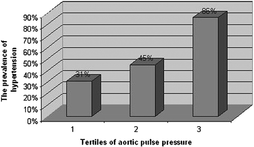
All patients were classified into tertiles of aortic pulse pressure level to evaluate whether aortic pulse pressure was associated with the presence of cardiac syndrome X in the study. We found that aortic pulse pressure was positively and significantly associated with the presence of cardiac syndrome X (). Forty‐two per cent of patients in the lowest tertile, 78% in the middle and 88% in the highest tertile were found to have cardiac syndrome X. The crude OR of the risk of cardiac syndrome X was 4.99 (95% CI 2.40–10.37) for the middle and 10.20 (95% CI 3.70–28.12) for the highest tertile of aortic pulse pressure level compared with the lowest tertile. The multiple‐adjusted OR of the risk of cardiac syndrome X was 6.72 (95% CI 2.76–16.37) for tertile 2 and 29.94 (95% CI 5.59–160.44) for tertile 3 of the aortic pulse pressure level compared with the lowest tertile, tertile 1 after adjustment for age, sex, BMI, the presence of hypertension, diabetes mellitus, smoking habit, family history of coronary artery disease, serum levels of total, LDL‐cholesterols and triglycerides. Therefore, aortic pulse pressure was significantly associated with the risk of cardiac syndrome X (for a 10‐mmHg increase in aortic pulse pressure: OR 20.47; 95% CI 1.38–304.57). The prevalence of hypertension also varied between tertiles of aortic pulse pressure ().
Table II. OR of cardiac syndrome X according to aortic pulse and fractional pulse pressures.
Figure 1. The prevalence of hypertension and tertiles of aortic pulse pressure.
In addition, all patients were divided into two groups as lower and higher levels of aortic fractional pulse pressure (). Crude cumulative incidence rates of the presence of cardiac syndrome X were 46% for the lower and 79% for the higher group of aortic fractional pulse pressure level (p for trend <0.001). After adjustment for the same variables mentioned above, the multiple‐adjusted OR of the risk of cardiac syndrome X was 4.09 (95% CI 2.05–8.15) for the higher group compared with the lower group of aortic fractional pulse pressure level.
Discussion
In our study, we found that patients with cardiac syndrome X had significantly increased aortic pulse and fractional pulse pressures compared with control subjects. In addition, these significant and positive correlations were independent of risk factors for coronary artery disease.
Cardiac syndrome X has been considered as a subgroup of cardiac metabolic syndrome that affects a large portion of the population. However, as compared with the metabolic syndrome, the prognosis of this syndrome is generally excellent Citation[12]. Several possible causes for this clinical entity have been suggested. Although microvascular angina has been proposed to be the main reason, the exact mechanism of the syndrome is not known. Recently, more evidence of ischemia has been shown with the use of new imaging techniques like magnetic resonance and nuclear scintigraphic imaging Citation[13,14].
Increased pulse pressure has been shown to be independently associated with adverse cardiovascular events in large cohorts of the population Citation[5,6]. Although these studies have included male subjects largely, it has been shown that increased aortic pulse and fractional pulse pressures are significantly associated with the presence of angiographic coronary artery disease in women Citation[15]. Although measured brachial pulse pressure has been shown to be significantly associated with the risk of coronary heart disease Citation[6], invasively measured aortic pulse pressure and evaluated fractional pulse pressure via catheterization gives more accurate and reliable values compared with sphygmomanometer‐measured brachial blood pressure. Pulse pressure also increases from central to peripheral arteries. Increased aortic pulse pressure is associated with increased aortic stiffness. When the aortic wall has stiffened due to atherosclerotic process, systolic blood pressure increases and the diastolic blood pressure decreases relative to mean pressure because of shortened time constant of diastolic blood pressure decay resulted in increased aortic pulse pressure. Therefore, central aortic pulse pressure is a more reliable and accurate indicator of aortic stiffness. Aortic fractional pulse pressure has also been shown to be strongly associated with angiographic coronary heart disease in men and women Citation[9],Citation[15]. Because aortic stiffness has been shown to be related with coronary atherosclerosis Citation[16], aortic pulse pressure is more strongly associated with coronary atherosclerosis than the peripheral pulse pressure. In addition, other than aortic pulse pressure, it has been also shown that fractional pulse pressure is independently associated with coronary atherosclerosis Citation[9],Citation[15].
In the present study, we studied patients with normal coronary angiograms. Therefore, which mechanism is responsible for the significant increase in aortic pulse and fractional pulse pressures in patients with cardiac syndrome X compared with control subjects? The endothelium of the vascular tissue has many functions for the continuation of the normal vascular physiology. It plays a key role in maintaining vascular homeostasis through balancing endothelial‐derived relaxing and contracting factors. The role of the endothelium in controlling the vascular tone, especially vasodilatation, has been shown via the endothelial‐derived relaxing factor, which was later named as nitric oxide (NO) Citation[17,18]. Endothelial dysfunction occurs when NO activity in the vascular tissue is decreased, resulting in decreased endothelium‐dependent vasodilatation. Therefore, higher aortic pulse and fractional pulse pressures in patients with cardiac syndrome X might be the result of endothelial dysfunction caused by impaired vascular resistance. Although in our study we conclude that higher aortic pulse and fractional pulse pressures in patients with cardiac syndrome X were due to endothelial dysfunction, in the literature, pulsatile flow and endothelial function are usually positively related Citation[19].
In addition, autonomic imbalance in the central and peripheral nervous system, which is another underlying pathophysiological mechanism in cardiac syndrome X, might cause increased aortic pulse and fractional pulse pressures in this clinical entity. Sympathetic nervous hyperactivity results in increased heart rate and systemic vascular resistance. Thus elevated sympathetic tonus causes increased systolic and unchanged or decreased diastolic blood pressures. Therefore, aortic pulse pressure and so fractional pulse pressures are increased.
Study limitations
Since contrast angiography is a type of lumenography, diffusely diseased coronary arteries might not be detected properly, resulting in underestimation of lesions and overestimation of angiographically normal coronary arteries. In addition, although a rather large number of the study population was used in our study, we believe it is still too limited in number to generalize the results because of invasive nature of the study. Lastly, the use of a Millar conductance catheter for aortic pressure measurement is more appropriate than the use of a pigtail system catheter.
Conclusion
We have demonstrated that aortic pulse and fractional pulse pressures, indicators of the risk of coronary artery disease, in patients with cardiac syndrome X was significantly and independently elevated compared with control subjects. Although prognosis is good, prospective studies with large sample sizes using multivariate survival analysis are needed for prognostic evaluation of increased aortic pulse and fractional pulse pressures in patients with syndrome X in real‐life practice on long‐term follow‐up.
Acknowledgement
There are no conflicts of interest regarding the authors.
References
- Kemp H. G., Jr., Vokonas P. S., Cohn P. F., Gorlin R. The anginal syndrome associated with normal coronary arteriograms. Report of a six year experience. Am J Med 1973; 54: 735–742
- Chauhan A., Mullins P. A., Taylor G., Petch M. C., Schofield P. M. Both endothelium‐dependent and endothelium‐independent function is impaired in patients with angina pectoris and normal coronary angiograms. Eur Heart J 1997; 18: 60–68
- Mohri M., Koyanagi M., Egashira K., Tagawa H., Ichiki T., Shimokawa H., et al. Angina pectoris caused by coronary microvascular spasm. Lancet 1998; 351: 1165
- Pasceri V., Lanza G. A., Buffon A., Montenero A. S., Crea F., Maseri A. Role of abnormal pain sensitivity and behavioral factors in determining chest pain in syndrome X. J Am Coll Cardiol 1998; 31: 62–66
- Benetos A., Safar M., Rudnichi A., Smulyan H., Richard J. L., Ducimetieere P., et al. Pulse pressure: A predictor of longterm cardiovascular mortality in a French male population. Hypertension 1997; 30: 1410–1415
- Franklin S. S., Khan S. A., Wong N. D., Larson M. G., Levy D. Is pulse pressure useful in predicting risk for coronary heart disease? The Framingham Heart Study. Circulation 1999; 100: 354–360
- Danchin N., Benetos A., Lopez‐Sublet M., Demicheli T., Safar M., Mourad J. J., on behalf of the ESCAPP Investigators. Aortic pulse pressure is related to the presence and extent of coronary artery disease in men undergoing diagnostic coronary angiography: A multicenter study. Am J Hypertens 2004; 17: 129–133
- Jankowski P., Kawecka‐Jaszcz K., Czarnecka D., Brzozowska‐Kiszka M., Styczkiewicz K., Styczkiewicz M., et al. Ascending aortic, but not brachial blood pressure‐derived indices are related to coronary atherosclerosis. Atherosclerosis 2004; 176: 151–155
- Nishijima T., Nakayama Y., Tsumura K., Yamashita N., Yoshimaru K., Ueda H., et al. Pulsatility of ascending aortic blood pressure waveform is associated with an increased risk of coronary heart disease. Am J Hypertens 2001; 14: 469–473
- Friedewald W. T., Levy R. I., Fredrickson D. S. Estimation of the concentration of low density lipoprotein cholesterol in plasma, without use of the preparative ultracentrifuge. Clin Chem 1972; 18: 499–502
- Jankowski P., Kawecka‐Jaszcz K., Czarnecka D., Bryniarski L. Ascending aortic blood pressure waveform may be related to the risk of coronary artery disease in women, but not in men. J Hum Hypertens 2004; 18: 643–648
- Kemp H. G., Kronmal R. A., Vlietstra R. E., Frye R. L. Seven year survival of patients with normal or near normal coronary arteriograms. A CASS registry study. J Am Coll Cardiol 1986; 7: 479
- Panting J. R., Gatehouse P. D., Yang G. Z., Grothues F., Firmin D. N., Collins P., et al. Abnormal subendocardial perfusion in cardiac syndrome X detected by cardiovascular magnetic resonance imaging. N Engl J Med 2002; 346: 1948–1953
- Buchthal S. D., den Hollander J. A., Merz C. N., Rogers W. J., Pepine C. J., Reichek N., et al. Abnormal myocardial phosphorus‐31 nuclear magnetic resonance spectroscopy in women with chest pain but normal coronary angiograms. N Engl J Med 2000; 342: 829–835
- Guray Y., Guray U., Altay H., Cay S., Yilmaz M. B., Kisacik H. L., et al. Aortic pulse pressure and aortic pulsatility are associated with angiographic coronary artery disease in women. Blood Press 2005; 14: 293–297
- Weber T., Auer J., O'Rourke M. F., Kvas E., Lassnig E., Berent R., et al. Arterial stiffness, wave reflections, and the risk of coronary artery disease. Circulation 2004; 109: 184–189
- Furchgott R. F., Zawadzki J. V. The obligatory role of endothelial cells in the relaxation of arterial smooth muscle by acetylcholine. Nature 1980; 288: 373–376
- Moncada S., Higgs A. The L‐arginine‐nitric oxide pathway. N Engl J Med 1993; 329: 2002–2012
- Hwang J., Ing M. H., Salazar A., Lassegue B., Griendling K., Navab M., et al. Pulsatile versus oscillatory shear stress regulates NADPH oxidase subunit expression: Implication for native LDL oxidation. Circ Res 2003; 93: 1225–1232