
ABSTRACT
Functional Magnetic Resonance Imaging (FMRI) is a noninvasive test to analyze several medical ailments by using magnetic resonance imaging (MRI) to detect the abnormalities in the active part of the brain and evaluate the minute changes in the blood flow, which cannot otherwise be accomplished with other imaging techniques. With its vast applications in healthcare, it has become one of the most explored studies by the researcher’s community, therefore, the current paper aims to address a comprehensive systematic literature review (SLR) of the application of FMRI in healthcare. The SLR scrutinized and assessed the currently available literature using inclusion and exclusion criteria. The chief motive of conducting SLR on the current research was to eradicate the biases and make it more systematic as compared to the informal literature review. The outcomes of the review state that due to accessibility of the public datasets and the data augmentation practices, the application of FMRI in Healthcare has remarkably raised from the last five years and its application is practically available for every disease diagnosis. The performance of the diagnosis of the disease is more effectual and proficient as equal to the human experts performing it manually.
Introduction
Artificial Neural Network (ANN) is the network of neurons which are connected together. Deep Learning is the study of ANN (LeCun, Bengio, and Hinton Citation2015). Deep Learning is considered as the fourth industrial revolution (4IR) due to availability of huge amount of data (labeled or unlabeled) and data generation techniques such as GANs (Goodfellow et al. Citation2014).
The major application of the Deep Learning is the Healthcare. With the help of Deep learning, we can not only diagnose the disease but also predict the future occurrence of diseases. The rises in the application are due to availability of lots of public datasets such as ChestX-ray14 datasets (Rajpurkar et al. Citation2017) for pneumonia detection, Autism Brain Imaging Data Exchange (ABIDE) (Martino et al. Citation2014) for the detection of autism spectrum disorder (ASD), Alzheimer’s Disease Neuroimaging Initiative (ADNI) (Petersen et al. Citation2010) for the detection of Alzheimer disease and many more. Similarly, there are many challenges like Grand Challenges in Medical Image Analysis (Grand Challenge, 2021), Automatic Non-rigid Histological Image Registration (ANHIR) (Anhir Grand Challenge, 2021) challenge and Challenges in Global Health and Development (Grand Challenge Maps, 2021) Problems was held for increasing the performance of diseases diagnosis on respected datasets. Among the different modalities of images for disease diagnoses Functional Magnetic Resonance Imaging (FMRI) is important and most widely used neuroimaging technique which is specifically used for understanding the brain activity.
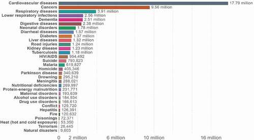
Healthcare is risky field which is directly concerned with the life of human beings. In one year, lots of people lost their life due to certain disease and also in developing countries like Pakistan where many peoples even do not able to diagnose the disease earlier due to poor health facilities and financial issues illustrated in . Artificial Intelligence provides the facility of effective and efficient diagnosis, prognosis and treatment as well. The below graph shows that huge amount of population died due to cardiovascular diseases, cancer, and lung diseases, respectively, and early diagnosis and prognosis increases the chances of patient survival rate.
Figure 1. World overall causes of death in 2017 (Our world in data, 2021).
So, it is observed that Healthcare is important field of research and spending therefore in this review we are going to address the comprehensive application of FMRI in Deep Learning for medical disease diagnosis.
Medical image classification is an important research topic and is widely used in the treatment of various diseases. In the past few years, traditional machine learning classifiers have been used extensively for the diagnosis of such diseases, but they are not speed and space efficient (Razzak, Naz, and Zaib Citation2018). Therefore, researchers have moved toward the deep neural network models as their performance is relatively very high (Zhao et al. Citation2016). Convolutional Neural Network (CNN) gives the best result when used for different classification tasks. A wide variety of research has been conducted in this area where researchers have used CNN for effective classification and diagnosis of different diseases using medical images. In CNN, the step of feature selection is done by the algorithm itself because of which it increases the performance of the algorithm (Hijazi, Kumar, and Rowen Citation2015). Transfer learning is a technique in which already trained model is used for classification of other problems. Some of the layers from the trained models are used for solving or classifying the new problem of interest. Most of the problems related to CNN are solved using method of transfer learning which saves the time and reduces the complexity as well. Different architectures of CNN can be used and with the help of them high accuracies can be achieved (Jmour, Zayen, and Abdelkrim Citation2018).
Human brain is considered as the most intricate organ of the human body which subsumes into hundreds of billion nerve cells that are further linked with thousands of other nerve cells. Therefore, a minute injury or disease can lead to drastic impairments (Mohsen et al. Citation2018). However, the neuroscientists and brain researchers have mastered a colossal amount of physical linkage in the brain with addition to how the complex nervous system supplies and processes the information (Pennese et al. Citation2015). Currently, most of the human brain research, circles around noninvasive methodologies which limits the analysis of the brain at cellular level, i.e., Magnetic Resonance Imaging (MRI). To overcome the limitations of noninvasive diagnostic approaches, FMRI, a medical imaging technique, was brought to light by Seiji Ogawa in 1990, which escorted novel insights into the advancement and treatment of copious neurological disorders (Cohen et al. Citation2017). It assesses the brain activity by utilizing the Blood Oxygen Level-Dependent (BOLD) signals over time that accord cognizance of local actions of the neurons. The interdisciplinary study of FMRI is highly influenced by signal processing, graph theory and machine learning (Liu Citation2016). In recent times machine learning has acquired quite an emerging interest of brain researchers utilizing several algorithms to train the classifiers to decode the activities of the stimuli and other variables of interest from FMRI data Holzinger Citation2016). Techniques in supervised learning are used for the prediction and classification of various aspects for an example, patient’s disease prognosis and his prediction of its treatment. Similarly, unsupervised learning techniques focuses on the structure of multiple functional networks within the brain and divulge its structure and dynamics. Pattern recognition has also aided in recognizing the patterns of the brain (Garg Citation2017). Both these studies have offered a series of powerful tools and techniques to neuroimaging data analysis. In 1990, FMRI was initially been used which is a medical imaging technique that assesses the activities and behaviors of the brain (Kwong et al. Citation1992), (Ogawa et al. Citation1992). The study of FMRI deals with the associations between the activation of the brain and the responsibilities accomplished by the participants during a brain scan (Amaro and Barker Citation2006). It evaluates the brain activity by consuming the Blood Oxygen Level Dependent (BOLD) signals over time that accord cognizance of local actions of the neurons. With the progression in the fields of image processing, machine learning, virtual reality, and artificial intelligence, the extension of the applications of FMRI to routine and nonclinical situations has brought a great breakthrough currently (Frackowiak et al. Citation2004), (Toga and Mazziotta Citation1996). The applications encompassed the techniques for the subject stimuli generation, experiment control, and the combined activation of stimuli tracing . MediaStim framework (Ford et al. Citation1998) was initiated for the multimedia data analysis and authoring in neuroimaging. Numerous computational methodologies have extensively been used (Shereena and Raju Citation2016) that comprised of copious statistical methods (Mahmoudi et al. Citation2012) and preprocessing techniques for the reduction of noise and enhancing quality (CitationSomkuwar and Bhargava). FMRI is broadly applied in various subjects that include economics, neuroscience, computer science, psychology, and political science (Lindquist et al. Citation2020). The software used most extensively in the study of image analysis and segmentation with FMRI data include, SMP and FSL of MatLab (Pinho et al. Citation2013).
Based on the previous prominent literature review we are going to answer the following questions:
RQ1) Is proposed method detecting disease accurately?
RQ2) What will be the future of Healthcare and How it will affect the human being?
RQ3) Is performance of Automatic disease diagnosis being better than human experts?
RQ4) Proposed method contribute to medical world as a life savior.
The rest of the paper is organized as follows. The next Section II will include comprehensive review of some literature of application of FMRI in Biomedical Imaging. The answer of the above asked questions is mentioned in discussion Section III. Finally, the last Section IV will conclude our review with future work.
Research Methodology
In the current research our aim is to perform a systematic literature review (SLR) on the application of FMRI in disease diagnosis. In the following SLR we had scrutinized, organized and assessed the current available literature related to the disease diagnosis with FMRI by using inclusion and exclusion criteria. The chief motive of conducting SLR on the current research is to eradicate the biases and make it more systematic as compared to the informal literature review. Following are the three core phases of our SLR methods that include, planning the review, conducting the review and then reporting the review. . Represents the phases of SLR.
Phase 1: Planning the Review
The first step of our proposed research is to derive research questions related to extensive applications of FMRI in disease diagnosis. We selected a suitable data warehouse, enlisted the search strings, stated the inclusion, and exclusion criteria and then defined the quality evaluation criteria. One by one all these steps are defined in detail below.
Research Questions
The current research is focused on the comparative literature review of the application of FMRI in the disease diagnosis. Based on the previous prominent literature review we will answer the following research questions:
RQ1: Is the proposed method detecting the disease accurately?
RQ2: What will be the future of healthcare and how it will affect human beings?
RQ3: Is the performance of Automatic disease diagnosis better than human experts?
RQ4: Is the proposed method contributing to the medical world as a life savior?
Data Sources
An opposite data warehouse was acknowledged in the light of previous research works. List of data warehouses are specified in .
Table 1. Data search sources
Search Strings
The comprehensive search strings were developed using the key search terms used in the title. These terms were based on the existing research articles of disease diagnosis using FMRI. The search strings along with their repositories that were searched for the current research are listed in .
Table 2. Research string used
Inclusion Criteria
Following are the criteria that was used to select which type of literature explored by the search strings will be effective for data extraction.
Each paper must be in journal or conference.
Papers should be related to FMRI.
Papers that have our search strings in their titles are selected.
Then these paper’s introduction and conclusion are studied.
In next phase all these papers’ introduction and conclusion is read.
In the next phase all these papers are thoroughly read.
Papers should be in English.
Papers published between January 2014 to December 2020 are considered.
Exclusion Criteria
The exclusion criteria disregard the irrelevant findings from the total selected literature associated with the search strings. Following are the exclusion criteria for the current research:
Eliminate the papers that do not have our search strings.
Papers in case of replication, the most complete and latest version of paper published are included.
Study Quality Evaluation
Based on the inclusion and exclusion criteria, we have selected the papers for the current research review. The QE checklist comprises four questions (QE1–QE4). For every provided item (QE1–QE4), the quality evaluation score (QES) was assigned in . The questions of quality evolution criteria (QE1–QE4) are useful to measure the extent to which a paper is effective and appropriate for addressing the research question of the current research. Furthermore, the aim of study quality assessment is to ensure that a particular study finding will make a valuable contribution to our SLR.
Table 3. Quality evaluation criteria of selected studies
Primary Study Selection
Initially, 70 research works were explored using the search strings from the mentioned online repositories in by following an inclusion criteria and exclusion criteria. The selected research papers were processed to improve using the tollgate approach. This method followed five phases shown in and .
Table 4. Tollgate approach
Figure 2. Tollgate approach for paper selection.
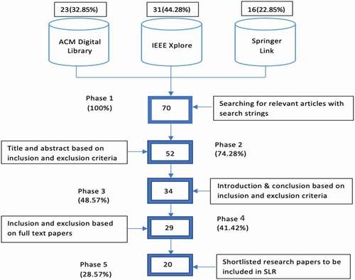
After the tollgate method was applied, 20 research papers got shortlisted out of 50. The selected studies were evaluated with the mentioned QE criteria. A list of the finally selected articles is given in Appendix A. Every selected primary study was labeled as [SP] to indicate it as the SLR study.
Data Extraction and Synthesis
The process of data extraction investigates the gained information from the selected studies that relates to our research questions. For data extraction firstly, we develop a formal form by using Microsoft Excel Sheet to record the study-wise concepts, ideas, contributions and findings. The following data was extracted by using this form.
Study title
Publication year
Used research methods
The chosen studies were studied carefully to excerpt the concepts, notions and contributions related to the application of FMRI in biomedical imaging by the first two authors. The remaining two authors carefully reviewed the data extracted from selected studies to reduce the biases of researchers thus enhancing the validity of the application of FMRI in disease diagnosis.
Phase 3: Reporting the Review
Quality Attributes
The quality evaluation (QE) score for all the shortlisted research papers which were determined based on four QE questions. This analysis of the QE score indicated to which extent the selected papers are effective to answer the research questions of the current study.
Temporal Distribution of the Selected Primary Studies
For the selected primary articles, the publication years were selected from January 2014 to December 2020.
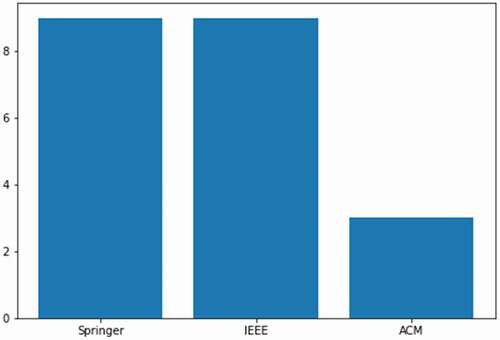
The bar plot of the final selected papers is presented in . The figure shows that out of overall 21 papers selected nine were from springer, nine from IEEE, and three from MDPI.
Figure 3. Total number of final selected papers.
Literature Review
The selection criteria in term of keyword searching and Repositories are shown in .
Table 5. Research paper selection criteria
(Ramzan et al. Citation2020) trained a deep residual neural network combined with other transfer learning techniques for Alzheimer disease classification into cognitively normal (CN), significant memory concern (SMC), early mild cognitive impairment (EMCI), mild cognitive impairment (MCI), late mild cognitive impairment (LMCI), and Alzheimer’s disease (AD), respectively. Subsequently, the performance of proposed study was compared with other Alzheimer detection strategies. The datasets used in the experimentation was Alzheimer Disease Neuroimaging (ADNI) and ImageNet. The performance of proposed method was evaluated by using precision, recall, f1 measure, AUC and ROC metrics. The accuracy of CN, SMC, EMCI, LMCI, MCI, and AD were 100%, 96.85%, 97.38%, 97.43%, 97.4%, and 98.01%, respectively.
Similarly, (Duc et al. Citation2020) proposed a Convolutional Neural Network (CNN) method for the detection of Alzheimer disease then the Mini Mental State Examination was predicted by using different machine learning techniques such as linear least square regression, Support Vector Machine, bagging based methods and tree-based method. The datasets used in the experimentation was obtained from Chosun University National Dementia Research Center (Gwangju, South Korea). The AD detection accuracy of proposed CNN was 85.27%. By applying ensemble of all machine learning method, the lowest root mean square error was (3.27 + 0.58) and highest R2 was (0.620 + .02).
(Bi et al. Citation2020) proposed a deep feature extraction method for the AD detection. Specifically, the CNN was used to learn deep regional connectivity features and Recurrent Neural network method was used to learn adjacent positional features. Then the performance of AD detection was improved by using Extreme Learning Machine (ELM). The ADNI datasets was used in the experimentation. The AUC, accuracy, and recall were the evaluation criteria used in the experimentation.
(Aghdan et al. Citation2019) proposed a CNN based model for the detection of ASD. The proposed CNN was the combination of different classifiers. Additionally, the comparison among the techniques was also performed. The performance of proposed method was evaluated on Autism Brain Imaging Data Exchange (ABIDE I and ABIDE II) datasets. The ABIDE I datasets is composed of 116 entries out of which 62 are typical control (TC) and 54 are ASD entries. Similarly, the ABIDE II is composed 343 entries out of which 187 are TC and 156 are ASD. The accuracy, sensitivity and specificity on ABIDE I was 0.72, 0.712, and 0.73, respectively. Subsequently, the accuracy, sensitivity and specificity on ABIDE I was 0.7, 0.582, and 0.804, respectively. Similarly, the accuracy, sensitivity, and specificity on combination of ABIDE I and ABIDE II were 0.7045, 0.679, and 0.7421, respectively. Similarly, (Aghdan et al. Citation2018) they also proposed a Deep Belief Network (DBN) for the detection of ASD in children by using the combination of rs-FMRI and structured-MRI. A DBN is defined by the stack of Restricted Boltzmann Machine (RBM). Therefore, multiple stacks of RBM were trained in the proposed method. The performance of proposed method was validated by using ABIDE I and ABIDE II. The accuracy, sensitivity, specificity and F1-score of the proposed method were 65.56%, 84%, 32.96%, and 74.76%, respectively.
(Bi et al. Citation2020) performed the data analysis of Parkinson disease. Specifically, a multiple statistical strategy was evaluated to predict brain pathological areas and also to detect the discriminative genes related to Parkinson disease. The datasets used in the analysis were ADNI and Parkinson Progression Markers Initiative (PPMI). The PPMI was specifically used to discriminate genes. The experimentation results were evaluated by using accuracy. The results indicate that combination of clustering evolution random forest (CERF) and Pearson were better than other techniques in term of accuracy which was 88.1%.
(Oh et al. Citation2019) proposed CNN for AD detection. Specifically, an unsupervised convolutional autoencoder network was used for the classification of AD and normal control (NC) additionally, transfer learning technique was used for the classification of progressive mild cognitive impairment (pMCI) and stable mild cognitive impairment. The validation of proposed method was performed on ADNI datasets and classification accuracies of AD and PMCI were 86.60% and 73.95% respectively.
Similarly, (Khazaee, Ebrahimzadeh, and Babajani-Feremi Citation2016) proposed a combination of graph theory and machine learning for the classification of AD and Healthy control. Precisely, statistical analysis of graph measurement of brain signals and multiclass Support Vector Machine (SVM) algorithms were proposed for the classification HC, MCI, and AD. The datasets used for the validation were ADNI datasets. The accuracies for classifying HC from AD and MCI, AD from HC and MCI, and MCI from HC and AD, were 87.3%, 97.5%, and 72.0%, respectively.
Recently, (Sheng et al. Citation2020) used a basic machine learning technique for distinguishing the AD, cognitive impairment (MCI) from HC. Specifically, a joint human connectome project multimodal parcellation (JHCPMMP) was used form Data preprocessing then logistic regression recursive feature elimination (LR-RFE) was used for feature selection and finally SVM, logistic regression, and KNN was applied for classification. The datasets used by them was ADNI. The maximum accuracy was obtained by using SVM, i.e., 89% for AD vs. MCI vs. HC.
(Shi and Liu Citation2020) proposed a classification framework for the detection MCI. First of all, HilbertHuang transform (HHT) was used for decomposing rs-FMRI into intrinsic mode functions. Then, Hilbert weighted frequencies (HWFs) was applied to detect Region of Interest (ROI) and finally, SVM was implemented to classify MCI into different stages. The validation of proposed method was performed on ANDI datasets. The accuracy of the proposed method was 87.94% and decrease to 81.56% when noise was introduced.
(Bruening et al. Citation2015) proposed an algorithm named partial voluming effects correction (PVEc) to produce CBF images. These images have independence theoretically of associated PVE and the tissue content. Tissue information has been taken from structural images having high resolution by using that tissue information CBF has been computed. Limitation of their proposed algorithm is inherent smoothing of functional data has been introduced in this PVEc method.
(Moti et al. 2008) presented a model based on FMRI that helps in early detection of liver metastases. To characterizing colorectal hepatic metastases the proposed method is using FMRI statistics, model helps in following their early hemodynamical changes. To differentiate metastatic and healthy tissues of liver a classification model has been presented with accuracy of 84.38% having precision of 80%. This approach is not helping in completely automatic detection of metastases, classification model has accuracy lesser than 90% more achievement required in this regard.
(Neehal et al. Citation2020) used SVM classifier to classify with prediction of Parkinson’s disease their goal is to detect it in early stages. In proposed model they have achieved 99.53% of sensitivity whereas model is accurate upto 99.76% specificity of 100%. Limitation in proposed solution is accuracy is only dependent on 8 subjects, accuracy may vary if there are more subjects.
(Al-sharoa, Al-khassaweneh, and Aviyente Citation2018) proposed a framework that shows evolution of FMRI connectivity of networks that are constructed across different region of interests. By using tucker decomposition of tensor, the proposed method is based on determination of subspace that describe the community structure. Results showing dynamic connectivity of brain.
(Guo and Zhang Citation2020) used neural network functionality approach that allows detection of Alzheimer’s disease recognition. As compared to R-FMRI results the proposed learning method is significantly much improved and standard deviation has been reduced by 45%. Methodology includes classification of R-FMRI model, Brain networks development, autoencoder pretraining. Due to variation in classification based on random data sample production the experimentation of model should be repeated ten times which is a limitation of proposed method.
(Manokar et al. Citation2012) present decomposition and classification of FMRI images that belongs to patient having Alzheimer’s and Tumors positive, the proposed method is based on neural networks. Cannys edge detection algorithm has been used to find optimal edges of any image. However, the percentage of accuracy is 50 to 60 which could be enhanced.
(Li et al. Citation2018) proposed method to detect Alzheimer’s disease using smaller datasets in perspective of knowledge transfer. In proposed method disparity among different datasets has been diminished and accuracy increased by 20% which is quite significant. Proposed method is intended toward addressing the problem due to smaller datasets. Methods seems qualitative but measurements that shows quality should be quantitative and discrete, it is unknown whether change in dataset cause variation in accuracy or not.
(Dachena et al. Citation2020) shows application of MRI, FMRI and cognitive data for Alzheimer’s disease detection. A method has been proposed to classify or discriminate Alzheimer’s disease using support vector machine. Multimodel approach has been used in order to get satisfactory results, the model provides accuracy of 95.65% whereas specificity is 97.22% and 93.39% of sensibility. Proposed method is only tested on a single data set which is considerably limitation of method.
(Hanson, Westlye, and Lundervold Citation2014) proposed a data-driven approach that address graphs which are subject specific as well as threshold on different level. The study illustrated on simple synthetic graphs and tested on state FMRI of healthy people that have varying genetic risks of Alzheimer’s disease.
(Huang, Liu, and Tan Citation2020) proposed a multitask learning that enables diagnose of ASD and attention deficit hyperactivity disorder by using resting state FMRI data sets. Proposed method includes graph-based feature selection to filter out irrelevant features. The classification performance achieved are a) mean accuracy of 0.687 with tolerance of 0.005 b) accuracy in ASD and attention deficit hyperactivity disorder is 0.650 with 0.014 of tolerance.
Discussion
A comprehensive literature review of F-MRI for disease detection is performed. In this review, total 20 paper from IEEE, ACM and Springer is selected by using different variant of F-MRI keywords. We examined 20 latest research papers and individual proposed research method is explained in details with their corresponding datasets and results. Based on the review, we observed that Convolutional Neural Network is the most widely used technique in F-MRI modalities. Subsequently, transfer learning is also widely used for disease diagnosis in F-MRI. Similarly, we also observed that F-MRI is mostly used for ADNI detection with maximum accuracy of more than 90%. The review also reveals the fact that with the development of powerful Deep learning libraries (Keras and TensorFlow) and more computation power, the application of Artificial Intelligence in medical imaging is tremendously rises.
In introduction section four question were coined;
Is proposed method detecting disease accurately?
To the best of our knowledge the maximum classification accuracy of ADNI was 95% and classification accuracy of Parkinson disease was approximately 97% which shows the effectiveness of disease detection using Deep learning for F-MRI modality. Similarly, maximum sensitivity and specificity was between 97% and 99% for ADNI and 95% and 98% for Parkinson disease.
What will be the future of Healthcare and How it will affect the human being?
In future, due to availability of deep learning libraries and more computation power we are not only able to diagnosis the disease efficiently but also able to find the complex patterns in the datasets. Nowadays, particularly in developing countries the manually diagnosis and prognosis of diseases are expensive, time consuming and inefficient but in future we world not able to diagnosis easily but also able to find the complex pattern in F-MRI datasets.
Is performance of Automatic disease diagnosis being better than human experts?
With the help of Artificial Intelligence specifically machine learning, we can achieve approximate or more accuracy for disease diagnosis and prognosis with little resources and low time complexity as compared to human experts. Therefore, we conclude that performance of automatic disease diagnosis is better than human performance.
Is proposed method contribute to medical world as a life savior?
Efficient diagnosis of any disease plays important role in the savior of life. As the literature review reveals the accuracy of disease diagnosis is more than 95% therefore, we conclude that the efficient disease diagnosis through F-MRI using machine learning is consider as life savior.
The summary of the findings is listed in .
Table 6. Summary of literature review
Table 7. Summary discussion
Conclusion and Future Work
In this paper, a comprehensive literature review of the application of FMRI in disease diagnosis is performed. Based on the review, it is concluded that, due to availability of public datasets learning and data augmentation techniques the application of F-MRI in Healthcare tremendously rises from last five years and almost every disease is diagnosis easily. The performance of disease diagnosis is more effective and efficient as compared to human experts However, the prognosis of the disease is not efficient at this time due to insufficient of patterns data but we can able to predict the cause of certain disease in near future efficiently. In future, we are interested to review the more categories of the Medicals with more focus on the technical content of the respective architecture because this effort is more toward theoretical contents. For instance, in addition to current research columns another column which contain information about the numbers of hidden layer should be added.
Disclosure Statement
No potential conflict of interest was reported by the author(s).
Additional information
Funding
References
- Aghdam, M. A., A. Sharifi, and M. M. Pedram. 2018. Combination of rsfMRI and sMRI data to discriminate autism spectrum disorders in young children using deep belief network. Journal of Digital Imaging 31 (6):895–903. doi:https://doi.org/10.1007/s10278-018-0093-8.
- Aghdam, M. A., A. Sharifi, and M. M. Pedram. 2019. Diagnosis of autism spectrum disorders in young children based on resting-state functional magnetic resonance imaging data using convolutional neural networks. Journal of Digital Imaging 32 (6):899–918. doi:https://doi.org/10.1007/s10278-019-00196-1.
- Al-sharoa, E., M. Al-khassaweneh, and S. Aviyente. 2018. Tensor based temporal and multilayer community detection for studying brain dynamics during resting state fMRI. IEEE Transactions on Biomedical Engineering 66 (3):695709.
- Amaro, E., and G. J. Barker. 2006. Study design in fMRI: Basic principles. Brain and Cognition 60 (3):220–32. doi:https://doi.org/10.1016/j.bandc.2005.11.009.
- Bi, X., X. Zhao, H. Huang, D. Chen, and M. Yuliang. 2020. Functional brain network classification for Alzheimer’s disease detection with deep features and extreme learning machine. Cognitive Computation 12 (3):513–27.
- Bi, X.-A., H. Wu, Y. Xie, L. Zhang, X. Luo, and Y. Fu. 2020. The exploration of Parkinson’s disease: A multi-modal data analysis of resting functional magnetic resonance imaging and gene data. Brain Imaging and Behavior 1–11.
- Bruening, D. E., S. Dharssi, R. M. Lazar, R. S. Marshall, and I. Asllani. 2015. Improved partial volume correction method for detecting brain activation in disease using Arterial Spin Labeling (ASL) fMRI. In 2015 37th Annual International Conference of the IEEE Engineering in Medicine and Biology Society (EMBC), Milano, Italy, 5441–5444. IEEE.
- Cohen, J. D., N. Daw, B. Engelhardt, U. Hasson, K. Li, Y. Niv, K. A. Norman, J. Pillow, P. J. Ramadge, N. B. Turk-Browne et al. 2017. Computational approaches to fMRI analysis. Nature Neuroscience 20(3):304–13. doi:https://doi.org/10.1038/nn.4499.
- Dachena, C., S. Casu, M. B. Lodi, A. Fanti, and G. Mazzarella. 2020. Application of MRI, fMRI and cognitive data for Alzheimer’s disease detection. In 2020 14th European Conference on Antennas and Propagation (EuCAP), Copenhagen, Sweden, pp. 1–4. IEEE.
- de Pinho Queirós, G., G. Coelho, S. Wang, Y. Zhang, T. Zhan, P. Phillips, Y. D. Zhang, G. Liu, S. Lu, and X. Wu. 2016. Pathological brain detection by artificial intelligence in magnetic resonance imaging scanning (invited review). Progress in Electromagnetics Research 156:105–33. https://doi.org/10.2528/PIER16070801.
- Di Martino, A., C.-G. Yan, Q. Li, E. Denio, F. X. Castellanos, K. Alaerts, J. S. Anderson, M. Assaf, S. Y. Bookheimer, M. Dapretto et al. 2014. The autism brain imaging data exchange: Towards a large-scale evaluation of the intrinsic brain architecture in autism. Molecular Psychiatry 19 (6):659–67. doi:https://doi.org/10.1038/mp.2013.78.
- Duc, N. T., S. Ryu, M. N. I. Qureshi, M. Choi, K. H. Lee, and B. Lee. 2020. 3D-deep learning based automatic diagnosis of Alzheimer’s disease with joint MMSE prediction using resting-state fMRI. Neuroinformatics 18 (1):71–86. doi:https://doi.org/10.1007/s12021-019-09419-w.
- Ford, J., F. Makedon, C. Owen, S. Johnson, and A. J. Saykin. 1998. Stimulus tracking in functional magnetic resonance imaging (fMRI). In Proceedings of the sixth ACM international conference on Multimedia (MULTIMEDIA ‘98), Association for Computing Machinery, New York, NY, USA, 445–54. DOI: https://doi.org/10.1145/290747.290819
- Frackowiak R. S. 2004 Jan 26. Human brain function. Elsevier.
- Garg, H. 2017. Some new biparametric distance measures on single-valued neutrosophic sets with applications to pattern recognition and medical diagnosis. Information 8 (4):162. doi:https://doi.org/10.3390/info8040162.
- Goodfellow, I., J. Pouget-Abadie, M. Mirza, X. Bing, D. Warde-Farley, S. Ozair, A. Courville, and Y. Bengio. 2014. Generative adversarial nets. In Advances in neural information processing systems, 2672–80.
- Guo, H., and Y. Zhang. 2020. Resting state fMRI and improved deep learning algorithm for earlier detection of Alzheimer’s disease. IEEE Access 8:115383–92. doi:https://doi.org/10.1109/ACCESS.2020.3003424.
- Hanson, E. A., E. Westlye, and A. Lundervold. 2014. A PCA-based thresholding strategy for group studies of brain connectivity-with applications to resting-state fMRI. 2014 Southwest Symposium on Image Analysis and Interpretation, California, USA, IEEE.
- Hijazi, S., R. Kumar, and C. Rowen. 2015. Using convolutional neural networks for image recognition, 1–12. San Jose, CA, USA: Cadence Design Systems Inc.
- Holzinger, A. 2016. Interactive machine learning for health informatics: When do we need the human-in-the-loop? Brain Informatics 3 (2):119–31. doi:https://doi.org/10.1007/s40708-016-0042-6.
- Huang, Z.-A., R. Liu, and K. C. Tan. 2020. Multi-Task learning for efficient diagnosis of ASD and ADHD using Resting-State fMRI data. 2020 International Joint Conference on Neural Networks (IJCNN), Glasgow, USA. IEEE.
- Jmour, N., S. Zayen, and A. Abdelkrim. 2018. Convolutional neural networks for image classification. 2018 International Conference on Advanced Systems and Electric Technologies (IC_ASET), Hammamet, Tunisia, IEEE.
- Khazaee, A., A. Ebrahimzadeh, and A. Babajani-Feremi. 2016. Application of advanced machine learning methods on resting-state fMRI network for identification of mild cognitive impairment and Alzheimer’s disease. Brain Imaging and Behavior 10 (3):799–817. doi:https://doi.org/10.1007/s11682-015-9448-7.
- Kwong, K. K., J. W. Belliveau, D. A. Chesler, I. E. Goldberg, R. M. Weisskoff, et al. 1992. Dynamic magnetic resonance imaging of human brain activity during primary sensory stimulation. Proceedings of the National Academy of Sciences of the United States of America, USA 89:5675–79.
- Last Accessed, March 31, 2021. http://www.eiu.com/industry/healthcare
- Last Accessed, March 31, 2021. https://anhir.grand-challenge.org.
- Last Accessed, March 31, 2021. https://grand-challenge.org.
- Last Accessed, March 31, 2021. https://grandchallenges.org/#/map.
- Last Accessed, March 31, 2021. https://ourworldindata.org/grapher/annual-number-of-deaths-by-cause.
- LeCun, Y., Y. Bengio, and G. Hinton. 2015. Deep learning. nature 521 (7553):436–44. doi:https://doi.org/10.1038/nature14539.
- Li, W., Y. Zhao, X. Chen, Y. Xiao, Y. Qin et al. 2018. Detecting Alzheimer’s disease on small dataset: A knowledge transfer perspective. IEEE Journal of Biomedical and Health Informatics 23(3):1234–42. doi:https://doi.org/10.1109/JBHI.2018.2839771.
- Lindquist, M. A., Y. D. Zhang, Z. Dong, W. Sh, X. Yu, X. Yao, Q. Zhou, H. Hu, M. Li, C. Jiménez-Mesa, et al. 2020. Advances in multimodal data fusion in neuroimaging: Overview, challenges, and novel orientation. Information Fusion December1 64:149–87.
- Liu, T. T. 2016. Noise contributions to the fMRI signal: An overview. NeuroImage 143:141–51. doi:https://doi.org/10.1016/j.neuroimage.2016.09.008.
- Mahmoudi, A. H., S. Takerkart, F. Regragui, D. Boussaoud, and A. Brovelli. 2012. Multivoxel pattern analysis for fMRI data: A review. Hindawi Publishing Corporation Computational and Mathematical Methods in Medicine 2012:14. Article ID 961257. doi:https://doi.org/10.1155/2012/961257.
- Manokar, N. V., et al. 2012. Wavelets based decomposition and classification of diseased fMRI brain images for inter-racial disease types of Alzheimer’s Vs tumors using SOFM and enhancement by LVQ neural networks. 2012 2nd IEEE International Conference on Parallel, Distributed and Grid Computing, Himachal Pradesh, India, IEEE.
- Mohsen, H., E.-S. A. El-Dahshan, E.-S. M. El-Horbaty, A.-B. M. Salem et al. 2018. Classification using deep learning neural networks for brain tumors. Future Computing and Informatics Journal 3(1):68–71. doi:https://doi.org/10.1016/j.fcij.2017.12.001.
- Neehal, A. H., et al. 2020. Prediction of Parkinson’s disease by analyzing fMRI data and using supervised learning. 2020 IEEE Region 10 Symposium (TENSYMP), Dhaka, Bangladesh. IEEE.
- Ogawa, S., D. W. Tank, R. Menon, J. M. Ellermann, S. G. Kim, et al. 1992. Intrinsic signal changes accompanying sensory stimulation: Functional brain mapping with magnetic resonance imaging. Proceedings of the National Academy of Sciences of the United States of America, USA 89:5951–55.
- Oh, K., Y.-C. Chung, K. W. Kim, W.-S. Kim, and O. Il-Seok. 2019. Classification and visualization of Alzheimer’s disease using volumetric convolutional neural network and transfer learning. Scientific Reports 9 (1):1–16. doi:https://doi.org/10.1038/s41598-019-54548-6.
- Pannese, E. 2015. Neurocytology: Fine structure of neurons, nerve processes, and neuroglial cells. Neurocytology, Springer.
- Petersen, R. C., P. S. Aisen, L. A. Beckett, M. C. Donohue, A. C. Gamst, D. J. Harvey, C. R. Jack, W. J. Jagust, L. M. Shaw, A. W. Toga et al. 2010. Alzheimer’s disease neuroimaging initiative (ADNI): Clinical characterization. Neurology 74(3):201–09. doi:https://doi.org/10.1212/WNL.0b013e3181cb3e25.
- Rajpurkar, P., J. Irvin, K. Zhu, B. Yang, H. Mehta, T. Duan, D. Ding et al. Chexnet: Radiologist-level pneumonia detection on chest x-rays with deep learning. arXiv preprint arXiv:1711.05225 (2017).
- Ramzan, F., M. U. Ghani Khan, A. Rehmat, S. Iqbal, T. Saba, A. Rehman, and Z. Mehmood. 2020. A deep learning approach for automated diagnosis and multi-class classification of Alzheimer’s disease stages using resting state fMRI and residual neural networks. Journal of Medical Systems 44 (2):37. doi:https://doi.org/10.1007/s10916-019-1475-2.
- Razzak, M. I., S. Naz, and A. Zaib. 2018. Deep learning for medical image processing: Overview, challenges and the future, 323–50. Springer, Cham: Classification in BioApps.
- Sheng, J., M. Shao, Q. Zhang, R. Zhou, L. Wang, and Y. Xin. 2020. Alzheimer’s disease, mild cognitive impairment, and normal aging distinguished by multi-modal parcellation and machine learning. Scientific Reports 10 (1):1–10. doi:https://doi.org/10.1038/s41598-020-62378-0.
- Shereena, V. B., and G. Raju. 2016. Literature review of fMRI image processing techniques. 2016 International Conference on Wireless Communications, Signal Processing and Networking (WiSPNET), Chennai, India, 1473–76.
- Shi, J., and B. Liu. 2020. Stage detection of mild cognitive impairment via fMRI using Hilbert Huang transform based classification framework. Medical Physics 47 (7):2902–15. doi:https://doi.org/10.1002/mp.14183.
- Somkuwar, A., and S. Bhargava, Noise reduction techniques in medical imaging data-A review, 2nd International Conference on Mechanical, Electronics and Mechatronics Engineering (ICMEME’2013), June 17-18, 2013 London (UK).
- Toga, A. W., and J.-C. Mazziotta, Eds. 1996. Brain mapping: The methods. Academic Press.
- Zhao, H., O. Gallo, I. Frosio, J. Kautz, et al. 2016. Loss functions for image restoration with neural networks. IEEE Transactions on Computational Imaging 3 (1):47–57. doi:https://doi.org/10.1109/TCI.2016.2644865.