Abstract
Internal jugular venous catheters (IJVC) for hemodialysis are a commonly employed temporary vascular access for hemodialysis. Most hospitals still follow the use of blind technique, which uses anatomical landmarks. Even in the most experienced hands this procedure has a variable success rate. Ultrasound guidance can decrease the incidence of periprocedural complications and improve the success rate. In this randomized study we compared the procedure success rate and periprocedural complications in patients undergoing ultrasound guided vs. nonultrasound guided IJVC insertion for a temporary hemodialysis access. Methods. All patients subjected to insertion of an IJVC between March 2004 and June 2004 were enrolled into the study, randomized to either the blind (group A) or ultrasound guided (group B) procedure, which uses a portable ordinary ultrasound machine without a needle guide. The aseptic Saldinger technique was used for catheterization in both the groups. Baseline characteristics of patient and periprocedural events were recorded. Results. A total of 60 patients were randomized, 30 patients each in two groups. First attempt venous cannulation success rate was 56.7% in group A compared to 86.7% in group B. Chance of occurrence of adverse outcome was significantly more in the blind procedure (P = 0.020). A post-procedure chest radiograph done in all patient showed no complications. Conclusion. Ultrasound guided procedure for internal jugular vein catheter insertion using an ordinary ultrasound machine was significantly safer and more successful as compared to the blind technique.
Background
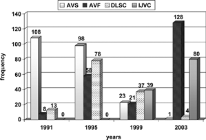
Success of hemodialysis depends on establishing and maintaining a good vascular access. Permanent vascular access in the form of an AV fistula or AV graft is commonly employed for this purpose. But not all patients are fortunate enough to have a functional permanent vascular access on the day they need their first hemodialysis. Some patients first come to obtain medical assistance when they are already in advanced renal failure and need dialysis; some may have a late referral to a nephrologist; some who are waiting for their vascular access to mature, might develop acute deterioration of renal function and present with a need for hemodialysis. In these cases there is an immediate need for a safe vascular access. Various central venous catheters have been used for this purpose. Internal jugular venous catheters (IJVC) are a commonly employed temporary vascular access for hemodialysis. Data from our center shows an increasing trend of IJVC use over the last decade ().
Most hospitals still follow the use of blind technique for IJVC catheter insertion, using anatomical landmarks. Even in the most experienced hands this procedure has a variable success rate and may be associated with periprocedural complications. Ultrasound guidance may decrease the incidence of periprocedural complications and improve the success rate. Ultrasound machines with a Site-Rite Probe have been used, but they are not widely available. Ordinary ultrasound machines without a needle guide are widely available to treating nephrologists. Therefore, we undertook this randomized study to compare the procedure success rate and periprocedural complications in patients undergoing ultrasound-guided vs. nonultrasound-guided IJVC insertion for a temporary hemodialysis access.
Methods
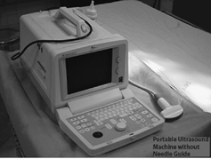
All patients subjected to insertion of an IJVC for temporary vascular access at our dialysis unit, from March 2004 to June 2004, were enrolled in the study. Patients were randomized to either the blind (group A) or the ultrasound-guided (group B) procedure. A portable ordinary ultrasound machine with a 3.5 MHz curved probe without a needle guide or any color doppler facility was used (). The aseptic Saldinger technique was used for catheterization in both the groups. All procedures were performed by nephrologists without any involvement from a radiologist. All nephrologists of our unit who had done at least 25 cases by either method were eligible to perform the procedure in the study population. Only a single catheter make of uncuffed double-lumen IJVC was used for all patients. Baseline characteristics of patient and periprocedural events were recorded. All patients were subjected to routine postprocedure chest radiograph.
Steps of Ultrasound Guided Catheter Insertion
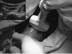
After cleaning and draping, the area is anesthetized with a local anesthetic agent; the appropriately disinfected ultrasound probe is kept parallel to the clavicle in the Selldot triangle (). The internal jugular vein can be easily identified as a round nonechogenic area that can be compressed by pressure on the ultrasound probe. It should be differentiated from the carotid artery that is pulsatile and lying medially and slightly deeper to the vein. Once the vein is identified, the introducer needle in inserted, puncturing the skin slightly superior to the probe directed at its midpoint and 45 degree from horizontal plane, in the direction of the vein. Once the vein has been punctured the ultrasound probe can be removed and further procedure is the same for both techniques.
Periprocedural events in terms of number of attempts to cannulate the vein were recorded. Each push of the needle was counted as an attempt and the change of direction of the needle, even without coming out of the skin puncture, was counted as a separate attempt. More than three attempts or inability to cannulate was counted as a failed procedure. Complications like carotid artery puncture, hematoma formation, or any others were recorded.
Results
A total of 60 patients were randomized during the study period, 30 in each group. These included 38 males and 22 females. All had a diagnosis of chronic renal failure (CRF). Eleven (18.3%) patients were diabetics. Seventy percent of patients were initially begun on hemodialysis within 2 weeks prior to the procedure. Eight (13.3%) patients had history of previous neckline insertion. All procedures were done on the right side.
Baseline characteristics between the two groups were comparable (). A special note was made for any factors that could adversely affect or increase the difficulty of the procedure. Group B had a greater number of patients with a short neck and an altered sensorium.
Table 1. Baseline characteristics
Among the periprocedural events (), the first attempt venous cannulation success rate was 86.7% in the ultrasound-guided procedure (group B) as compared to 56.7% in the blind-technique (group A) (P = 0.01). Chance of occurrence of adverse outcome was significantly more in the blind procedure (P = 0.020). A postprocedure chest radiograph done in all patient showed no complications.
Table 2. Periprocedural events
Discussion
With the increasing use of IJVC as temporary dialysis access it becomes important to learn safe, efficient, and easy to perform ways of cannulation. The landmark-guided approach has limitations. The location of the internal jugular vein is known to show variations.Citation[1] This can lead to difficulty in cannulation and other complications. Ultrasound guidance to cannulate the internal jugular vein has been reported earlier.Citation[2&3] Studies have described ultrasound guidance using a Site-Rite probe.Citation[4] This is expensive and may not be available at all centers. We utilized an ordinary probe, which is used for abdominal ultrasonography, and found that it can easily serve the purpose.
Previous studies have employed a single expert to perform all the procedure to maintain uniformity and eliminate any bias. But, if we look at the practical application, the procedure is performed by different doctors with variable clinical skill in day-to-day practice and, therefore, the results of a study based on procedures done by single expert may not predict the actual risk of adverse events to which the patient is exposed. Therefore, we included all nephrologists at our center that had done a minimum of 25 procedures with each method as participants in the study.
Ultrasonography is an important tool in the hands of a nephrologist and, as much as possible, it should be used for the management of patients. No radiologist was involved in the study. Randolph et al.Citation[5] point out that a variable definition of failed catheterization was used across the studies and possibly in the same study, which pose a limitation on interpreting these prior studies. In our study we have chosen more than three attempts or inability to cannulate as a failed procedure.
So what is the impact of ultrasound guided central venous catheterization on improving the success rate and reducing complications?
Cannulation of the internal jugular vein was achieved in all patients (100%) using ultrasound and in 93.3% using the landmark-guided technique. The vein was entered on the first attempt in 86.7% of patients using ultrasound and in 56.7% using the landmark technique (p < 0.010). Using ultrasound, puncture of the carotid artery did not occur and no other complications were noted in our study. Denys et al.Citation[2] have reported 100% success using ultrasound and 88.1% success using the landmark-guided technique. They found a first attempt success of 78% with ultrasound-guided technique (USG) and 38% using the landmark technique. They reported carotid artery puncture occurred in 1.7% of patients, brachial plexus irritation in 0.4%, and hematoma in 0.2% when using the ultrasound-guided approach. A chest radiograph was done postprocedure in all patients to note the position of the catheter tip. It did not show any other complications in our patients. These results show that even with use of an ordinary ultrasound machine without a needle guide, a significant number of patients benefit in terms of fewer periprocedural complications and better first-pass success rate.
Thus, the ultrasound-guided procedure of internal jugular vein catheter insertion using an ordinary ultrasound machine is significantly better in its safety and success as compared to the blind technique; and this procedure should be offered to all patients.
References
- Caridi, J G.; Hawkins, I F., Jr.; Wiechmann, B N.; Pevarski, D J.; Tonkin, J C. Sonographic guidance when using the right internal jugular vein for central venous access.Am. J. Roentgenol. 1998, 171:1259–1263. [CSA]
- Denys, B G.; Uretsky, B F.; Reddy, P S. Ultrasound-assisted cannulation of the internal jugular vein. A prospective comparison to the external landmark-guided technique.Circulation 1993, 87:1557–1562. [PUBMED], [INFOTRIEVE], [CSA]
- Teichgraber, U K.; Benter, T.; Gebel, M.; Manns, M P. A sonographically guided technique for central venous access.Am. J. Roentgenol. 1997, 169(3):731–733. [CSA]
- Muhm, M. Ultrasound guided central venous access.BMJ 2002, 325 (7377), 1373–1374. [PUBMED], [INFOTRIEVE], [CSA], [CROSSREF]
- Randolph, A G.; Cook, D J.; Gonzales, C A. Ultrasound guidance for placement of central venous catheters: a meta-analysis of the literature.Crit. Care Med. 1996, 24:2053–2058. [PUBMED], [INFOTRIEVE], [CSA], [CROSSREF]


