Abstract
“Watermelon stomach” is a common name for gastric antral vascular ectasia (GAVE syndrome). This endoscopic finding is characterized by the appearance of parallel longitudinal red columns along mucosal folds, along with capillars dilatation and hemorrhagy. Finding reliable method for its recognition is of paramount importance. Patient B.D., a 54-year-old woman, developed renal failure, which led to hemodialysis treatment, on the basis of pyelonephritis chronica. As a consequence of the gastrointestinal bleeding, the patient had black stools and developed severe anemia. The endoscopic finding showed the existence of visible columns of vessels transversing the antrum in longitudinal folds and converging in the pylorus, with clear red spots and surrounding hyperemy covered by drops of fresh blood. The diagnosis of “watermelon stomach” was confirmed after the pathohistological examination of the tissue taken at the biopsy, followed by total gastrectomy. Postoperative status was normal, without gastrointestinal hemorrhagia, and she went on with hemodialysis. Before the surgery she received 105 blood transfusions, and after surgical treatment she has received only 18 so far. At the moment she is in good health condition, and on hemodialysis. The reason we have reported this case of “watermelon stomach” syndrome in patient with chronic renal failure is to indicate that this rare anomaly of gastric blood vessels can lead to gastrointestinal blood loss in these patients. Since it is often the reason for many wrong diagnoses, it should be also taken into consideration in cases like these.
Introduction
“Watermelon stomach” is a common name for gastric antral vascular ectasia, the so-called GAVE syndrome. This syndrome is characterized by the appearance of parallel longitudinal red columns along mucosal folds, as a consequence of submucosal and mucosal capillaris dilatation as well as hemorrhagy.Citation[1&2] The etiology of this vascular anomaly is still unknown. The term “watermelon stomach” was first introduced by Jabarri et al. in 1984, as an explanation of the endoscopic finding, which resembled the exterior of a watermelon.Citation[3]
Gastric antral vascular ectasia is often the misdiagnosed cause of recurrent upper gastrointestinal bleeding because of its resemblance to gastritis and the diagnosis cannot be made solely based on endoscopic finding. Still, this disorder has been found in association with several clinical conditions, such as hepatic cirrhosis, some autoimmune diseases, and very rare chronic renal failure.Citation[1&2] Although chronic uremia, hemorrhagic gastritis, and bleeding peptic ulcer are presently often the reason for gastrointestinal blood loss, a connection between renal failure and GAVE syndrome is rarely found.
The disease usually appears in elderly women, with clinical manifestations that include sudden and acute hemorrhages leading to severe sideropenic anemia Although spontaneous termination of bleeding is possible, followed by numerous blood transfusions, for the time being this condition is treated by pharmacotherapy (estrogen-progesterone combination) or more often by bipolar electrocoagulation or argon plasma coagulation, which are considered very efficient.Citation[4-8] Surgical antrectomy is a final method to terminate the bleeding, with the risk of 7%.Citation[2]
Case
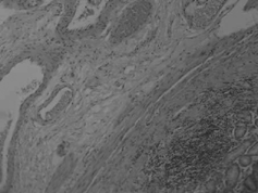
Patient B.D., a 54-year-old woman, has been on a chronic program of hemodialysis from April 1993 in The University Hospital Zemun, Belgrade. The patient was admitted with the diagnosis of chronic pyelonephritis. After the three years of treatment, it developed into renal failure and led to hemodialysis treatment. Markers for HBV and HCV infection were negative and she had no other chronic diseases (diabetes mellitus, heart failure, other autoimmune diseases, nor liver cirrhosis). The upper gastroscopy finding from May 1997 showed hypertrophic gastropathy. As a consequence of the gastrointestinal blood loss, the patient had a few black stools and after April 2000 she developed severe anemia. In order to clarify etiology of anemia, therral endoscopic examination of the upper gastrointestinal tract was conducted. The finding showed existence of visible columns of vessels transversing the antrum in longitudinal folds and converging in the pylorus, with clear red spots and surrounding hyperemy covered by drops of fresh blood (). A possible diagnosis was the “watermelon stomach” syndrome, which was confirmed after the pathohistological examination of the tissue taken at the biopsy. Namely, the histopathological finding showed dilated and thrombosed submucosa and mucose capillars and hemorrhages in mucosa and reactive fibromuscular hyperplasia of lamina propria, without inflammatory infiltrates ().
Figure 1 Endoscopic finding of Watermelon stomach—Endoscopic finding showed presence of erythematous, slightly elevated plaques above the gastric mucosa, confluent and radially disposed towards pylorus, resembling a watermelon bark (Watermelon stomach).
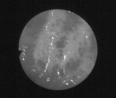
Figure 2 Histopathological finding of Watermelon stomach—On histopathologic remedy dyed with HE, a thicker lamina muscularis mucosae and thicker blood vessels in submucosus can be seen.
Before the diagnosis was confirmed, the patient underwent endoscopic examination of the jejunum, ileum, and colon, and selective angiography, but no pathological changes were found, nor could the place of bleeding be identified. The treatment began endoscopically with 1% ethoxysclerol, which caused sclerosis of described changes and that partially stopped the bleeding. Since the patient's condition worsened again and bipolar electrocoagulation or argon plasma coagulation were not available to us, total gastrectomy was a surgent's treatment of choice. The operative risk was estimated to be low, because of the patient's normal cardiovascular status at the time. The patient reacted well to surgical treatment. Postoperative status was normal, with fast correction of anemia and ferremia (before: RBC 1.38 × 10¹²/L, Hgb 40 g/L, HCT 13%, Fe 5.6 µmol/L, and after: RBC 3.6 × 10¹²/L, Hgb 105 g/L, HCT 34 %, Fe 18.5 µmol/L). She had no more gastrointestinal hemorrhagia and she has continued with hemodialysis treatment up to now.
From April 2000 till December 2001, when she underwent total gastrectomy, she received 105 blood transfusions. Yet, in period after surgical treatment, and up to now, she received only 18 transfusions (the last one in December 2003).
Latest biochemical findings showed satisfactory red blood cells count (3.10 × 10¹²/L, Hgb 92 g/L, HCT 29%), with serum creatinine values of 408 nmol/L and urea of 19 mmol/L. Additional therapy consisted of IV amino-acids, IV iron supplementation, and vitamin B12 once a week.
Discussion
The GAVE syndrome is a rare, but significant, cause of gastrointestinal blood loss. It is usually present in elderly women like in our patient. Clinically it is manifested as sideropenic anemia (88%), positive hemoccult (42%), melena (15%), hematemesis (3%), and seldom hematohesis (1%). This condition may also be associated with various autoimmune diseases (primary biliary cirrhosis, diabetes mellitus, hypothyreosis, sclerodermia).Citation[1], Citation[9] Achlorihydia and hypergastrinemia are considered important for development of this type of antral mucosis disorder, often present in liver cirrhosis.Citation[10] It is proposed that “watermelon stomach” is a consequence of repeated antral mucosus prolapses through the pylorus with trauma and mucosal ischemia Still our patient had no liver disease nor any of the mentioned autoimmune disorders.
In chronic renal failure, apart from GAVE syndrome which can be the cause for digestive bleeding, hemorrhagic gastritis and bleeding peptic ulcer may often be present as a consequence of uremia. In the literature we can find data indicating possible vascular anomalies as the cause of chronic bleeding in the jejunum and ileum in the form of angiodysplasia in patients with chronic renal failure.Citation[11&12]
Often GAVE is the reason for many wrong diagnoses, since it is a rarely reported cause of occult upper gastrointestinal blood loss.Citation[7] In our case, it took 3 months before the correct diagnosis was made, and in the meantime the patient received numerous blood transfusions. It is often mistaken for hemorrhagic gastritis, since the oozing bleeding prevents distinguishing of the correct diagnosis. This syndrome seems much like portal hypertensive gastropathy, which is only seen in liver cirrhosis, in which red spots are diffusely spread, just like the sneak skin, all over the fundus but also on antral mucosa.Citation[13]
In order to histopathologically confirm the GAVE syndrome it is necessary to detect the presence of dilated capillars within submucosis and mucosis, partially thrombosed, followed by fibromuscular hyperplasia in lamina propria and thickening of the submucosa with dilated tortuous venous channel.Citation[3] Portal hypertensive gastropathy has similar histological characteristics, but without fibromuscular hyperplasia and with a lower number of dilated capillars then in GAVE syndrome. This shows that these are two different histological entities, with probably different etiopathogenesis.Citation[13]
Nardone et al. have indicated the importance of octreoide usage in termination of bleeding in cases of gastrointestinal tract angiodysplasia and «watermelon stomach», especially in patients who could not undertake surgical intervention because of their age or cardiovascular status.Citation[14] Hormone estrogen-progesterone therapy, proposed by Moss SF et al., is shown to be efficient in final termination of bleeding in GAVE syndrome, and many authors are in favor of this approach.Citation[4], Citation[15&16] Still, most authors are in favour of argon plasma coagulation and bipolar electrocoagulation.Citation[5-7]
Surgical treatment with Billroth I anastomosis was the treatment of choice in 88% of presented surgical patients.Citation[1] Repeated bleedings, according to their data, were stopped in 100% of cases, which was not the case in patients who underwent nonsurgical treatment, after the two and a half year follow-up period. According to these authors, antrectomy represents the definite and best therapy.
The reason we have reported this case of “watermelon stomach” syndrome in patient with chronic renal failure is to indicate that this rare anomaly of gastric blood vessels can lead to gastrointestinal blood loss in patients on chronic hemodialysis. Since it is often the reason for many wrong diagnoses it should be also taken into consideration in cases like these.
References
- Gostout, C J.; Viggiano, T R.; Ahlquist, D Q.; , et al. The clinical and endoscopic spectrum of the watermelon stomach. J. Clin. Gastroenterol. 1992, 15, 256–263. [PUBMED], [INFOTRIEVE], [CSA]
- Gretz, J E.; Achem, S R. The watermelon stomach: clinical presentation diagnosis and treatment. Am. J. Gastroenterol. 1998, 93, 890–895. [PUBMED], [CSA], [CROSSREF]
- Jabbari, M.; Cherry, R.; Lough, J O.; , et al. Gastric antral vascular ectasia: the watermelon stomach. Gastroenerology 1984, 87, 1165–1170. [CSA]
- Moss, S F.; Ghosh, P.; Thomas, D M.; , et al. Gastric antral vascular ectasia: maintenance treatment with estrogen-progesterone. Gut 1992, 33, 715–717. [PUBMED], [INFOTRIEVE], [CSA]
- Binomoeller, K F.; Katon, R M. Bipolar electrocoagulation for watermelon stomach. Gastroint. Endosc. 1989, 36, 399–402. [CSA]
- Gostout, C J.; Ahlquist, D A.; Radeford, C M.; , et al. Endoscopic laser therapy for watermelon stomach. Gastroenterlogy 1989, 96, 1462–1465. [CSA]
- Tomori, K.; Nakamoto, H.; Kotaki, S.; , et al. Gastric angiodysplasia an patients undergoing maintenance dialysis. Adv. Perit. Dial. 2003, 19, 136–142. [PUBMED], [INFOTRIEVE], [CSA]
- Hermans, C.; Goffin, E.; Horsmans, Z.; , et al. An unusual cause of reccurent upper GI tract bleeding in the uraemic patient: efficient treatment with oestrogen-progesterone therapy. Nephrol. Dial. Transplant. 1996, 11, 871–874. [PUBMED], [INFOTRIEVE], [CSA]
- Manolis, N.; Eliades, C.; Duncombe, V.; Spenser, D. Scleroderma and watermelon stomach. J. Rheumatol. 1996, 23, 776–778. [CSA]
- Takeda, Z.; Okumara, S.; Sakamoto, N.; , et al. A case gastric antral vascular ectasia (GAVE) with chronic renal failure. Nippon Jinzo Gakki Shi. 2001, 42, 82–87. [CSA]
- Zuckerman, G R.; Cornette, G L.; Clouse, R E.; Hartel, H R. Upper gastrointestinal bleeding in patients with chronic renal failure. Ann. Intern. Med. 1985, 102, 588–592. [PUBMED], [INFOTRIEVE], [CSA]
- Qintero, E.; Pique, J M.; Bombi, J A.; , et al. Gastric mucosal vascular ectasis causing bleeding in cirrhosis. A distinct entity associated with hypergastrinemia and low serum levels of pepsinogen I. Gastroenterology 1987, 93, 1054–1061. [CSA]
- Payen, J L.; Cales, P.; Voigt, J J.; , et al. Severe portal hypertensive gastropathy and antral vascular ectasia are distinct etites in patients with cirrhosis. Gastroenterology 1995, 108, 138–144. [PUBMED], [INFOTRIEVE], [CSA], [CROSSREF]
- Nardone, G.; Rocco, A.; Balzano, T.; Budillon, G. The efficacy of octreotide therapy in chronic bleeding due to vascular abnormalities of the gastrointestinal tract. Aliment. Pharmacol. Ther. 1999, 13, 1429–1436. [PUBMED], [INFOTRIEVE], [CSA], [CROSSREF]
- Bronner, M.; Pate, M.; Cunningham, J.; , et al. Estrogen-progesterone treatment therapy for bleeding gastrointestinal telangiectasias in chronic renal failure. Ann. Intern. Med. 1986, 105, 371–374. [PUBMED], [CSA]
- Moreiras, M.; Rodriguez Goyanes, G.; Cuina, L.; , et al. More about watermelon stomach: a case report in a CAPD patient. Nephrol. Dial. Transplant. 1998, 13 (1), 230–231. [PUBMED], [INFOTRIEVE], [CSA], [CROSSREF]