Abstract
Progression of kidney damage was studied in 18 patients with Balkan endemic nephropathy (BEN), with a mean 15-year follow-up after renal biopsy. According to kidney function, estimated by 99mTc-DTPA clearance, patients were divided into three groups: with apparently normal kidney function (clearance 103.5 ± 21.3 mL/min/1.73 m2), with incipient renal failure (clearance 65.5 ± 11.3), and with advanced renal failure (clearance 28.0 ± 6.2). The mean yearly decrease of glomerular filtration rate was 2.74 mL/min. In two patients, an increase of kidney function was recorded. Six patients become dialysis dependent, two from the group with incipient renal failure, but all four from the group with advanced renal failure. Three patients died after 8 to 12 years of follow-up, one from causes unrelated to kidney disease and two from end-stage renal failure. This study has shown that BEN is characterized by a slow course and prolonged evolution, modified by medical supervision and treatment.
Introduction
Balkan endemic nephropathy (BEN) is a familial chronic tubulointerstitial disease with insidious onset and slow progression to terminal renal failure. It was first described in Serbia by Danilovic et al. in 1957.Citation[1] It affects people living in the alluvial plains along the tributaries of the Danube River in Serbia, Bosnia, Croatia, Bulgaria, and Rumania.Citation[2]
The disease usually affects adults in their forth or fifth decade with eventual end-stage renal failure (ESRF) in their sixth decade. No cases have been documented in children and adolescents. The age distribution is increasingly skewed toward the elderly.Citation[3] The median age of incident cases has increased by 5.5 years between the 1970s and 1980s, and by another 7.5 years between the 1980s and 1990s, a trend paralleled by the associated urothelial carcinoma.Citation[4] The incidence of ESRF and BEN death rates has decreased between 1978 and 1997 in the South Morava region, as well as in some areas in Serbia, Bosnia, Croatia, and Bulgaria.Citation[5] Data on survival of BEN patients and their kidneys are scanty. At the time when BEN was described in 1957, the clinical course of the disease was very rapid, with ESRD within months.Citation[1] After 12 years of follow-up of 265 BEN patients, diagnosed in 1971 in endemic villages of the municipality of Lazarevac, Velimirovic reported the average survival of 7.4 years for patients in the early clinical phase of BEN and 5.7 years for patients with chronic renal failure.Citation[6] In a retrospective cohort study of 97 BEN patients discovered in 1971 in the field examination in the village of Sopic, the analysis performed 27 years later has revealed the mean survival time of 16.4 years for all patients, 23.1 for suspected, and 13.3 for manifested patients.Citation[7] The aim of our study was to establish progression of kidney damage, measured by 99mTc-DTPA clearance, in 18 BEN patients after an average of 15 years after renal biopsy.
Methods
Eighteen patients form the South Morava River basin, which underwent kidney biopsy from 1985 to 1991, were prospectively followed until 2003. The individuals were diagnosed having BEN according to the current diagnostic criteria for BEN—place of birth and residence in an endemic village, family history of the disease, unremarkable urinary sediment, low-grade tubular proteinuria, sterile urine, anemia, and tubulointerstitial changes on renal biopsy.Citation[8] According to kidney function, estimated by 99mTc-DTPA clearance, patients were divided into three groups: apparently normal kidney function (6 patients), incipient renal failure (8), and advanced renal failure (4).
Morphology analysis was based on the percutaneous kidney biopsy material taken from 18 BEN patients and on kidney tissue of 7 control persons obtained during surgery for renal carcinoma. Periodic Acid Schiff (PAS)-stained, 5-µm thick sections were used for light microscopy studies. Semithin sections were used for the analysis of capillaries network of the cortical interstitium. The sections were analyzed using a projection microscope (magnification × 400) on 20 consecutive fields in a subcapsular-corticomedullary row. Number of interstitial capillaries per 0.1 mm2, area of interstitial capillaries per 0.1 mm2 ( × 103 µm2), main area of capillaries, and length density of capillaries were determined.Citation[9]
Renal size was estimated by ultrasonography. Kidney function was measured by 99mTc-DTPA clearance.
The results are expressed as mean ± standard deviation. Statistical significance was estimated using Student's t test.
Results
Basic Clinical Data
The mean age at biopsy was 54.8 ± 7.8 in patients with apparently normal renal function, 54.1 ± 3.7 in patients with incipient renal failure, and 59.0 ± 3.5 in those with advanced renal failure (). Proteinuria was mostly in the limits of normal; the highest (280 ± 95 mg/L) was found in patients with incipient renal failure at the last examination, whereas the lowest was found in patients with ESRF. Moderate anemia was observed even in patients with apparently normal renal function, but marked anemia was observed in BEN patients with renal failure, especially in those with ESRF (). Mean systolic and diastolic pressure was increased in BEN patients with incipient and advanced renal failure.
Table 1. Basic data of BEN patients
Kidney Size and Function
Kidney size in BEN patients without renal failure was apparently in the limits of normal; however, it was reduced in patients with renal failure (). This was followed by a marked reduction in glomerular filtration rate (GFR) estimated by 99mTc-DTPA clearance. In two patients, from the first group, an increase of kidney function by 4 and 7 mL/min, respectively, 15 years after renal biopsy, was recorded. Both patients received treatment with angiotensin-converting enzyme (ACE) inhibitors. The mean overall yearly decrease of GFR was 2.74 mL/min, 1.89 mL/min in patients without renal failure, 3.10 mL/min in patients with incipient renal failure, and 3.31 mL/min in patients with advanced renal failure ().
Table 2. Renal size and function of BEN patients
Kidney Morphology
Capillary network of kidney interstitium was demonstrated markedly reduced in BEN patients, significantly in those with renal failure (). The surface of capillaries in 0.1 mm2 was found significantly decreased, as well as the average surface of one capillary, which indicates not only reduction of capillaries, but also reduction of the size of their lumen. The number of interstitial capillaries per 0.1 mm2 and its length density (length of capillaries in the unit of volume tissue) significantly decreased ().
Table 3. Renal interstitial capillaries in different stages of BEN
Clinicopathologic Correlation
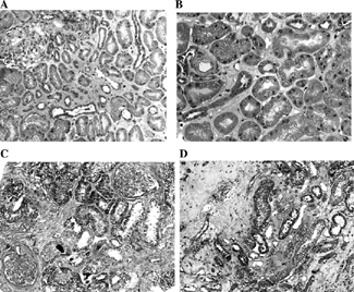
Clinicopathologic correlation of the two patients with extensive family history of BEN, one with normal kidney function and another one with renal failure at the time of biopsy, will be presented. Kidney disease of the first patient was discovered in 1981; renal biopsy performed in 1986. GFR measured by 99mTc-DTPA clearance was 110 mL/min/1.73 m2, arterial blood pressure 120/80 mmHg. Renal biopsy ( and ) shows discrete focal interstitial sclerosis and apparently normal tubuli. Discrete arteriolar narrowing and hyalinosis of the vessel walls were demonstrated. Glomeruli were apparently normal, but 2 out of 13 showed global sclerosis. Focal granular C3 deposits were noted along tubular basement membrane, as well as in interstitial arterioles and small arteries. Glomerular findings were negative. This has already been described.Citation[17] Seventeen years later, GFR was reduced to 22.5 mL/min/1.73 m2, with a serum creatinine of 220 µmol/L and serum urea of 11.6 mmol/L. Marked anemia with hemoglobin of 9.6 g/dL was noted. Arterial blood pressure was 150/90 mmHg. Kidney size was 10.2 × 4.0 × 1.12 cm (right) and 10.5 × 4.6 × 1.14 (left) by ultrasonography. GFR was reduced by an average 5.1 mL/year; however, he had no regular medical controls and treatment. Kidney disease of the second patient was discovered at the age of 50, renal biopsy was performed in 1992. GFR measured by 99mTc-DTPA clearance was 28 mL/min/1.73 m2, with a serum creatinine of 168 µmol/L and a hemoglobin of 11.2 g/dL. Arterial blood pressure was 130/80 mmHg. Renal biopsy ( and ) has revealed extensive interstitial sclerosis and tubular atrophy. More than half of glomeruli showed global sclerosis. Sclerosis of arterioles and small interlobular arteries, with an increase of thickness of the vessel wall, was found. Eleven years from biopsy, she entered ESRF requiring dialysis. From biopsy until ESRF, GFR decreased on average 2.1 mL/year. She was under regular medical supervision and treatment, but died of lung cancer 11 months after the start of hemodialysis.
Figure 1. Varying extent of kidney cortex damage in two BEN patients. (A) Small scattered areas of discrete interstitial sclerosis with apparently normal tubuli. Segmental widening of the mesangium and discrete periglomerular sclerosis. Trichrom Masson × 100. (B) Discrete interstitial sclerosis and apparently normal proximal tubular epithelial cells on a larger magnification. A small arterial blood vessel with a thickened, partly hyalinized wall and narrowed lumen, similar to that in benign nephroangiosclerosis. Trichrom Masson × 200. (C) Larger areas of acellular interstitial sclerosis accompanied by tubular atrophy. Two glomeruli are seen: one in advanced sclerosis and the other with widening of the mesangium. Marked periglomerular sclerosis. Trichrom Masson × 100. (D) Juxtamedullary cortex with advanced acellular interstitial sclerosis and tubular atrophy. Trichrom Masson × 100.
Follow-Up and Treatment
Regular medical supervision was instituted after renal biopsy and was strictly followed by the majority of patients. Patients have become familiar with the disease, including prevention and treatment. Hypertension, when present, was treated with diet and ACE inhibitors. Urinary tract infection was treated with semisynthetic penicillins and cephalosporins. Nephrotoxic drugs were avoided. In patients with advanced renal failure, a moderate hypoprotein diet was prescribed. Detection and treatment of potentially reversible aggravating factors was performed by a qualified nephrologist. Replacement therapy was offered timely to patients in ESRF.
Outcomes
Six patients become dialysis dependent, 2 from the group with incipient renal failure, but all 4 from the group with advanced renal failure. Three patients died after 8 to 12 years of follow-up, one from causes unrelated to kidney disease and two from ESRF.
Discussion
This prospective study confirms that BEN is a slowly progressive kidney disease leading to ESRF. Although some symptoms seem to be more specific, there is no clear-cut clinical or pathologic definition that is solely sufficient for disease diagnosis. The onset is without any acute episode. The disease progresses slowly with the occurrence of nonspecific signs (lassitude, fatigue, headache, weight loss and reduced appetite, xanthochromic or pale skin) and specific signs of kidney damage (reduced tubular transport, intermittent proteinuria with low molecular weight proteins such as β-2 microglobulin, and a gradual rise in blood nitrogen).Citation[2] Usually, no increased blood pressure is found, even in the more advanced phases of the disease due to the early impairment of the juxtaglomerular apparatus. Anemia (normocytic, normochromic, or, in rare cases, hypochromic) is one of the major symptoms, and it seems to develop well before other signs of BEN become manifest. Although in the advanced stages of the disease anemia is proportional to the degree of kidney insufficiency and, thus, could be attributed to the increased blood urea level and to the reduction of erythropoietin synthesis (as a result of the destruction of the erythropoietin-secreting peritubular endothelium of the kidney by the etiologic agent), it usually appears before significant kidney damage takes place.
Two recent epidemiologic studies carried out in Serbia and Croatia have suggested that the incidence of BEN may decrease in the near future.Citation[3], Citation[10] In a study performed to investigate the incidence of BEN patients on dialysis, and BEN-associated mortality in endemic areas around the South Morava River in Serbia from 1978 to 1997, a marked decrease in the incidence of ESRD and BEN-induced mortality in the last 10 years has been documented.Citation[5]
Detailed morphologic studies of kidney biopsies in different stages of BEN have permitted to define the initial lesion, target nephron structures, and evolution of the kidney damage.Citation[11-14] On light microscopy there is a diffuse cortical, mainly subcapsular, hypocellular interstitial fibrosis with tubular atrophy. The columns of Bertin and the medulla are usually spared. Most glomeruli show obsolescence of the collaptic type and are packed together in the vascular lesions in most cases. They include multifocal arteriolar hyalinosis, mainly interlobular arterial mural sclerosis and thickened peritubular capillary basement membranes. At early stages, the previously mentioned changes are multifocal, mainly in medullary rays and outer medulla. They are associated with areas of interstitial edema, together with peritubular capillary wall thickening, as well as proximal tubule epithelial cell degenerative lesions ().
Findings on immunofluorescent microscopy are nonspecific.Citation[15] Glomerular immune deposits are either absent or limited to scanty, nonsignificant, granular segmental deposits of mainly IgM and/or C3. In a few cases, tubular basement membranes displaying granular or linear deposits of C3 have been reported. Damaged proximal tubule epithelial cells overexpress cytokeratin and vimentin.Citation[16] Collagen IV and laminin deposits have been observed in tubular basement membranes. Arterial walls frequently contain C3 deposits sometimes associated with IgM, and peritubular capillaries contain laminin.Citation[17&18]
Kidney size was apparently normal in patients without renal failure, but decreased markedly in patients with advanced renal failure. Renal function decreased significantly by the progression of kidney damage. This is in accordance with our previous results.Citation[2], Citation[8]
Clinicopathologic correlation has shown progression from marked tubulointerstitial damage and advanced renal failure (99mTc-DTPA clearance 28 mL/min/1.73 m2) until ESRF could be prolonged by 11 years (GFR decrease of 2.1 mL/min per year) with the appropriate treatment. If no regular medical supervision was accepted by the patient, progression is much faster (GFR decrease of 5.1 mL/min per year). Treatment with ACE inhibitors and diet has been described to slow progression of kidney damage in BEN.Citation[18&19]
In two patients, an increase of kidney function by 4 and 7 mL/min, respectively, 15 years after renal biopsy was found. We speculate that the increase of GFR in these patients may be due, at least in part, to the ACE inhibitor treatment.
This study, based on initial renal biopsy and GFR estimation by 99mTc-DTPA clearance, has shown that BEN is characterized by a slow course and prolonged evolution, possibly modified by medical supervision and treatment.
References
- Danilovic V, Djurisic M, Mokranjac M, Stojimirovic B, Zivojinovic J, Stojakovic P. Nephrites chroniques provoquees par l'intoxication au plomb par voie digestive (farine).Presse Med. 1957;65:2039–2040. [PUBMED], [INFOTRIEVE], [CSA]
- Stefanovic V, Polenakovic M. Balkan nephropathy. Kidney disease beyond the Balkans? [Editorial].Am J Nephrol. 1991;11:1–11. [PUBMED], [INFOTRIEVE], [CSA]
- Radovanovic Z. Epidemiological characteristics of Balkan endemic nephropathy in eastern regions of Yugoslavia.IARC Sci Publ. 1991;115:11–20. [PUBMED], [INFOTRIEVE], [CSA]
- Radovanovic Z. Balkan endemic nephropathy in Serbia: current status and future research.Facta Univ. 2002;9:26–30. [CSA]
- Cukuranovic R, Petrovic B, Cukuranovic Z, Stefanovic V. Balkan endemic nephropathy: a decreasing incidence of the disease.Pathol Biol. 2000;48:558–561. [PUBMED], [INFOTRIEVE], [CSA]
- Velimirovic D. Contribution to the study of clinical course of endemic nephropathy. In: Ph.D. thesis. University of Belgrade, 1984, [ in Serbian].
- Bukvic D, Jankovic S, Djukanovic L, Marinkovic J. Survival of Balkan endemic nephropathy patients.Nephron. 2000;86:463–466. [PUBMED], [INFOTRIEVE], [CSA], [CROSSREF]
- Stefanovic V. Diagnostic criteria for endemic (Balkan) nephropathy. In: Strahinjic S, Stefanovic V, eds. Current Research in Endemic (Balkan) Nephropathy.NisSerbia: University Press, 1983; 351–363.
- Bohle A, Gise H, Mackensen-Haen S, Stark-Jakob B. The obliteration of the postglomerular capillaries and its influence upon the function of both glomeruli and tubuli.Klin Wochenschr. 1981;59:1043–1050. [PUBMED], [INFOTRIEVE], [CSA], [CROSSREF]
- Ceovic S, Plestina R, Miletic-Medved M, Stavljenic A, Mitar J, Vukelic M. Epidemiological aspects of Balkan endemic nephropathy in a typical focus in Yugoslavia.IARC Sci Publ. 1991;115:5–10. [PUBMED], [INFOTRIEVE], [CSA]
- Dojcinov D, Strahinjic S, Stefanovic V. Pathology of the kidney in the early phases of endemic (Balkan) nephropathy. In: Strahinjic S, Stefanovic V, eds. Endemic (Balkan) Nephropathy.NisSiberia: University Press, 1979; 91–104.
- Mandal A K, Sindjic M, Sommers S C.. Kidney pathology in endemic nephropathy.Clin Nephrol. 1987;27:304–308. [PUBMED], [INFOTRIEVE], [CSA]
- Ferluga D, Hvala A, Vizjak A, Trnacevic S, Halilbasic A. Renal function, protein excretion, and pathology of Balkan endemic nephropathy. III. Light and electron microscopic studies.Kidney Int. 1991;40(Suppl. 34):S57–S67. [CSA]
- Cukuranovic R, Stefanovic N, Savic V, Stefanovic V. Quantitative analysis of the renal changes in Balkan endemic nephropathy.Int Urol Nephrol. 1998;30:229–236. [PUBMED], [INFOTRIEVE], [CSA]
- Vizjak A, Trnacevic S, Ferluga D, Halilbasic A. Renal function, protein excretion, and pathology of Balkan endemic nephropathy. IV. Immunohistology.Kidney Int. 1991;40(Suppl. 34):S68–S74. [CSA]
- Stefanovic V, Cukuranovic R, Dojcinov D, Savic V. Coexpression of vimentin and cytokeratin in damaged tubular epithelia of kidney in Balkan nephropathy.Nephron. 1996;72:119–120. [PUBMED], [INFOTRIEVE], [CSA]
- Cukuranovic R, Savic V, Dojcinov D, Stefanovic V. Immunohistochemical localization of laminin in renal lesions of Balkan nephropathy.Nephron. 1995;70:504–505. [PUBMED], [INFOTRIEVE], [CSA]
- Vizjak A, Trnacevic S, Halilbasic A, Ferluga D. Immunohistologic kidney biopsy study of Balkan endemic nephropathy.Facta Univ. 2002;9:88–91. [CSA]
- Stefanovic V, Cosyns J P.. Balkan nephropathy. In: Davison A M, Cameron J S, Grunfeld J P, , et al., eds. Oxford Textbook of Clinical Nephrology. 3rd Ed.Oxford: Oxford University Press, 2004;1095–1102.