Introduction
Because the first liposuction surgery performed in Italy in 1974, this therapy has been refined and developed into a cosmetic procedure with rare complications. Tumescent liposuction and various generations of ultrasound-assisted lipoplasty are currently available. In addition to the local complications such as bruising, swelling, pain, and temporary numbness at the surgical site, systemic complications such as sepsis,Citation[1&2] fat embolism,Citation[3-7] and visceral perforations have been occasionally reported.
Even though studies have failed to demonstrate changes in central venous pressure after large-volume fat aspirations in young healthy patients,Citation[8] liposuction may be associated with hypotensionCitation[9] and intravascular volume contraction. In these patients, the hemodynamic imbalance is usually offset by intense volume resuscitation. To our knowledge, there has been only a single report of acute renal failure (ARF) after liposuction that was attributed to acute tubular necrosis from excessive bleeding.Citation[10] We report an unusual cause of ARF that was possibly precipitated by the hemodynamic alterations after liposuction.
Case Report
A 58-year-old Caucasian woman, who was otherwise healthy, underwent liposuction and removal of breast implants at an outpatient surgery facility. No obvious complications were reported during the surgery or in the immediate postoperative period. She was prescribed oxycodone/acetaminophen, diazepam, and cephalexin at the time of hospital discharge. On the same day, she noted a significant decrease in urine output, and on the first postoperative day, the patient became completely anuric. Over the next few days, she drank increased amounts of fluid, as instructed by her surgeon, but remained anuric. On the fourth postoperative day, she was hospitalized for lethargy and confusion. Her previous medical history was unremarkable. Her past surgeries included breast implants, rhytidectomy (facelift surgery), and gastric bypass.
At the time of the present admission, blood pressure was 102/69 mmHg, pulse rate 100 beats/minute, and oxygen saturation was 88% on room air that improved to 95% saturation with supplemental oxygen by nasal cannula. Physical exam was consistent with fluid overload as evidenced by bibasilar crackles on lung auscultation and 1 + lower-extremity edema. There was no evidence of bleeding or hematoma formation at the liposuction sites (upper and lower backs). Laboratory tests showed elevated serum creatinine (7.4 mg/dL) and blood urea nitrogen (41 mg/dL) levels. Serum creatinine was 0.7 mg/dL, 6 months prior to surgery. Urine analysis was unremarkable, except for proteinuria of 100 mg/dL on dipstick. Specifically, urine microscopic exam did not reveal granular or cellular casts or eosinophils. Hemoglobin was significantly lower during this admission (8.3 g/dL) as compared with preoperative values (12.7 g/dL). Total serum creatine phosphokinase was 1000 mg/dL. Serum liver enzymes, antinuclear antibody, human immunodeficiency virus enzyme-linked immunoabsorbent assay, hepatitis panel, C3, and C4 were all normal. Serum albumin was 2.1 mg/dL. Erythrocyte sedimentation rate was elevated (93 mm/h).
Chest x-ray showed moderate pulmonary edema and bilateral pleural effusions. Renal ultrasound revealed normal-size kidneys (right 11.3 cm, left 11.7 cm) with normal echogenicity. There was no evidence of hydronephrosis. Computed tomography of the abdomen was normal and did not reveal retroperitoneal hematoma. The patient was initiated on hemodialysis for volume overload and ARF. She did not recover renal function and was continued on maintenance hemodialysis. A kidney biopsy was performed.
Renal Biopsy Findings
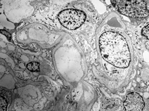
Nine glomeruli were present, one of which was globally sclerotic and the others were normal (). There were several tubular casts involving various tubular segments. These casts were large, fragmented, variegated, and were associated with histiocytes or multinucleated giant cells (). The tubules without casts showed focal changes, including cytoplasmic vacuolization, flattening, or disruption of tubular cells. There was focal interstitial fibrosis, edema, and inflammation, with mostly lymphocytes and rare mature plasma cells. Immunofluorescent studies showed four open glomeruli, which were negative for IgG, IgA, IgM, C3, C4, C1q, kappa light chain, lambda light chain, and C1q. Most tubular casts were strongly positive for kappa light chain but negative for lambda light chain. There was no staining for the glomerular or tubular basement membrane. Electron microscopic examination showed no significant glomerular changes, but confirmed the tubular lesions described previously. Features of immunoglobulin fragment deposition were not seen. These changes were strongly suspicious for myeloma cast nephropathy.
Figure 1. Large and fragmented tubular casts associated with histiocytes or multinucleated giant cells are present (center and upper right). Two nonspecific hyalin casts are seen (left). A glomerulus shows only mild mesangial sclerosis (hemotoxylin & eosin, original magnification × 400).
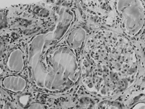
Figure 2. Electron microscopic examination shows normal glomerular capillaries. Electron dense deposits, light chain, or amyloid are not seen (original magnification × 2000).
Subsequent Course
The peripheral blood smear did not reveal abnormal plasma cells or blast cells. Bone marrow biopsy showed a diffuse infiltrate of plasma cells that comprised 25% to 30% of all nucleated cells. On immunohistochemical analysis, plasma cells stained positive for CD138 and cytoplasmic kappa chains and negative for cytoplasmic lambda chains. Skeletal survey did not reveal osteolytic lesions. Serum electrophoresis was positive for an M spike in the gamma region. The patient was diagnosed with IgG myeloma and started on chemotherapy. She received four cycles of high-dose dexamethasone with partial response and a decline in plasma cells in the bone marrow to 14%. Other chemotherapy regimens are being considered at this point. Six months after her initial presentation, she has remained dialysis dependent.
Discussion
Multiple myeloma is characterized by clonal proliferation of plasma cells and myeloma accounts for ∼ 10% of all hematologic malignancies. Renal involvement is very common in patients with multiple myeloma.Citation[11] In fact, kidney involvement assumes significance because the presence and severity of renal insufficiency and the response of renal disease to therapy influence overall patient survival.Citation[12] It is not uncommon for renal insufficiency to be the initial presentation for this disease.Citation[13] Our patient was completely asymptomatic until the development of ARF.
Even though multiple myeloma is associated with a myriad of renal syndromes, cast nephropathy is the most common cause of ARFCitation[11] in this population. Other renal diseases include amyloidosis (immunoglobulin fragment), light-chain deposition disease, proximal renal tubular acidosis, hypercalcemia-mediated renal injury, and cryoglobulinemia. Excess synthesis of a monoclonal immunoglobulin fragment, most frequently light chain, by neoplastic plasma cells results in high urinary concentration of these light chains, and the interaction of these light chain molecules and Tamm-Horsfall protein leads to their precipitation as casts within the lumens of distal tubules and collecting ducts. Light-chain casts can precipitate under various circumstances. Volume contraction is a major risk factor for myeloma cast nephropathy by virtue of increasing the concentration of light chains in the tubular lumen and by slowing the flow of urine. Because our patient became oliguric immediately after surgery, we believe the tubular casts were precipitated by the hemodynamic and renal circulation imbalance that occurred during surgery. Hypotension and tissue ischemia,Citation[14&15] induced by a combination of anesthetic agent, blood loss, sequestration of plasma at surgical site, and lidocaine use, have been described. Rao et al. identified five deaths after tumescent liposuction and directly attributed three deaths to intraoperative hypotension and bradycardia.Citation[16] Although controversy still lingers as to whether liposuction was directly culpable for these hypotensive episodes, the fact remains that this procedure has inherent risks of anesthesia and surgery.
In myeloma patients with renal involvement, the severity of renal insufficiency and the response to therapy have a significant impact on patient survival. In addition to observing a high prevalence (22%) of renal insufficiency in 423 myeloma patients, Blade et al.Citation[12] showed that patients who recovered their renal function had a median survival of 28 months versus 4 months for those with irreversible renal failure. Even though the overall renal function recovery rate was only 26%, the subgroup of patients with serum creatinine < 4 mg/dL had a higher chance of recovery. Our patient presented with a higher serum creatinine, has not responded to therapy yet, and remains dialysis dependent.
In summary, we have presented a patient with ARF after liposuction that was related to myeloma kidney. Our presentation reiterates the need for kidney biopsy in patients with ARF when etiology is unclear.
References
- Nagelvoort R W, Hulstaert P F, Kon M, Schuurman A H. Necrotising fasciitis and myositis as serious complications after liposuction. Ned. Tijdschr. Geneeskd. 2002;146(50):2430-2435. [PUBMED], [INFOTRIEVE], [CSA]
- Umeda T, Ohara H, Hayashi O, Ueki M, Hata Y. Toxic shock syndrome after suction lipectomy. Plast. Reconstr. Surg. 2000;106(1):204-207, discussion 8–9. [PUBMED], [INFOTRIEVE], [CSA]
- Abbes M, Bourgeon Y. Fat embolism after dermolipectomy and liposuction. Plast. Reconstr. Surg. 1989;84(3):546-547. [PUBMED], [INFOTRIEVE], [CSA]
- Dillerud E. Re: fat embolism after liposuction. Ann. Plast. Surg. 1991;26(3):293. [PUBMED], [INFOTRIEVE], [CSA]
- Fourme T, Vieillard-Baron A, Loubieres Y, Julie C, Page B, Jardin F. Early fat embolism after liposuction. Anesthesiology. 1998;89(3):782-784. [PUBMED], [INFOTRIEVE], [CSA], [CROSSREF]
- Scroggins C, Barson P K. Fat embolism syndrome in a case of abdominal lipectomy with liposuction. Md. Med. J. 1999;48(3):116-118. [PUBMED], [INFOTRIEVE], [CSA]
- Foroozan R, Varon J. Bilateral anterior ischemic optic neuropathy after liposuction. J. Neuro-ophthalmol. 2004;24(3):211-213. [CSA], [CROSSREF]
- Kenkel J M, Lipschitz A H, Luby M, , et al. Hemodynamic physiology and thermoregulation in liposuction. Plast. Reconstr. Surg. 2004;114(2):503-513, discussion 14–15. [PUBMED], [INFOTRIEVE], [CSA], [CROSSREF]
- Trott S A, Beran S J, Rohrich R J, Kenkel J M, Adams W P, Jr, Klein K W. Safety considerations and fluid resuscitation in liposuction: an analysis of 53 consecutive patients. Plast. Reconstr. Surg. 1998;102(6):2220-2229. [PUBMED], [INFOTRIEVE], [CSA]
- Lee J G, Lee J S, Lee Y K, Song C S, Cho J M. Acute renal failure after thrombotic agent therapy due to excessive bleeding after cosmetic liposuction. Ren. Fail. 2002;24(1):103-107. [PUBMED], [INFOTRIEVE], [CSA], [CROSSREF]
- Sakhuja V, Jha V, Varma S, , et al. Renal involvement in multiple myeloma: a 10-year study. Ren. Fail. 2000;22(4):465-477. [PUBMED], [INFOTRIEVE], [CSA], [CROSSREF]
- Blade J, Fernandez-Llama P, Bosch F, , et al. Renal failure in multiple myeloma: presenting features and predictors of outcome in 94 patients from a single institution. Arch. Intern. Med. 1998;158(17):1889-1893. [PUBMED], [INFOTRIEVE], [CSA], [CROSSREF]
- Irish A B, Winearls C G, Littlewood T. Presentation and survival of patients with severe renal failure and myeloma. QJM. 1997;90(12):773-780. [PUBMED], [INFOTRIEVE], [CSA]
- Cohen A, Kishore K, Wolansky L, Frohman L. Pituitary apoplexy occurring during large volume liposuction surgery. J. Neuro-ophthalmol. 2004;24(1):31-33. [CSA], [CROSSREF]
- Minagar A, Schatz N J, Glaser J S. Liposuction and ischemic optic neuropathy. Case report and review of literature. J. Neurol. Sci. 2000;181(1–2):132-136. [PUBMED], [INFOTRIEVE], [CSA], [CROSSREF]
- Rao R B, Ely S F, Hoffman R S. Deaths related to liposuction. N. Engl. J. Med. 1999;340(19):1471-1475. [PUBMED], [INFOTRIEVE], [CSA], [CROSSREF]