Abstract
Principles. Coumadin-induced renal and retroperitoneal hemorrhages are rare. Clinical and laboratory findings are not specific for the diagnosis. Computed tomography (CT) has some advantages in the evaluation of these patients. The aim of this study is to report our experience regarding renal and retroperitoneal hemorrhage due to Coumadin, and describe clinical and CT findings, treatment, and prognosis of the patients. Methods. We reviewed our CT archive to search patients with renal and retroperitoneal hemorrhage caused by Coumadin treatment retrospectively. A total of seven patients with Coumadin-induced renal and retroperitoneal hemorrhages were included in this study. Results. Four patients had abdominal pain, two patients had hematuria, and one patient had abdominal pain and hematuria. There was retroperitoneal hemorrhage in three patients on abdominal CT. One patient had hemorrhage in the renal pelvis and the jejunum, another had hemorrhage in the renal pelvis and the perirenal area, and another had hemorrhage in the perirenal area and the retroperitoneal region. In the last patient with hematuria, there was no hemorrhage. None of the patients had a lesion-causing hemorrhage. Coumadin was stopped, and vitamin K and fresh-frozen plasma were given to patient. One patient with massive retroperitoneal hemorrhage died, whereas other patients were successfully treated using conservative approaches. Conclusions. Hematuria and abdominal pain are the most common complaints in Coumadin-induced renal and retroperitoneal hemorrhage. CT can be the first imaging modality in these patients due to its ability to directly evaluate all peritoneal and retroperitoneal structures. It also allows evaluation of any underlying lesion that can cause hemorrhage from the renal area and the urinary tract. Conservative treatment is the first choice, and prognosis is good when diagnosed early.
INTRODUCTION
Nontraumatic retroperitoneal hemorrhage is a rare entity. Renal tumors, mainly renal cell carcinoma and angiomyolipoma, comprise the majority of traumatic kidney ruptures. The other causes of retroperitoneal hemorrhage include rupture of the abdominal aorta, blood dyscrasias, vasculitis, and oral anticoagulation. Oral anticoagulant therapy could be responsible for a few cases.[Citation[1]]
This study reports our experience regarding renal and retroperitoneal hemorrhage due to Coumadin treatment, and describes clinical manifestations, computed tomography (CT) findings, and treatment and prognosis of these patients.
METHODS
We reviewed our CT archive to search patients with renal and retroperitoneal hemorrhage caused by Coumadin treatment retrospectively. Abdominal CT scan was performed within 24 h in all patients following history and physical examination. Toshiba X press GX model (Tochigi-Ken) helical scanner was used in all CT examinations. Abdominal and pelvic examinations were performed from the diaphragm to the pelvic floor. Nonionic iodinated 100-mL contrast material (Ultravisit, Schering 370/100 mg/mL) was administered by antecubital vein. Oral contrast material (950 mL water and 50 mL ionic iodinated contrast material [Urographin, Schering]) was also used in six patients. Table speed and slice thickness were 7 mm in all examinations. All CT examinations were evaluated by two radiologists. Hemorrhages were seen as heterogeneous hyperdense areas on CT scans. International normalized ratio (INR) levels were also obtained for all patients at admission.
RESULTS
Seven patients with renal and retroperitoneal hemorrhage were found in this survey. presents location of hemorrhage, complaints of the patients, and INR levels. Three patients were female, and three were male. The patients' ages were between 30 and 72 (mean = 58). Five patients received anticoagulant therapy for prosthetic cardiac valve replacement, 1 patient for atrial fibrillation, and 1 patient for deep venous thrombosis. All patients had abnormal INR levels.
Table 1 Summary of the patient informations
In the patient with hematuria and abdominal pain, hemorrhage was seen in both the jejunum and the right renal pelvis. Retroperitoneal hemorrhage was detected in four patients. One patient with abdominal pain and vomiting had a very large hemorrhage in the left anterior pararenal region. This lesion extended from the upper pole of the left kidney to the psoas and iliac muscles. One patient with hematuria had hemorrhage in the left renal pelvis and the perirenal area (). In the patient with retroperitoneal hemorrhage, the lesion was between the left kidney and the psoas muscle, and the other patient with retroperitoneal hemorrhage had massive left retroperitoneal hemorrhage (). The last patient with retroperitoneal hemorrhage had massive hemorrhage in the right perirenal and retroperitoneal areas, including the muscles (). There was no hemorrhagic area and lesion-causing hemorrhage on CT in the patient with hematuria.
Figure 1. On contrast-enhanced abdominal CT image at level renal hilum, hemorrhage in the left renal pelvis and perirenal area.
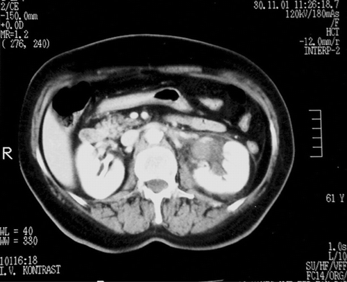
Figure 2. On contrast-enhanced pelvic CT image, large retroperitoneal hemorrhage involving all retroperitoneal structures.
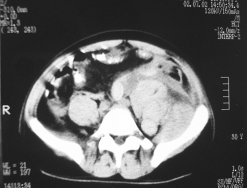
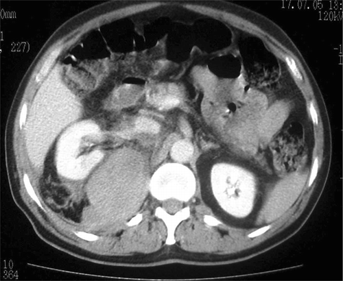
Figure 3. On contrast-enhanced abdominal CT image, right perirenal and retroperitoneal hemorrhage.
Anticoagulant was stopped, and fresh-frozen plasma and vitamin K were given to five patients. In one patient who had normal CT findings, anticoagulant was stopped and followed. Erythrocyte suspension was also given to the patients with low hemoglobin values.
One patient died 1 day later, but the prognosis was favorable in the remaining six patients.
DISCUSSION
Oral anticoagulants are commonly used for medical conditions; however, on occasion, the appropriate anticoagulation range is difficult to maintain due to patient noncompliance and/or drug interactions. Coumadin drug interactions are common and may affect anticoagulation levels very rapidly. Common medications that may increase warfarin effect are cimetidine, clofibrate, alcohol, nonsteroidal anti-inflammatory drugs, and many antibiotics, such as ciprofloxacin, erythromycin, fluconazole, ketoconazole, metronidazole, and sulfonamides.[Citation[2]]
Anticoagulation overdose can be associated with severe bleeding complications. Bleeding occurs in particular in the form of nose bleeds, bleeding into the skin, from the kidneys, in the gastrointestinal tract, into the eyes and muscles, or from the respiratory tract.[Citation[3]]
The majority of significant complications is abdominal and includes bleeding into the lumen, wall, or mesentery of the gut; retroperitoneal bleeding; and bleeding into abdominal wall hematoma.[Citation[4]] Acute abdominal pain in the patient receiving anticoagulants poses a unique diagnostic and therapeutic challenge. The causes are from rectus sheath hematoma to intraperitoneal, retroperitoneal hemorrhage and intestinal wall hematoma.[Citation[5]]
Patients on anticoagulant therapy may have urologic complications.[Citation[6]] The three types of hemorrhage that may occur in and around the urinary tract are retroperitoneal, intraluminal, and intrarenal.[Citation[7]] Renal hemorrhage may be suburothelial, intraparenchymal, subcapsular, perinephric, or pararenal in location or may involve the renal sinus. Suburothelial and renal sinus hemorrhage occur most commonly in anticoagulated patients.
The most common complication is hematuria. Less common, but more serious, is spontaneous hemorrhage arising from urologic structures, such as spontaneous perirenal hematoma.[Citation[6]]
Conventional physical signs are not adequate for making the diagnosis because the patients with simple hematoma may present with high fever, leukocytosis, and rebound tenderness.[Citation[5]]
CT should be used early in the diagnostic evaluation of abdominal pain occurring in patients receiving anticoagulants.[Citation[8]] CT offers some specific advantages: direct evaluation of the bowel wall and adjacent mesentery; observation of vascular structures in the peritoneal cavity and retroperitoneal area; and when intravenous contrast material is used, physiological information about blood flow.[Citation[9]]
CT is the most valuable examination in the evaluation of patients with suspected acute renal hemorrhage. On unenhanced CT, recent renal hemorrhage is characterized by high-attenuation areas. Postcontrast scan should also be obtained to facilitate identification of disorders such as small neoplasm-causing spontaneous renal hemorrhage.
The occurrence of perirenal hematoma in a patient on anticoagulant therapy does not necessarily indicate underlying renal pathology and has a favorable prognosis. The literature suggests that some of these cases occur in association with renal tumor.[Citation[7]] Hematuria can lead to early detection of malignancy in anticoagulant patients. Evaluation of hematuria in the patients taking oral anticoagulant is necessary to identify and treat clinically important pathology.[Citation[10]]
Hematuria and abdominal pain were the most common complaints in the patients with renal and retroperitoneal hemorrhage in our study. CT could show all peritoneal cavity and retroperitoneal area and organs. One patient had hemorrhage in the renal and jejunal segment, and CT showed hemorrhage in two different regions. In addition to location of hemorrhage, extension of hemorrhage in renal and retroperitoneal areas and amount of hemorrhage could be evaluated by CT scan. There was no underlying lesion causing hemorrhage in renal and retroperitoneal area on CT. One patient with hematuria had normal CT findings. In this patient, a limited intraluminal hemorrhage might occur. If there is a lesion-causing hemorrhage, treatment can be changed according to lesion location and characterization.
It has been suggested that the diagnosis can be made preoperatively and that conservative management is the first choice for perirenal hematoma without a definite etiology.[Citation[6],Citation[11]]
Conservative treatment was preferred in our patients. There was no another complication in six patients, but one patient with massive retroperitoneal hemorrhage died 1 day later. We believe that there was a delay in the admission of the patient to the hospital.
Our conclusions are that hematuria and abdominal pain are the most common complaints in Coumadin-induced renal and retroperitoneal hemorrhage. CT can be the first imaging modality in these patients due to its ability to directly evaluate all peritoneal and retroperitoneal structures. CT also allows evaluation of the underlying lesion that can cause hemorrhage from the renal and urinary tract. Conservative treatment is the first choice, and prognosis is good when diagnosed early.
REFERENCES
- Chang SY, Ma CP, Lee SK. Spontaneous retroperitoneal hemorrhage from kidney causes. Eur Urol 1988; 15: 281–284, [PUBMED], [INFOTRIEVE]
- Jimenez J. Abdominal pain in a patient using warfarin. Post-grad Med J 1999; 75: 747–748
- Botzler R, Wagner TH, Ritter U. Endoscopic finding of an intramural hemorrhage in the duodenum under anticoagulant therapy with phenprocoumon. Endoscopy 1986; 18: 64, [PUBMED], [INFOTRIEVE]
- Ashley S. Spontaneous mesenteric hematoma and small bowel infarction complicating oral anticoagulant therapy. RJ Soc Med 1990; 83: 116
- Euhus DM, Hiatt JR. Management of the acute abdomen complicating oral anticoagulant therapy. Am Surgeon 1990; 56: 581–585, [PUBMED], [INFOTRIEVE]
- Mabjeesh NJ, Matzkin H. Spontaneous subcapsuler renal hematoma secondary to anticoagulant therapy. J Urol 2001; 165: 1201, [PUBMED], [INFOTRIEVE], [CROSSREF]
- Hiratsuka Y, Takeuchi F, Tsunoda Y, Ishii T. Anticoagulant induced submucosal hemorrhage mimicking a renal pelvic tumor. J Urol 2000; 163: 231, [PUBMED], [INFOTRIEVE], [CROSSREF]
- Scott WW, Fishman EK, Siegelman SS. Anticoagulants and abdominal pain. The role of computed tomography. JAMA 1984; 252: 2053–2056, [PUBMED], [INFOTRIEVE], [CROSSREF]
- Bartnicke BJ, Balfe DM. CT appearance of intestinal ischemia and intramural hemorrhage. Radiol Clin North Am 1994; 32: 845–858, [PUBMED], [INFOTRIEVE]
- Ripley TL, Havrda DE, Blevins S, Culkin D. Early evaluation of hematuria in a patient receiving anticoagulant therapy and detection of malignancy. Pharmacotherapy 2004; 24: 1638–1640, [PUBMED], [INFOTRIEVE], [CROSSREF]
- Mita K, Kobukata Y. Conservative management of non-traumatic subcapsular renal hematoma: a case report. Int J Urol 1994; 2: 181–182