Abstract
Objective. Paroxysmal atrial fibrillation (AF) observed in patients undergoing chronic hemodialysis program with higher rates is an important morbidity and mortality cause that negatively influences the hemodynamics and leads to thromboembolic complications. It is known that P wave dispersion (PWD) facilitating the development of paroxysmal atrial fibrillation is increasing during intradialytic process. This study researched the influence of various amounts of ultrafiltration that applied in the various hemodialysis sessions in the same patient cohort on PWD. Materials and Methods. 25 patients in a chronic hemodialysis program undergoing four hours bicarbonate hemodialysis three times a week were included in the study. The patient cohort was divided into four groups regarding the amount of ultrafiltration (UF) performed during a four-hour standard hemodialysis session as following: UF up to 1 liter, UF of 1–2 liters, UF of 2–3 liters, and UF of 3–4 liters. Notes were kept until each patient had been included once into each group regarding the amount of ultrafiltration performed parallel to intradialytic weight gain in different hemodialysis session. A 12-lead ECG was taken from the patients immediately before the hemodialysis and within 20 minutes after completion of the session, and maximum P wave duration (Pmax), minimum P wave duration (Pmin) and PWD values (i.e., the difference between Pmax and Pmin) were measured. The inter-group data was assessed with a one-way ANOVA, and the within-group assessments were performed with paired samples test. Mann Whitney U test was used for the evaluations performed according to the presence of diabetes. Findings. The mean age of 25 patients (15 male and 10 female) was 62.7 ± 20.2 (range: 21–89). PWD after UF was decreased significantly in group 1 (56.12 ± 15.26 vs. 46.60 ± 18.45 ms, p = 0.018) and were increased in groups with UF more than 1 liter: group 2 (39.68 ± 21.26 vs. 47.12 ± 21.20 ms, p = 0.020), group 3 (41.60 ± 23.99 vs. 65.92 ± 31.04 ms, p = 0.001), and group 4 (17.52 ± 14.67 vs. 26.80 ± 15.52 ms, p = 0.007). Furthermore, while PWD before UF was significantly higher in a diabetic group compared to a nondiabetic group (68.85 ± 10.44 vs. 51.16 ± 14.06 ms, p = 0.007), it was seen that PWD difference had disappeared after UF application (57.14 ± 17.99 vs. 42.50 ± 17.40 ms, p = 0.065). Conclusion. UF application of more than 1 liter during hemodialysis session increases the PWD value significantly. Hypervolemia exceeding 1 liter between two dialysis sessions should be avoided in all patient groups, especially in diabetics, and an effective UF planning should be arranged because of a decrease in PWD values with UF observed in diabetics.
INTRODUCTION
The P wave occurring by the spread of excitation from the sinoatrial node over the atrial musculature represents the depolarization (electrical activation) of the atria (two small chambers) of the heart. Prolongation in P wave dispersion (PWD), the difference between maximum P wave duration (Pmax) and minimum P wave duration (Pmin), is known as the paroxysmal atrial fibrillation (AF) facilitating electrocardiographic finding.Citation[1],Citation[2] The effects of the diseases leading to a predisposition towards developing AF on PWD remains to be research topic.Citation[3–8]
The high prevalence of paroxysmal atrial fibrillation (AF) in patients undergoing chronic hemodialysis program with changing hemodynamic conditions compared to normal population is an important morbidity and mortality cause.
In this study, the influence of various amounts of ultrafiltration applied in the various hemodialysis sessions in the same patient cohort on P wave duration was researched.
MATERIALS AND METHODS
In this study, the patient cohort was divided into four groups regarding the amount of ultrafiltration performed during a four-hour standard hemodialysis session as follows: group 1, with a UF up to 1 liter; group 2, with a UF of 1–2 liters; group 3, with a UF of 2–3 liters; and group 4, with a UF of 3–4 liters. Notes were kept until each patient had been included into one of the four groups according to the amount of ultrafiltration performed parallel to weight gain between two dialysis in different hemodialysis sessions. A 12-lead ECG on which the measurements would be performed was taken from each patient belonging to four groups before UF and after (i.e., within the first 20 minutes after completion) UF. A total of 120 ECGs were taken into consideration for evaluation. Durations of P wave (Pmax, Pmin, and P wave dispersion) were recorded by measuring from the ECGs taken during entering the dialysis (before UF) and after completion of the dialysis (after UF). Durations of P wave were measured with calipers in three consecutive complexes of each lead by one observer.
25 patients in a chronic hemodialysis program undergoing four hours bicarbonate hemodialysis three times a week were included into the study. In hemodialysis, “hollow fiber” polysulfone dialyzers (F6, Fresenius, Germany) and hemodialysis solutions containing 140 mmol/L Na, 2 mmol/L K, 35 mEq/L bicarbonate (Renasol BA-140/ BB-8.4, Fresenius Medical Care, Ankara, Turkey) were used. Arterial blood pressures obtained before and after hemodialysis were recorded. Blood samples for Na and K were taken twice (i.e., when entering dialysis and after session). Other biochemical parameters were measured from the blood samples taken during entering the dialysis. The patients with following criteria were not included into the study: patients undergoing dialysis shorter than six months, unstable patients requiring hemodynamic intervention within intradialytic process, patients with chronic atrial fibrillation, patients with acute coronary syndrome symptoms, patients using digitalis, and patients with Class 3–4 heart failure according to New York Heart Association classification. The distribution of the antihypertensive agent usage of the patients were as follows: two patients, angiotensin converting enzyme (ACE) inhibitors; one patient, angiotensin receptor antagonist; two patients, dihydropyridine group calcium channel blocker; one patient, alpha-adrenergic blocking agents; and three patients, beta-adrenergic blocking agents. No changes was made regarding antihypertensive agent choices during the studies.
A SPSS for Windows 10.0 program was used for statistical analysis during the evaluation of the data obtained in the study. One-way ANOVA test and post hoc Tukey test were used for comparing descriptive statistical methods (mean, standard deviation) as well as qualitative data between groups. Paired Samples t test was used to compare the parameters within the groups. Mann Whitney U test was used for the evaluations performed according to the presence of diabetes. Results were assessed with 95% confidence interval and a significance level of p < 0.05.
FINDINGS
Twenty-five chronic hemodialysis patients (15 male and 10 female) with a mean age of 62.72 ± 20.26 (range: 21–89) and receiving treatment in Nephrology Department of GATA Haydarpaşa Training Hospital were included into the study. The patients are in chronic hemodialysis program for an average of 5.2 ± 3.8 years, and seven of them are diabetic. When the data of the echocardiographic studies performed at baseline were evaluated, the parameters were determined as following: mean ejection fractions (EF), 58.08 ± 5.89%; mean left ventricle internal diastolic diameter (LVIDD), 52.08 ± 2.62 mm; mean left ventricle internal systolic diameter (LVISD), 32.36 ± 2.56 mm; mean left ventricle end diastolic volume (LVEDV), 124.96 ± 10.58 mL; mean right atrium diameter (RAD), 37.08 ± 2.28 mm; and mean left atrium diameter (LAD), 39.24 ± 3.46 mm. General features of the patients and mean UF amounts of the groups are shown in and , respectively.
Table 1 General characteristics of patients
Table 2 The distribution of the groups according to UF amounts
Pmax durations were not significantly different according to ECG data obtained before and after UF (p > 0.05). Pmin measurements performed before UF, however, were significantly different between groups (p < 0.01). Pmin measurements of group 4 were significantly higher than groups 1–3 in Tukey HSD test, which was one of the post hoc tests performed to determine the group from which the significance originated (p = 0.001 for all three groups). Group 1 measurements was found to be significantly lower than group 2 (p = 0.022; see ).
Table 3 The distribution of P measurements obtained from ECG before and after UF according to group
Inter-group Pmin measurements performed after UF were also significantly different (p < 0.01). In significance analysis, Pmin measurements of group 4 were significantly higher than groups 1–3 (p = 0.001 for each group).
Inter-group Pmin measurements performed before UF were also significantly different (p < 0.01). PWD measurements of group 4 were significantly lower than groups 1–3 in the evaluation performed to determine the group from which the significance originated (p = 0.001 for all three groups). PWD measurements of group 1 performed before UF were found to be significantly higher than groups 2 and 3 (p = 0.017 and p = 0.043), respectively (see ).
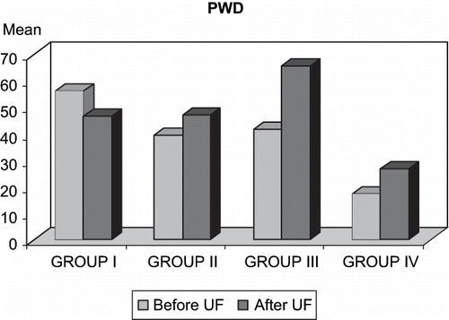
Inter-group PWD values after UF were also significantly different (p < 0.01). In significance analysis, PWD measurements of group 4 were found to be significantly lower than groups 1–3 (p = 0.012; p = 0.009; p = 0.001, respectively). The decrease in PWD levels of group 1 values after UF considering the values before UF was also statistically significant (p < 0.01). Again, the increase in groups 2–4 values after UF considering the values before UF was statistically significant (p < 0.05; see and ) .
The difference between inter-group serum Na levels before and after UF was not significant (p > 0.05). Serum K levels before UF between groups were significantly different (p < 0.01). K measurements of group 1 were found to be significantly lower than the other groups in Tukey HSD test, which was one of the post hoc tests performed to find out the group from which the significance originated (p = 0.007; p = 0.003, respectively). The decrease in serum Na and K levels observed in all groups was found to be statistically significant (p < 0.01) (see ) . When the effects of the difference in serum Na and K levels observed with UF on P wave were evaluated, it was seen that the difference in serum Na and K levels had no effect on PWD.
Table 4 Change in electrolyte levels before and after UF according to group
Mean arterial blood pressures before and after UF were determined as following: 134.2/81.2 mmHg and 127.6 / 72.8 mmHg in group 1; 145.4 / 92.4 mmHg and 124.8 / 74.6 mmHg in group 2; 158 / 90.36 mmHg and 129.52 / 68 mmHg in group 3; and 161.36 / 90.2 mmHg and 116.6 / 65.2 mmHg in group 4.
No significant correlation was determined in any group between mean systolic and diastolic blood pressures obtained before and after UF and mean hemoglobin levels of the patients and PWD measurements (p > 0.05).
In the assessment performed in diabetic patient group, while Pmin levels before UF were found to be significantly lower than nondiabetic patient group, PWD measurements were determined higher than nondiabetic patient group (68.85 ± 10.44 vs. 51.16 ± 14.06 ms, p = 0.007). While PWD after UF decreased, the difference from the nondiabetic patient group was disappearing (57.14 ± 17.99 vs. 42.50 ± 17.40 ms, p = 0.065; see ) .
Table 5 The evaluation of P wave changes according to the presence of diabetes
DISCUSSION
Hypervolemia observed in chronic hemodialysis patients proportional to the decrease of urine amount was meant to be avoided with UF performed during dialysis session. Either uncontrolled hypervolemia or UF planning in high quantities may be concluded with hemodynamic abnormalities. AF that might have a mortality or morbidity cause with these changing hemodynamic conditions is a very important arrhythmia encountered in clinical practice with frequency, increasing from 10.9% to 27%.Citation[9],Citation[10] The objective of this study was to investigate the effects of hypervolemia findings before dialysis and UF amounts performed during dialysis session on PWD leading to a predisposition to AF.
Yildirir et al. compared 44 hypertensive patients having a history of paroxysmal AF with 50 hypertensive patients not having a history of paroxysmal AF. In their study, they researched the effect of hypertension on PWD and showed that PWD was determined to be higher in patients with a positive history of AF. Additionally, it was emphasized that further research was required for this objective.Citation[11] In this study, average maximum blood pressure was 161.36 / 90.2 mmHg, and it was determined that both systolic and diastolic blood pressure changes before and after UF had no effect on PWD.
Szabo et al. researched the effect of hemodialysis on PWD in nondiabetic patients and emphasized that PWD was increased after dialysis.Citation[12] Similarly, Tezcan et al. published that Pmax and PWD measurements were increased after dialysis compared to measurements taken before dialysis in their study performed on 32 patients.Citation[13] In these studies, it was reported that changes in serum Na levels were ineffective on P wave duration, but there was a negative correlation between serum potassium level after dialysis and PWD. In the present study, it was seen that PWD was not affected by electrolyte changes observed during dialysis and hemoglobin levels of average 11.62 ± 0.81 g/dL. It was considered that the difference was originated from variation of UF amounts applied during dialysis. Two study groups did not mention the possible effects of various UF amounts applied during dialysis. Again, in the present study, the relationship between various UF amounts applied in different sessions on the same patient group and P wave durations was inspected. Pmin measurements before UF was determined to be significantly higher in group 4 compares to groups 1–3, but after UF, a significant decrease in Pmin levels was observed in all groups except group 1. A prolongation in Pmin duration was noticed in group 1. PWD before UF was significantly lower in group 4 compares to groups 1–3. After UF, PWD measurements were significantly increased in all groups except group 1. PWD measurements were significantly decreased in group 1.
From these results, it was understood that a favorable effect was occurred, as UF performed up to 1 liter in the session decreased PWD significantly, but UF performed over 1 liter in the session increased a predisposition towards paroxysmal AF because of a significant prolongation in PWD.
Seyfeli et al. reported that there was no significant relation between the presence of diabetes and PWD in patient group without renal failure symptoms, while no data could be found in the literature that included the effects of the presence of diabetes on P wave duration in the hemodialysis population.Citation[3] However, in the present study, it was seen that Pmin levels were significantly lower and PWD levels significantly higher before UF in the diabetic group. Thus, hypervolemia in diabetic patients increases PWD amount, the predictive value of which for paroxysmal AF significantly compared to nondiabetic patients. By applying effective UF, PWD amounts decrease in this group, in which the compliance to fluid is difficult and the difference with nondiabetic group disappears.
CONCLUSIONS
While ultrafiltration application up to 1 liter during a four-hour standard hemodialysis session is safe against AF risk, as it provides a significant decrease in PWD, ultrafiltration application greater than 1 liter leads to a predisposition toward paroxysmal AF by creating a significant prolongation in PWD. Hypervolemia in diabetic patients causes a significant increase in PWD compared to the nondiabetic group. Hypervolemia exceeding 1 liter between two dialysis sessions should be avoided in all patient groups, especially in diabetics, and an effective UF planning should be arranged because of a decrease in PWD values with UF observed in diabetics.
REFERENCES
- Dilaveris PE, Gialafos EJ, Sideris SK, Theopistou AM, Andrikopoulos GK, Kyriakidis M, Gialafos JE, Toutouzas PK. Simple electrocardiographic markers for the prediction of paroxysmal idiopathic atrial fibrillation. Am Heart J. May, 1998; 135: 733–738
- Dilaveres PE, Gialafos EJ, Andrikopoulos GK, Richter DJ, Papanikolaou V, Poralis K, Gialafos JE. Clinical and electrocardiographic predictors of recurrent atrial fibrillation. Pacing Clin Electrophysiol Mar, 2000; 23(3)352–358
- Seyfeli E, Duru M, Kuvandik G, Kaya H, Yalcin F. Effect of obesity on P-wave dispersion and QT dispersion in women. Int Obes (Lond). Jun, 2006; 30(6)957–961
- Kocer A, Karakaya O, Kargin R, Barutcu I, Esen AM. P wave duration and dispersion in multiple sclerosis. Clin Auton Res Dec, 2005; 15(6)382–386
- Yilmaz R. Effects of alcohol intake on atrial arrhythmias and P-wave dispersion. Anadolu Kardiyol Der Dec, 2005; 5(4)294–296
- Aytemir K, Amasyali B, Kose S, Kilic A, Abali G, Ato A, Isik E. Maximum P-wave duration and P-wave dispersion predict recurrence of paroxysmal atrial fibrillation in patients with Wolff-Parkinson-White syndrome after successful radiofrequency catheter ablation. J Interv Card Electrophysiol Aug, 2004; 11(1)21–27
- Jolda-Mydlowska B, Kobusiak-Prokopowicz M. Estimation of the P wave and PQ interval dispersion in patients with the recent myocardial infarction. Pol Merkuriusz Lek May, 2005; 18(107)499–502
- Perzanowski C, Ho AT, Jacobson AK. Increased P-wave dispersion predicts recurrent atrial fibrillation after cardioversion. J Electrocardiol Jan, 2005; 38(1)43–46
- Atar I, Konas D, Acikel S, Kulah E, Atar A, Bozbas H, Gulmez O, Sezer S, Yildirir A, Ozdemir N, Muderrisoglu H, Ozin B. Frequency of atrial fibrillation and factors related to its development in dialysis patients. Int J Cardiol Jan 4, 2006; 106(1)47–51
- Genovesi S, Pogliani D, Faini A, Valsecchi MG, Riva A, Stefani F, Acquistapace I, Stella A, Bonforte G, DeVecchi A, DeCristofaro V, Buccianti G, Vincenti A. Prevalence of atrial fibrillation and associated factors in a population of long-term hemodialysis patients. Am J Kidney Dis Nov, 2005; 46(5)897–902
- Yildirir A, Batur MK, Oto A. Hypertension and arrhythmia: blood pressure control and beyond. Europace Apr, 2002; 4(2)175–182
- Szabo Z, Kakuk G, Fulop T, Matyus J, Balla J, Karpati I, Juhasz A, Kun C, Karanyi Z, Lorincz I. Effects of haemodialysis on maximum P wave duration and P wave dispersion. Nephrol Dial Transplant Sep, 2002; 17(9)1634–1638
- Tezcan UK, Amasyali B, Can I, Aytemir K, Kose S, Yavuz I, Kursaklioglu H, Isık E, Demirtas E, Oto A. Increased P wave dispersion and maximum P wave duration after hemodialysis. Ann Noninvasive Electrocardiol Jan, 2004; 9(1)34–38
