Abstract
Renal cortical nephrocalcinosis is a rare condition in which there is calcification within the renal parenchyma. This article reports a 33-year-old patient who developed acute renal failure following multiple injuries leading to hemorrhagic shock. His renal failure improved gradually, though he required dialysis support for two months. Serial ultrasounds showed a progressive decrease in renal size, and a radiograph of the abdomen showed speckled calcification in both renal areas. A CT scan of abdomen showed diffuse cortical calcification involving the entire right kidney and upper half of the left kidney.
INTRODUCTION
Renal cortical nephrocalcinosis is a rare condition in which there is calcification within the renal parenchyma. We report a case of acute renal failure secondary to polytrauma and severe sepsis that developed into renal cortical nephrocalcinosis.
CASE REPORT
A 33-year-old patient presented in the emergency with a history of sustaining multiple injuries due to a fall from a height of about 20 feet. Examination showed marked pallor, cold extremities, hypotension (systolic BP 60 mm of Hg), tachypnea, multiple abrasions over the trunk, contusion over the right hypochondrium, and diminished air entry on the right side of the chest with subcutaneous emphysema. He was resuscitated with Haemaccel, Ringer lactate, and blood transfusions, and was operated upon the same day. Exploratory laparotomy revealed a large retroperitoneal hematoma extending from top to bottom in the right flank of abdomen. From the hemopneumothorax on the right side, 400 mL of blood was drained after chest tube insertion. He also received antibiotics (cefotaxime and metronidazole) and seven units of blood transfusions.
Investigations in the initial three days of hospitalization showed Hb 5gm/dL (improved to 9.6gm/dL after transfusions), TLC 8800/cmm, polymorphs 70%, platelets 2,74,000/cmm, prothrombin time 13/17 seconds, normal bleeding and clotting times, blood urea 36mg/dL, serum creatinine 1.1mg/dL, Na 136mEq/L, K 4.8mEq/L, serum bilirubin 0.5mg/dL, AST 48 IU/L, ALT 39 IU/L, alkaline phosphatase 267 IU/L, blood sugar (fasting and PP) 78 and 101mg/dL, serum calcium 9.6mg/dL, serum phosphorus 3.8mg/dL. Urinalysis showed 1+ albuminuria with 4–6 WBCs/HPF and no RBCs. Urine culture was sterile, and urine spot sodium was 95mEq/L. An ultrasound of the abdomen and chest showed both kidneys normal in size and echogenicity (right kidney 11.5 cm and left kidney 12.1 cm), bilateral pleural effusions, and ascites. He was negative for HBsAg, anti-HCV, and HIV. Blood cultures were sterile. A CT scan of abdomen and chest showed hydropneumothorax right, pleural effusion left, small fluid collection around the liver, bilateral normal kidneys, multiple fractures of appendages (spines and transverse processes) of 11th dorsal to 5th lumbar vertebrae, fractures of the right 11th and 12th ribs, injury to right psoas, and comminuted fracture right ilium. CT scan of the head was normal. Post-operatively, he improved hemodynamically but developed oliguria and azotemia and was managed conservatively for acute renal failure with diet and fluid restrictions. In view of worsening azotemia, he was taken up for hemodialysis on the ninth post-operative day. While in the hospital, the course was also complicated by right-sided pneumonia, which resolved with antibiotics (ceftazidime and teicoplanin). Blood cultures were sterile, but bronchial lavage grew pseudomonas. He remained oliguric (urine output 300–450mL/day) until the twelfth post‐operative day; thereafter, his urine output improved, and he made a good recovery. However, azotemia persisted, and he received a total of 12 sessions of hemodialysis until the 58th post-operative day. He remained stable without dialysis thereafter with urine output of 1.5–2 liters per day.
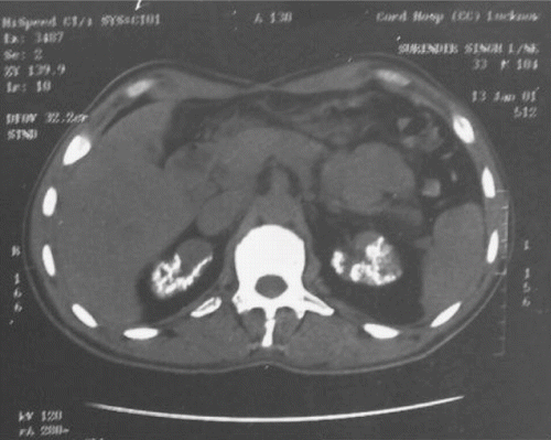
Serial ultrasounds showed a progressive decrease in renal size (right kidney 11.5 cm, 9.5 cm, and 8.1 cm, and left kidney 12.1 cm, 9.8 cm, and 6.5 cm at 3rd, 45th and 75th post-operative days). A radiograph of the abdomen showed speckled calcification in both renal areas. A CT scan of the abdomen done on the 75th post-operative day showed a diffuse cortical calcification involving the entire right kidney and upper half of left kidney (see ). The pelvicaliceal system was normal with an extra-renal pelvis on the left side. Follow-up at 16 months after the initial injury showed a blood urea of 68mg/dL and creatinine of 2.3mg/dL.
Figure 1. CT scan of abdomen showing bilateral renal cortical nephrocalcinosis.
DISCUSSION
Acute cortical necrosis (ACN) represents a rare cause of acute renal failure (ARF), occurring in approximately 2% of cases and which may be complete or patchy. ACN is more common following severe shock; it is the type rather than severity of shock which is of greatest importance in determining the risk. A majority of cases used to follow obstetric shock, particularly from abruptio placentae, but with improved obstetric care this is now extremely rare.Citation[1] Hemorrhage occurring due to non-obstetric causes rarely leads to ACN. It must be suspected in cases of ARF, when oliguria or anuria persists for a long period. Histological confirmation may not be possible in all cases because patients' condition may not always allow for a biopsy, and in cases with a patchy form of ACN, the specimen may not detect the typical changes. Some cases of ACN develop the calcification of the renal parenchyma, a condition called renal cortical nephrocalcinosis (RCN). The risk factors for the development of RCN and its effect on the outcome of the case are not well defined. The case presented above developed ARF following severe hemorrhage due to polytrauma, and a plain x-ray and CT scan of the abdomen showed calcification in both kidneys consistent with RCN.
The most common cause of cortical nephrocalcinosis is acute renal cortical necrosis caused by sepsis, concealed accidental hemorrhage, toxemia of pregnancy, drugs, snake bite, arsenic poisoning, extra-corporeal shock wave lithotripsy, and hemolytic uremic syndrome. The other causes include primary and secondary oxalosis, chronic glomerulonephritis, intrarenal infections in HIV seropositive patients (mycobacterium avium, pneumocystis carinii), amphotericin B, chronic pyelonephritis, acute and chronic renal allograft rejection, autosomal recessive polycystic kidney disease, and benign nodular cortical nephrocalcinosis.Citation[2–4]
In most instances, nephrocalcinosis is discovered many weeks after the onset of acute renal failure while the patient is kept alive by dialysis. In the present case, RCN was detected 10 weeks after the onset of ARF. Until the general introduction of clinical ultrasound, virtually all cases of macroscopic nephrocalcinosis were demonstrated by contrast radiography. The ultrasound is more sensitive than contrast radiography in detecting mild cases of nephrocalcinosis, particularly in children. However, an ultrasound provides less satisfactory pictures for demonstration purposes and yields occasionally false positive results from highly echogenic papillary cysts or deposits of hilar fat. Both ultrasonography and computed tomographic (CT) scan can detect nephrocalcinosis earlier than plain abdominal x-ray.Citation[5],Citation[6] A CT scan has been used less but clearly distinguishes a medullary from cortical nephrocalcinosis and shows the relationship of either to other structures in the kidney, particularly the renal capsule, cysts, and calyceal system. However, calcification is difficult to detect on MR imaging. Due to a signal void, calcification will appear hypointense both on T1- and T2-weighted images, and small calcifications are therefore easily missed on MR imaging.
Three different patterns of cortical nephrocalcinosis have been described. The most common pattern is of a thin peripheral band of calcifications, often with an extension into the septal cortex. The second type is represented by a double line of calcification along the two sides of necrotic zone in the cortex, the so-called “tram line” calcification, a term used by Lloyd Thomas et al. to describe the pattern of nephrocalcinosis seen in obstetric cases of cortical necrosis.Citation[7] The least common form consists of multiple punctate calcifications with a random distribution in the renal cortex. This is the pattern seen in the present case. However, none of these patterns is pathognomonic and may be recognized in all causes of cortical nephrocalcinosis.Citation[8]
REFERENCES
- Chugh KS, Jha V, Sakhuja V, Joshi K. Acute renal cortical nephrocalcinosis. Renal Failure. 1994; 16: 37–47
- Schepens D, Versvijwel G, Kuypers D, Vanrenterghem Y. Renal cortical nephrocalcinosis. Nephrol Dial Transplant. 2000; 15: 1080–1082
- Wrong O. Nephrocalcinosis. Oxford Textbook of Clinical Nephrology, S Cameron. Oxford University Press, New York 1998; 1380–1381
- Hoffbrand BI, Oppenheimer SM, Sachs ML, Wrong OM. Nodular cortical nephrocalcinosis: a benign and hitherto undescribed form of renal calcification. Nephron. 1987; 46: 370–372
- Manz F, Jaschke W, Van Kaick G, Waldherr R, Willich E. Nephrocalcinosis in radiographs, computed tomography, renography and histology. Pediat Radiol. 1980; 9: 19–26
- Arons WL, Christensen WR, Sosman MC. Nephrocalcinosis visible by x-ray associated with Chronic glomerulonephritis. Ann Intern Med. 1995; 42: 260–282
- Lloyd Thomas HG, Balme RH, Key JJ. Tram line calcification in RCN. Br Med J. 1962; 1: 909–911
- Banner MP. Nephrocalcinosis. Clinical Urography, HM Pollck. WB Saunders Company, Philadelphia 1990; 1768–1769