INTRODUCTION
The association of Mantle cell lymphoma (MCL) and renal disease is very rare. This article reports a case of the successful treatment of acute renal failure and nephrotic syndrome secondary to Focal Segmental Glomerulosclerosis (FSGS) and its novel association with MCL.
CASE HISTORY
In December 2003, a 68-year-old gentleman presented with a six-week history of lethargy and increasing swelling of his lower legs. He was previously fit and well. On admission, he was hypertensive 166/113 mmHg. He had clinical signs of lower leg pitting edema up to his groin, sacral edema, pleural effusion, and ascites. Urinalysis revealed proteinuria +3 but no active sediments. His plasma biochemistry revealed an elevated urea 36.1mmol/L, creatinine 362μmol/L, potassium 5.7, and decreased serum albumin. His hematological indices were Hb 12.2 g/dL, WCC 27.3 × 10⁹/L (neutrophils 18.2, lymphocytes 8.1, monocytes 0.8), and platelets 206 × 109/L. His autoantibody screen and cryoglobulin were negative, and he had normal levels of serum complement C3 and C4, immunoglobulin, and protein electrophoresis. A 24-hr urine collection confirmed nephrotic range proteinuria 18 g/day. Renal ultrasound showed normal size kidneys with no hydronephrosis. He underwent a kidney biopsy and was then given intravenous (IV) methylprednisolone 1 g OD for four days followed by oral azathioprine 50 mg OD with prednisolone 60 mg OD.
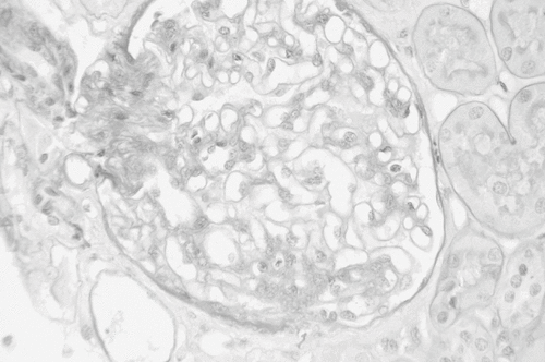
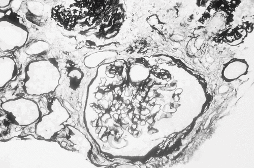
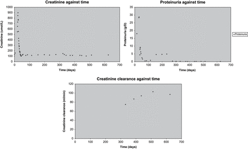
Despite this, his renal function continued to deteriorate, with a urea 57.3mmol/L and creatinine 899 μmol/L, and he was commenced on hemodialysis. Renal histology was consistent with FSGS in two out of eight glomeruli, which showed global sclerosis, minimal interstitial inflammation, hyaline sclerosis of arterioles, mild subtimal fibrosis (see and ), and immunofluorescence studies shown mesangial staining for IgM and C3. He was then commenced on IV cyclophosphamide 1 g and plasma exchange (PE). After a total of 10 PE over two weeks, his renal function gradually recovered until he became dialysis-independent six weeks after initial presentation with no edema, a plasma urea 11.6, creatinine 178, and creatinine clearance 23 mL/min, and his proteinuria had decreased to 4.4 g/day (as illustrated in ).
Figure 1. Renal histology illustrating segmental sclerosis at six o'clock (Periodic Acid Schiff stain).

Figure 2. Renal histology illustrating global sclerosis in 2 glomeruli and arteriolar hyalinosis at 2 o'clock (Methenamine silver stain).

After four weeks in the hospital, he was discharged home on azathioprine 50 mg OD and prednisolone 30mg OD. Subsequently, four weeks later, at the renal outpatient follow-up, his azathioprine was discontinued due to abnormal liver function tests (bilirubin 11, ALT 331, GGT 798, and albumin 38). His renal biochemistry profile, however, continued to improve with a plasma urea 9.1, creatinine 127, creatinine clearance of 77 mL/min, and proteinuria 0.6 g/day.
The results of cytogenetic analysis performed as a result of lymphocytosis discovered during his inpatient led to a suspicion of MCL. A computed tomography (CT) scan of chest, abdomen, and pelvis revealed only minimal mediastinal lymphadenopathy. Bone marrow trephine immuno-histochemistry showed one dense nodule in the lymphoid cells with positive staining of B cell marker CD20. Fluorescence in situ hybridization (FISH) analysis of blood revealed an 11:14 (q13,q32) translocation consistent with a diagnosis of MCL. Subsequently, the patient received six cycles of rituximab 375 mg/m2, cyclophosphamide 600 mg/m2, vincristine 1.4 mg/m2, and prednisolone 100mg for 5 days' (R-CVP) chemotherapy. He has responded well to this treatment with no further symptoms or edema, and at his last follow-up in September 2005, he had normal renal function with urea 8 umol/L, creatinine 124 umol/L, creatinine clearance 97 mL/min, and insignificant proteinuria 0.1 g/day.
DISCUSSION
Mantle cell lymphoma (MCL) is a rare type of lymphoma (5–8% of all non-Hodgkin's lymphomas). As in this case, virtually all of Mantle cell lymphomas are associated with a t(11;14) translocation resulting in overexpression of the cyclin D1 gene.Citation[1] Case reports of patients with MCL and glomerulonephritisCitation[2–5] have previously been described. All of the cases reported by previous groupsCitation[2–5] presented with acute renal failure, as did the present patient. In all patients previously reported, renal disease and function correlated well with the lymphoma activity, and when treated with chemotherapy, the renal disease and function improved. All of these cases not only illustrate the association of active MCL and renal disease but also support the hypothesis of a probable paraneoplastic phenomenon causing the glomerulonephritis and acute renal failure. In the two cases, reported by Karim et al. and our case, the immunosuppression given to treat glomerulonephritis may have accelerated the presentation and disease process of MCL.
In this patient, the lymphocytosis was the key to the diagnosis for MCL. There was no clinical evidence of lymphadenopathy or hepatosplenomegaly at the time of presentation. The cytogenetic studies of the lymphocytes (with cleaved nuclei) suggested the diagnosis, which led to further investigations and later confirmation of the diagnosis of MCL. Small mediastinal lymphadenopathy was detected by CT scan (in keeping with early stage of MCL). In this case, the positive staining of CD 20 on B cells and FISH analysis of blood revealing an 11:14 (q13,q32) translocation are pathognomonic features of MCL. However, MCL can also present with isolated proteinuria or hematuria or both, or more commonly with nephrotic syndrome and renal failure, as described in this case.
Non-Hodgkin lymphomas have been found to be associated with a spectrum of glomerulopathies.Citation[2] The present case is of interest and highlights three salient points. First, it is important to stress the need for a high clinical index of suspicion of lymphoma when a patient presents with either acute renal failure or nephrotic syndrome and lymphocytosis. This has to be distinguished from a neutrophilia associated with sepsis, which frequently co-exists in these patients. The present case describes the novel association of FSGS and MCL specifically, as there were reports of FSGS associated with other lymphomas, namely T cell lymphoma,Citation[6] lymphoblastic T cell lymphoma,Citation[7] and lymphoplasmacytoid lymphoma.Citation[8] The second is the successful use of plasma exchange in the treatment of FSGS, nephrotic syndrome, and acute renal failure in association with MCL. In this patient, the renal failure and nephrotic range proteinuria improved after two cycles of daily PE for five days. There may be an immune complex or immunoglobulin produced by the lymphoma B cells, and this was removed effectively by PE. In this case, the former is more likely, as there was no evidence of paraprotein or cryoglobulins detected in the serum and moreover the absence of lymphomatous infiltration in the renal biopsy. In contrast, others have tried to implicate many candidate compounds as the permeability factorCitation[9–11] or inhibitorsCitation[12],Citation[13] in patients with FSGS in their native kidneys (without lymphoma involvement) and recurrence of FSGS in their transplant kidneys. There is limited experience in the use of PE in the treatment of FSGS in native kidneys, but it may be justified when life-threatening nephrotic syndrome is present.Citation[14] However, this has more commonly been tried in renal transplant patients with the recurrence of their original diagnosis.Citation[14] Third, once rituximab, cyclophosphamide, vincristine, and prednisolone chemotherapy was instituted and his bone marrow and blood were clear of detectable disease, his renal function remained near normal, and proteinuria continued to be of insignificant magnitude.
In conclusion, the present case demonstrates a novel association of FSGS and MCL and supports the therapeutic approach of using plasma exchange and immunosuppression to treat secondary FSGS in native kidneys presenting with acute renal failure and nephrotic syndrome . If there is suspicion of lymphoma, this has to be investigated immediately with prompt diagnosis and chemotherapy treatment.
REFERENCES
- Williams ME, Densmore JJ. Biology and therapy of mantle cell lymphoma. Curr Opin Oncol Sep, 2005; 17(5)425–431
- Karim M, Hill P, Pillai G, Gatter K, Davies DR, Winearls CG. Proliferative glomerulonephritis associated with mantle cell lymphoma—natural history and effect of treatment in two cases. Clin Nephrol. 2004; 61: 422–428
- Wu Q, Jinde K, Yanagi H, Endoh M, Sakai H. Acute interstitial nephritis with polyclonal B cell infiltration and development of mantle cell lymphoma. Intern Med. 2002 Dec; 41(12)1158–1162
- Da'as N, Polliack A, Cohen Y, Amir G, Darmon D, Kleinman Y, Goldfarb AW, Ben-Yehuda D. Kidney involvement and renal manifestations in non-Hodgkin's lymphoma and lymphocytic leukemia: a retrospective study in 700 patients. Eur J Hematol. 2001; 67: 158–164
- Rerolle JP, Thervet E, Beaufils H, Vincent F, Rousselot P, Pillebout E, Legendre C. Crescentic glomerulonephritis and centrocytic lymphoma. Nephrol Dial Transplant. 1999; 14: 1744–1745
- Belghiti D, Vernant JP, Hirbec G, Gubler MC, Andre C, Sobel A. Nephrotic syndrome associated with T cell lymphoma. Cancer. 1981; 47: 1878–1882
- Herskowitz LJ, Gottlieb RP, Travis S. Nephrotic syndrome associated with non-Hodgkin's lymphoma: complete remission with chemotherapy. Clin Pediatr. 1982; 21: 441–443
- Schurgers M, Pauwels W, Praet M, Quatacker J, Lameire N. A 47-year-old man with lymphadenopathy and renal failure. Acta Clin Belg, 43: 149–157
- Savin VJ, Sharma R, Lovell HB, Welling DJ. Measurement of albumin reflection coefficient with isolated rat glomeruli. J Am Soc Nephrol. 1992; 3: 1260–1269
- Savin VJ, Sharma R, Sharma M, McCarthy ET, Swan SK, Ellis E, Lovell H, Warady B, Gunwar S, Chonko AM, Artero M, Vincenti F. Circulating factor associated with increased glomerular permeability to albumin in focal segmental glomerulosclerosis. N Engl J Med. 1996; 334: 878–883
- Sharma M, Sharma R, McCarthy ET, Savin VJ. The Focal segmental Glomerulosclerosis Permeability Factor: Biochemical characteristics and biological effects. Experimental Biology and Medicine. 2004; 229: 85–98
- Sharma R, Sharma M, McCarthy ET, Ge XL, Savin VJ. Components of normal serum block the focal segmental glomerulosclerosis factor activity in vitro. Kidney Int 2000; 58: 1973–1979
- Ghiggeri GM, Artero M, Carraro M, Perfumo F. Permeabilty plasma factors in nephrotic syndrome: more than one factor, more than one inhibitor. Nephrol Dial Transplant. 2001; 16: 882–885
- Savin VJ, McCarthy ET, Sharma M. Permeabililty factors in Focal Segmental Glomerulosclerosis. Seminars in Nephrology 2003; 23(2)147–160
