Abstract
We report a case of primary Sjögren's syndrome (SS) with cutaneous leukocytoclastic vasculitis and IgA nephropathy. The accurate diagnosis of SS was established based on objective signs and symptoms of ocular and oral dryness, a characteristic appearance of a biopsy sample from a minor salivary gland, and the presence of anti-SS-A autoantibody. A second autoimmune disorder was not present, so the diagnosis of primary SS was established. A histologic finding of skin biopsy of purpuric lesion was typical for leukocytoclastic vasculitis. Renal biopsy was performed for nephrotic range proteinuria. The pathologic finding of renal biopsy was IgA glomerulonephritis with crescent formation. The patient was treated with small doses of glucocorticoids and maintenance hemodialysis. Leukocytoclastic vasculitis is one of the most characteristic extraglandular manifestations of SS. However, IgA nephropathy associated with SS and leukocytoclastic vasculitis is a rare finding. SS patients with glomerulonephritis present a more diverse outcome, even requiring hemodialysis. Therefore, renal biopsy is warranted in SS with glomerulonephritis and systemic vasculitis.
INTRODUCTION
Sjögren's syndrome (SS) is a condition of autoimmune exocrinopathy with predominant salivary and lacrimal gland involvement. Cutaneous involvement of primary SS is also a characteristic extraglandular manifestation. Renal involvement is usually subclinical. Herein, we report a case of primary SS associated with IgA nephropathy and leukocytoclastic vasculitis.
CASE PRESENTATION
A 65-year-old female patient developed dry eye and dry mouth for one month. She also developed palpable skin rash and bilateral leg edema. She was the refereed to our hospital.
Upon admission, her temperature was 36°C, blood pressure 117/69 mmHg, pulse rate 127 beats per minute, and respiratory rate 22 breaths per minute. Her general appearance was ill-looking. Physical examination revealed pale conjunctiva, crackles in the both lung fields, pitting edema of lower extremities, and palpable skin lesions over bilateral lower legs.
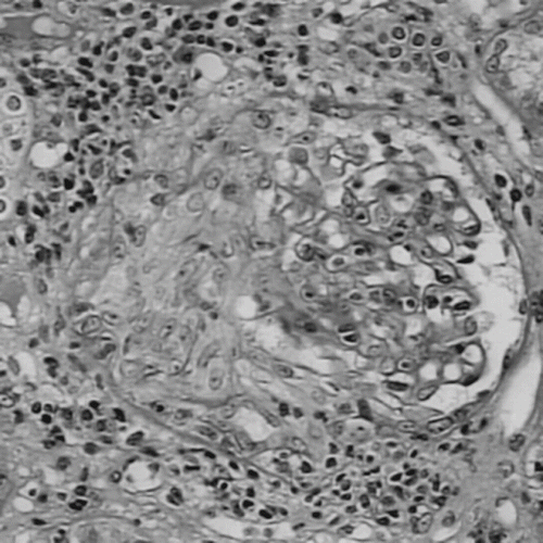
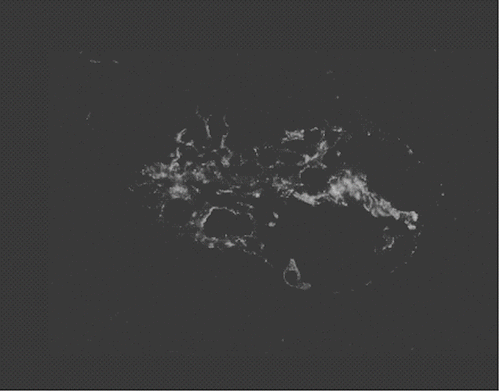
The laboratory data at admission were as follows: white blood cell 11320 (neu-segm70.0%, lympho 23.6%), red blood cell 3.03 × 10⁶/ul, hemoglobulin 9.1 g/dL, hematocrit 27.9%, MCV 92.1 fl, MCH 30.0 pg, MCHC 32.6 g/dL, platelet 547 × 103/ul, glucose 109 mg/dL, aspartate aminotransferase (AST) 34 IU/L, alanine aminotransferase (ALT) 35 IU/L, lactate dehydrogenase (LDH) 188 IU/L, BUN 87 mg/dL, creatinine 8.5 mg/dL, Na 133mmol/L, and K 3.6 mmol/L. The urine presented a protein concentration of 500 mg/dL, 15–20 erythrocytes, and 5–10 leukocytes per high power field with few granular casts. The urinary protein excretion level was 3.6 g/day. Rheumatoid factor was negative, ANA speckled positive, antids DNA antibody was 45 IU/L (normal <100 IU/L), C3 was 64 mg/dL (normal 60–120), C4 was 23.5 (normal 14–24), and cryoglobulins was negative. Anti-SS-A antibody was positive. A biopsy sample from a minor salivary gland confirmed the diagnosis of primary SS. Chest x-ray was normal. Renal echogram showed increased echogenicity of both renal parenchyma. Skin biopsy of lower leg lesions showed leukocytoclastic vasculitis period after vasculitis). Due to nephrotic range proteinuria, renal biopsy was performed on the third day of admission. Two of the 12 glomeruli obtained on PAS staining demonstrated circumferential and cellular crescent formation. Mononuclear cell infiltrate was found over tubules and interstitial area under light microscopy, which is compatible with interstitial nephritis (see ). Immunofluorescence staining demonstrated mesangial deposits of IgA and capillary wall deposits of IgA, IgM, and C3 (see ). Under electron microscopy, no electron-dense deposit was found.
Figure 1. PAS staining demonstrated circumferential and cellular crescent formation with interstitial nephritis.

The patient was treated with small doses of glucocorticoids and local symptomatic therapy for ocular and oral dryness. She also received maintenance hemodialysis but showed no improvement in her renal function.
DISCUSSION
Sjögren's syndrome (SS) is an autoimmune disorder. SS may be primary or associated with other connective tissue diseases, such as rheumatoid arthritis or systemic lupus erythematous. Cutaneous vasculitis is commonly involved in patients with primary SS; for example, Laktasic-Zerjavic et al.Citation[1] reported a case of primary SS with cutaneous leukocytoclastic vasculitis. In this case study, the accurate diagnosis of SS was established based on objective signs and symptoms of ocular and oral dryness, characteristic appearance of a biopsy sample from a minor salivary gland, and the presence of anti-SS-A autoantibody. No other autoimmune disorder was present, so a diagnosis of primary SS was established. A histologic finding of skin biopsy of purpuric lesion was typical for leukocytoclastic vasculitis. The patient was treated with small doses of glucocorticoids and local symptomatic therapy for ocular and oral dryness.
Ramos el al.Citation[2] reported different clinical and histologic types of cutaneous vasculitis in patients with primary Sjögren's syndrome. Clinical and immunologic characteristics of 558 consecutive patients with primary SS were investigated, and those with clinical evidence of cutaneous lesions were selected. The main cutaneous involvement was cutaneous vasculitis, present in 52 (58%) patients. Fourteen presented with cryoglobulinemic vasculitis, 11 with urticarial vasculitis, and the remaining 26 with cutaneous purpura not associated with cryoglobulins. A skin biopsy specimen was obtained in 38 patients (73%). Involvement of small-sized vessels was observed in 36 (95%) patients (leukocytoclastic vasculitis), while the remaining 2 (5%) presented with medium-sized vessel vasculitis (necrotizing vasculitis). Patients with cutaneous vasculitis had a higher prevalence of articular involvement, peripheral neuropathy, Raynaud phenomenon, renal involvement, antinuclear antibodies, rheumatoid factor, anti-Ro/SS-A antibodies, and hospitalization compared with SS patients without vasculitis. Six (12%) patients died, all of whom had multisystemic cryoglobulinemia. The author concluded that cutaneous involvement was detected in 16% of patients with primary SS, with cutaneous vasculitis being the most frequent process. The main characteristics of SS-associated cutaneous vasculitis were the overwhelming predominance of small versus medium vessel vasculitis and leukocytoclastic versus mononuclear vasculitis, with a higher prevalence of extraglandular and immunologic SS features. Small vessel vasculitis manifested as palpable purpura, urticarial lesions, or erythematosus maculopapules, with systemic involvement in 44% of patients in association with cryoglobulins in 30%. Life-threatening vasculitis was closely related to cryoglobulinemia.
Overt renal involvement in primary Sjögren's syndrome occurs in fewer than 10% of cases.Citation[3] The most frequently recognized histopathological renal lesion is interstitial lymphocytic infiltrates with tubular atrophy and fibrosis leading to tubular dysfunction. Tubulointerstitial changes and renal tubular acidosis are the prominent features of the disease. Mononuclear cell infiltration in the renal interstitium is also reported in patients with systemic vasculitis.Citation[4] On the other hand, glomerular nephropathy is not as common as tubulointerstitial nephropathy. Glomerular involvement and primary Sjögren's syndrome has been described previously only in isolated case reports, where membranous nephropathy and membranoproliferative glomerulonephritis have been presented.Citation[5–11] Dabadghao et al.Citation[12] reported proliferative glomerulonephritis leading to end stage renal disease in a patient with primary SS.
Mon et al.Citation[13] reported a case of minimal change disease and glomerular IgA deposition associated with Sjögren's syndrome. Sjögren's syndrome may be accompanied by a dysregulation of IgA system, implying the presence of increased serum polymeric IgA or circulating immune complexes and their consequent deposition within the kidney. IgA nephropathy may represent only one of the complications brought by IgA deposition. Watanabe et al.Citation[14] described the case of a 61-year-old woman diagnosed with primary Sjögren's syndrome (SS) after an eight-year history of IgA nephropathy and a three-year history of recurrent purpuric rashes. Her two daughters had previously been diagnosed with other autoimmune diseases. One daughter had Graves' disease and the other had Hashimoto's disease and systemic lupus erythematosus. The diagnosis of SS was made based on dryness of mucous membranes, Shirmer test, and parotid sialography. Thrombocytopenia, high platelet-aggregated IgG (PA-IgG) level, and normal megakaryocytes count in bone marrow suggested that her recurrent purpuric rashes were due to idiopathic thrombocytopenic purpura (ITP). Therefore, skin biopsy is necessary for recurrent skin rash in primary SS.
Goules et al.Citation[15] observed overt renal involvement in 4.2% of SS patients in a large cohort of 471 patients over 15 years. Interstitial involvement of the kidney was typically observed in younger patients with a short time of disease duration. This finding is compatible with the pathophysiology of the disease process, as SS is considered a systemic autoimmune epithelitis, and the autoreactive lymphocyte infiltrates can affect several extraglandular epithelial early in the disease process. In fact, the lymphocyte infiltrates around the renal tubules are similar to those observed in salivary glands, consisting mainly of activated T-lymphocytes bearing the CD4 phenotype and exhibiting junctional oligoclonality.
In contrast to interstitial nephropathy (IN), the pathogenesis of glomerular nephropathy (GMN) in SS is most probably attributed to the deposition of immune complexes, which are formed by cryoprecipitating monoclonal IgMk RF along with polyclonal IgG and IgA in a manner similar to that observed in renal involvement of hepatitis C-associated mixed monoclonal cryoglobulinemia. Subjective and objective sicca complex manifestations always precede the appearance of GMN. In the present study, 8 of 10 patients with GMN also had mixed monoclonal cryoglobulinemia and lower C4 levels. The late appearance of GMN in association with cryoglobulinemia denotes the oligoclonal or monoclonal B- cell activation, which occurs in SS. This monoclonal B-cell expansion is characterized by specific cross-reactive idiotypes of both heavy and light chains of IgMk monoclonal RF, in which somatic mutations are accumulated in a non-random fashion, suggesting an antigenic and most probably T-cell-dependent process that already exists in the affected epithelial tissues.
In nine patients who had renal biopsy performed, two distinct histologic types of GMN were observed: five patients had membranoproliferative GMN, four patients had mesangial proliferation GMN, and two patients had both IN and GMN. IN proved to be a rather benign condition, as none of the patients presented with end-stage renal failure. In contrast, patients with GMN present a more diverse outcome, sometimes requiring hemodialysis. Therefore, the prognosis of patients with GMN is more guarded, and biopsy is warranted.
In summary, we report a case of primary Sjögren's syndrome (SS) with cutaneous leukocytoclastic vasculitis and IgA nephropathy. Cutaneous involvement is occasionally detected in patients with primary SS, with cutaneous vasculitis being the most frequent process. The main characteristics of SS-associated cutaneous vasculitis were the small vessel leukocytoclastic vasculitis. In addition, tubulointerstitial nephropathy is more common than glomerulonephropathy in primary SS. The association of glomerular IgA nephropathy with Sjögren's syndrome (SS) and cutaneous leukocytoclastic vasculitis is not common. Further prospective study is warranted for understanding the pathogenesis of extraglandular and immunologic SS features.
DECLARATION OF INTEREST
The authors report no conflicts of interest. The authors alone are responsible for the content and writing of the paper.
REFERENCES
- Laktasic-Zerjavic N, Anic B, Curkovic B, Babic-Naglic D, Nola M, Loncaric D. Leukocytoclastic vasculitis in primary Sjögren syndrome: A case report. LiječNičKi Vjesnik. 2007; 129(5)134–137
- Ramos-Csals M, Anaya JM, Garcia-Carrasco M, Rosas J, Bove A, Claver G, Diaz LA, Herrero C, Font J. Cutaneous vasculitis in primary Sjögren syndrome: Classification and clinical significance of 52 patients. Medicine. 2004; 83(2)96–106
- Kassan SS, Talal N. Renal disease in Sjögren syndrome: Clinical and immunological aspects. Springer-Verlag, Berlin 1987; 96–102
- Yamada A. Tubulointerstitial nephropathy secondary to collagen-vascular diseases. Japan J Clin Med. 1995; 53(8)1969–1973
- Bnet SJ, Teixido PJ, Costa PB, Mayayo A, Carrera M. Sjögren syndrome and membranous glomerulonephritis. Rev Clin Esp. 1985; 17: 191–193
- Moutsopoulous HM, Balow JE, Cawley JJ, Stahl NI, Autonoych TT, Chused TM. Immune complex glomerulonephritis in sicca syndrome. Am J Med. 1978; 64: 955–960
- Siamopoulous KC, Mavridis AK, Elisaf M, Drosos AA, Moutsopoulous HM. Kidney involvement in primary Sjögren syndrome. Scand J Rheumatol. 1986; 61: 156–160
- Font J, Cervera R, Lopez-Soto A, Darnell A, Ingelmo M. Mixed membranous and proliferative glomerulonephritis in primary Sjögren syndrome. Br J Rheumatol. 1989; 28: 548–550
- Rodriguez MA, Tapanes FJ, Stekman IL, Pinto JA, Camejo O, Abadi I. Auticuklar and diffuse proliferative glomerulonephritis in primary Sjögren syndrome. Ann Rheum Dis 1989; 48: 683–685
- Khan MA, Akhtar M, Taher SM. Membranoproliferative glomerulonephritis in primary Sjögren syndrome. Report of a case with review of literature. Am J Nephrol. 1988; 8: 235–239
- Schilesinger I, Carlson TS, Nelson D. Type III membranoproliferative glomerulonephritis in primary Sjögren syndrome. Conn Med. 1989; 53: 629–632
- Dabadghao S, Aggarwal A, Arora P, Pandey R, Misra R. Glomerulonephritis leading to end stage renal disease in a patient with primary Sjögren syndrome. Clin Exp Rheumatol. 1995; 13: 509–511
- Mon C, Sanchez Hernandez R, Fernandz Reyes MJ, Estebanez C, Ortiz M, Alvarez-Ude F, Mampaso F. Minimal-change disease with mesangial IgA deposits associated with Sjögren syndrome. Nefrologia. 2002; 22(4)386–389
- Watanabe M, Fujimoto T, Iwano M, Shiiki H, Nakamura S. A report of a patient of primary Sjögren syndrome, IgA nephropathy and chronic idiopathic thrombocytopenic purpura. Jpn J Clinical Immunol. 2002; 25(2)191–198
- Goules A, Masouridi S, Tzioufas AG, Ioannidis JP, Skopouli FN, Moutsopoulos HM. Clinically significant and biopsy-documented renal involvement in primary Sjögren syndrome. Medicine. 2000; 79: 241–249
