Abstract
Emphysematous pyelonephritis (EPN) is a severe and complicated renal infection characterized by gas formation within the infected kidney and its surrounding tissues. Early diagnosis with a high index of suspicion and aggressive treatment are important for improving outcome. Bilateral involvement is rare, and surgical intervention is usually required because of its high mortality rate. A literature review found that EPN has rarely been noted in chronic dialysis patients, and those who show bilateral EPN have demonstrated no survival at all until now. Herein, we presented a 51-year-old diabetic uremic woman who developed right emphysematous pyelitis initially and then progressed to bilateral EPN when hospitalized. Percutaneous drainage (PCD) with simultaneous antibiotic therapy successfully eradicated her renal infection. In this study, all reported cases of EPN in chronic dialysis patients were also reviewed.
INTRODUCTION
Emphysematous pyelonephritis (EPN) is an uncommon renal infection with unique presentation of air production within the involved kidney.Citation[1] It occurs more frequently in patients with diabetes mellitus (70–90% of reported patients)Citation[2] and those with existing obstructive uropathy (25–40%).Citation[3] Treatment modalities of EPN include conservative antibiotics therapy, percutaneous drainage (PCD), and surgical intervention such as relief of associated obstruction or even nephrectomy. For most patients, combination therapy is usually needed and offered. Most reports revealed that the mortality of EPN is high because of the disease condition being severe and extensive renal tissues being involved. Bilateral EPN is rare in that the treatment for these patients might have several challenges. Yet few reports were found to deal with EPN in the dialysis patients. Thus, we report a chronic dialysis patient who survived from bilateral EPN by conventional therapies only and surgical intervention was not even introduced.
CASE PRESENTATION
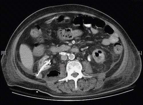
A 51-year-old woman with medical history of coronary artery disease, stroke, and type 2 diabetes mellitus had been on hemodialysis therapy for eight years. The dialysis therapy was three sessions per week, four hours per session. She was rather well prior to this admission, but began to experience febrile with disturbed consciousness and abdominal pain in April, 2007. She was sent to our emergency department immediately. Upon arrival, her body temperature was 36°C, heart rate 88 beats per minute, respiratory rate 20 breaths per minute, and blood pressure was 140/80 mm Hg. Physical examination found that her consciousness was clear but lethargic. The abdomen was soft, but mildly distended, and tenderness over right upper and lower quadrant area were noted. Other findings were also remarkable. Turbid urine was noted after Foley catheterization for urine sample collection. Laboratory tests revealed leukocytosis with left shift (white blood cell count 23,900 cells/uL, neutrophil 81%, platelet count 337,000 cells/uL), and a high CRP level (301.6 mg/L). Urinalysis showed marked pyuria (white blood cell > 100/HPF) and bacteriuria. Biochemical data showed blood urea nitrogen 52 mg/dL, creatinine 4.5 mg/dL, sodium 137 meq/L, and potassium 5.1 meq/L. Because of persisted fever and flank knocking pain, abdominal computed tomography (CT) was arranged and swollen kidney with gas formation within right renal calyces, right ureter, and urinary bladder were noted. The patient was treated by empiric broad-spectrum antibiotics (flumarin 1.0 gm per day), and a PCD tube was implanted subsequently. In the following three weeks, low grade fever and lower abdominal pain persisted. Pus-like fluid was drained continuously from PCD. However, the culture of blood and urine did not yield significant results. Laboratory test found marked leukocytosis (white blood cell count 20,600 cells/uL) and high CRP (284.6 mg/L) level. CT scan was performed for re-evaluation of her renal infection. Unexpectedly, left EPN and a right posterior pararenal gas-containing abscess developed (see ). Under this circumstance, surgical intervention was suggested but was not accepted unfortunately. Repeated urine culture yielded Klebsiella pneumoniae and Pseudomonas aeruginosa, and sensitive antibiotics (tienam 500 mg q12h) were administered thereafter. With continuous antibiotics therapy, fever subsided and her condition improved gradually. However, right pararenal and left renal abscess did not resolve completely in follow-up image study. A PCD tube was then inserted for left renal abscess. Only minimal residual abscess was found at one week interval in sonographic examination. The drainage tube was removed later and she was discharged, as image study demonstrated regressive change, and there was marked improvement in her clinical condition along with normalized CRP level.
DISCUSSION
Urinary tract infection is a common infectious disease, and the treatment response is usually excellent under appropriate antibiotics therapy. While complicated, urinary tract infection such as EPN, which we presented in this report, is associated with increased morbidity and mortality and may need further intervention such as drainage and operation in addition to antibiotics therapy. EPN was first described in 1898 by Kelly and MacCullum.Citation[4] Although not very common, this renal infection is characterized by specific disease entity, but an optimal treatment strategy remains unsettled. Pathogenesis of EPN involves gas-forming bacteria, high tissue glucose level, impaired tissue perfusion, and gas transport and underlying defective immunity.Citation[5] The most common isolated microorganism from EPN is Eschoerichia coli and Klebsiella pneumonia.Citation[6] The underlying diabetes and an obstruction of urinary tract are the most common factors for the development of EPN. Collectively, the overall mortality rate of EPN is about 25%, ranging from 11% to 42%.Citation[7] In a meta-analysis study, conservative treatment alone, type I EPN (renal parenchymal necrosis with absence of fluid content or presence of a streaky/mottled gas pattern), bilateral EPN, and thrombocytopenia are risk factors for mortality.Citation[7] Recently, Aswathaman et al. reported a successful treatment rate of 80% with combination of antibiotics and PCD, whereas nephrectomy was not needed in most patients.Citation[8]
The clinical features and image study of EPN have been investigated, as the CT scan becomes a very useful diagnostic tool and treatment was guided by image staging.Citation[9] Bilateral EPN is classified as the most severe form, and bilateral involvement is uncommon, accounting for only 6% of all cases in a large-scale analysis .Citation[7] Previous study reported that the clinical presentation of bilateral EPN is similar to unilateral EPN.Citation[10] Of note, the overall mortality rate of bilateral EPN was as high as 41%, and three-quarters of those patients may die if nephrectomy was not performed.Citation[9],Citation[10] It has been suggested that bilateral PCD be tried first and bilateral nephrectomy should be done if clinical response is poor.Citation[9] For our patient, the initial CT image study revealed right emphysematous pyelitis. Under antibiotics therapy in combination with PCD, however, this unilateral lesion progressed to ipsilateral pararenal abscess and left EPN. No previous report has described bilateral EPN evolving from unilateral to bilateral as noted in our patient. Aswathaman et al. have suggested that rescreening is helpful in detecting the need of drainage tube placement that contributes to more treatment success.Citation[8] It is recommended that a timely assessment on involved kidney is necessary while treating unilateral EPN, and an evaluation on the other kidney should be included. We assume that immediate PCD in association with appropriate antibiotics therapy might reduce the risk of systemic spread and prevent progression of EPN and contralateral involvement.
It is well recognized that urinary tract infection is not common in chronic dialysis patients, and EPN has rarely been noted in this population.Citation[11] Until now, only six patients have been reported.Citation[12–17] We summarized their clinical features, treatment modalities, and outcome in . All patients have had one kidney involvement, and three of them were transplant graft. The mean age was 58 years, 71% were women, and 57% of them had underlying diabetes mellitus. Two patients developed EPN after embolization of renal graft. Only one of these patients underwent peritoneal dialysis. Most of the pathogens were gram negative bacilli, and Eschoerichia coli and Klebsiella pneumonia constitute more than half of reported cases. Surgical resection of the infected kidney was performed in four patients. Except for one report, all patients survived. Without the need to preserve residual kidney function, nephrectomy will not be delayed when indicated. Removal of a non-functional kidney may prevent disease progression and improve outcome in these uremic patients. The pathogenesis of EPN in dialysis patients has not been elucidated yet. The impaired immunity and absence of residual urine flow may play a role in the development of this complicated urinary tract infection in dialysis population. Optimal treatment strategy of EPN in dialysis patients is still lacking, and further studies are required to establish a firm guideline.
Table 1 Summary of emphysematous pyelonephritis in chronic dialysis patients
In conclusion, this is the first report of chronic dialysis patient survived from bilateral EPN without nephrectomy. Appropriate antibiotics therapy in association with adequate drainage controls the kidney infection during unilateral to bilateral progression. A meticulous observation on clinical course and close monitoring of treatment response are mandatory in treating dialysis patients with this rare and complicated renal infection.
DECLARATION OF INTEREST
The authors report no conflicts of interest. The authors alone are responsible for the content and writing of the paper.
REFERENCES
- Michaeli J, Mogle P, Perlberg S, Heiman S, Caine M. Emphysematous pyelonephritis. J Urol. 1984; 131: 203–208
- Tseng CC, Wu JJ, Wang MC, Hor LI, Ko YH, Huang JJ. Host and bacterial virulence factors predisposing to emphysematous pyelonephritis. Am J Kidney Dis. 2005; 46: 432–439
- Bohlman ME, Sweren BS, Khazan RR, Minkin SD, Goldman SM, Fishman EK. Emphysematous pyelitis and emphysematous pyelonephritis characterized by computerized tomography. South Med J. 1991; 84: 1438–1443
- Kelly HA, MacCullum WG. Pneumaturia. JAMA 1898; 31: 375–381
- Chen KW, Huang JJ, Wu MH, Lin XZ, Chen CY, Ruaan MK. Gas in hepatic veins: A rare and critical presentation of emphysematous pyelonephritis. J Urol. 1994; 151: 125–136
- Picron B, Mauerhoff T, Farchakh E, Wese FX. Pyelonephrite emphysemateuse (PNE) chez une patiente diabetique. Revue de la litterature a propos d'un cas. Acta Clin Belg. 1991; 46: 94–99
- Falagas ME, Alexiou VG, Giannopoulou KP, Siempos II. Risk factors for mortality in patients with emphysematous pyelonephritis: A meta-analysis. J Urol. 2007; 178: 880–885
- Aswathaman K, Gopalakrishnan G, Gnanaraj L, Chacko NK, Kekre NS, Devasia A. Emphysematous pyelonephritis: Outcome of conservative management. Urology. 2008; 71: 1007–1009
- Huang JJ, Tseng CC. Emphysematous pyelonephritis: Clinico radiological classification, management, prognosis, and pathogenesis. Arch Intern Med. 2000; 160: 797–805
- McHugh TP, Albanna SE, Stewart NJ. Bilateral emphysematous pyelonephritis. Am J Emerg Med. 1998; 16: 166–169
- Fasolo LR, Rocha LM, Campbell S, Peixoto AJ. Diagnostic relevance of pyuria in dialysis patients. Kidney Int. 2006; 70: 2035–2038
- Goral S, Stone W. Emphysematous pyelonephritis in a nonfunctioning renal allograft of a patient undergoing hemodialysis. Am J Med Sci. 1997; 314: 354–356
- Chang CY, Shieh GS, Tong YC, Chow NH. Emphysematous pyelonephritis in a uremic patient with hemorrhagic renal cell carcionoma. J Urol R.O.C. 2001; 12: 131–134
- Anwar N, Chawla LS, Lew SQ. Emphysematous pyelitis presenting as an acute abdomen in an end-stage renal disease patient treated with peritoneal dialysis. Am J Kidney Dis. 2002; 40: E13
- Atar E, Belenky A, Neuman-Levin M, Yussim A, Bar-Nathan N, Bachar GN. Nonfunctioning renal allograft embolization as an alternative to graft nephrectomy: Report on seven years’ experience. Cardiovasc Intervent Radiol. 2003; 26: 37–39
- Wen YK, Chen ML. Xanthogranulomatous pyelonephritis complicated by emphysematous pyelonephritis in a hemodialysis patient. Clin Nephrol. 2007; 68: 422–427
- Ortiz A, Petkov V, Urbano J, et al. Emphysematous pyelonephritis in dialysis patient after embolization of failed allograft. Urology. 2007; 70: e17–e19
