Abstract
Background. Renal ischemia/reperfusion (I/R)-induced tubular epithelial cell injury, called ischemic acute renal failure, is associated with high mortality in humans. Protecting the kidney against I/R injury is very important during complicated renal operations, transplantation surgery, and anesthesia. Aim. The purpose of this study was to investigate and compare the efficiency of ketamine, thiopental, propofol, etomidate, and intralipid in reducing the injury induced by free radicals in a rat model of renal I/R. Method. Forty-two Wistar rats were divided into seven groups in our study. Rats in the sham group underwent laparotomy and waited for 120 minutes (min) without ischemia. Rats in the control group were given nothing with ischemia-reperfusion. Rats in the I/R groups were given ketamine (20 mg/kg), thiopental (20 mg/kg) propofol (25 mg/kg), etomidate (10 mg/kg) and 10% intralipid (250 mg/kg) intraperitoneally 15 min prior to the ischemia for 60 min, followed by reperfusion for 60 min. The blood samples and kidney tissues of the rats were obtained under anesthesia at the end of the reperfusion period. Biochemical malondialdehyde (MDA), superoxide dismutase (SOD), catalase (CAT), blood urea nitrogen (BUN), creatine (Cr), aspartate aminotransferase (AST) were determined, and histopathological analysis was performed with these samples. Results. MDA level was increased significantly in the control group (p < 0.05). Histopathological findings of the control group confirmed that there was renal impairment by tubular cell swelling, interstitial edema, medullary congestion, and tubular dilatation. MDA levels were lower in the ketamine, thiopental, and propofol groups compared to the control group (p < 0.05). In the thiopental and propofol groups, the levels of histopathological scores were significantly lower than control and etomidate groups in ischemia-reperfusion. Conclusion. Our results demonstrated that I/R injury was significantly reduced in the presence of propofol and thiopental. The protective effects of these drugs may belong to their antioxidant properties. These results may indicate that propofol and thiopental anesthesia protects against functional, biochemical, and morphological damage better than control in renal I/R injury.
INTRODUCTION
I/R damage can occur in clinical cases such as renovascular surgery, clamping of aorta, shock, and trauma.[Citation1] According to the severity of the damage, many clinical occasions varying from prerenal azotemia without tissue damage to the acute kidney failure related to tubular and cortical necrosis can be encountered. Acute tubular necrosis (ATN) and the development of renal failure related to I/R are main reasons behind morbidity and mortality.[Citation2] It is established that graft function delay developing after renal transplantation is related to ATN. ATN arises as a result of damage created by free oxygen radicals, which are formed in I/R.[Citation3] Free radical formation in ischemic tissue and its increase with reperfusion suggest the value in organ transplantation.[Citation4]
In the renal transplantation process, exposure of the graft for transplant to I/R damage in variable levels causes graft function delays and graft losses in post-operative period. Hence, in this procedure, it is an indisputable fact that approaches for preventing I/R damage have positive effects in graft functions and graft lifetime. Thus, before any important operation and renal transplantation, it would be wise to find a suitable prophylactic treatment in order to avoid I/R damage.
Anesthetic material should be chosen for patients with an increased risk for kidney failure, as well as kidney disease and/or undergoing operational implementation such as transplantation, in order to protect kidney's functions. However, no ideal anesthetic material has yet been developed to protect the functions of kidney.[Citation5] In this study, we sought to examine the possible effects of variable anesthetic materials against the damage created by ischemia and reperfusion.
MATERIALS AND METHODS
Animals
The experimental protocol used for this study was approved by the Animal Ethics Review Committee of the Faculty of Medicine, University of Kahramanmaras, and adhered to National Institutes of Health guidelines for the use of experimental animals. Forty-two Wistar rats were housed in individual cages in a temperature-controlled room with alternating 12-h light:dark cycles and acclimatized for a week before the study. Food was removed 8 h prior to the study, but all animals were allowed free access to water and rat chow diet.
Experimental Design
Animals were anesthetized with intramuscular injections of 60 mg/kg ketamine hydrochloride (Ketalar, Eczacıbaşı, Turkey). The abdominal region was shaved and sterilized with povidone iodine solution. A midline incision was made, and the abdominal viscera were retracted to the right side. The left renal hilus was dissected, the renal vascular pedicle was occluded using a microvascular clamp (Vascustatts II, midi 1001-532, Scatlan, Minnesota, USA), and the intestine was replaced into the abdominal cavity. At the end of the 60 minutes (min) ischemic period, 60 min reperfusion was established by removal of the clamp, and left nephrectomy was performed. Animals were randomly divided into seven groups, each consisting of six animals:
Group 1 (sham group, n = 6) rats were subjected to identical surgical procedures described above, except for renal I/R;
Group 2 (control group, n = 6) rats received 60 min of left renal ischemia, followed by 60 min of reperfusion;
Group 3 (I/R + ketamine group, n = 6) animals were administered ketamine hydrochloride (20 mg/kg, intraperitoneally [ip]) 15 min before the reperfusion phase;
Group 4 (I/R + thiopental group, n = 6) animals were administered thiopental sodium (20 mg/kg, ip; PENTAL, I.E. Ulagay Ilaç Sanayii Turk A.Ş. Topkapı, Istanbul, Turkey) 15 min before the reperfusion phase;
Group 5 (I/R + propofol group, n = 6) animals were administered propofol (25 mg/kg, ip; Propofol 1% Fresenius, Fresenius Kabi AB, Uppsala, Sweden) 15 min before the reperfusion phase;
Group 6 (I/R + etodimate group, n = 6) animals were administered etomidate (10 mg/kg, ip; Etomidate-Lipura, B. Braun Melsungen AG, Germany) 15 min before the reperfusion phase; and
Group 7 (I/R + intralipid group, n = 6) animals were administered intralipid (250 mg/kg, ip; intralipid 10% 500 ml Fresenius, Fresenius Kabi AB, Uppsala, Sweden) 15 min before the reperfusion phase.
At the end of the reperfusion period, the samples of blood and tissue were harvested for biochemical, serological, and histopathological evaluation. Blood (5 mL) was taken from the animals via cardiac puncture. Half of tissues were used for tissue biochemical analysis.
Biochemical Analysis
Serum samples were used for the measurement of serum urea levels, which were used to assess renal function, and as an indicator of renal I/R injury. The levels of serum urea were analyzed with DADE-BEHRING dimension-RLMax biochemical analyzer.
Renal Histology
The kidneys fixed in a 10% neutral buffered formalin solution were embedded in paraffin and were used for histopathological examination. Five micrometers (5 μm) thick sections were cut, deparaffinized, hydrated, and stained with hematoxylin and eosin. The renal sections were examined in blind fashion for tubular cell swelling, tubular dilatation, hyaline cast, interstitial edema, medullary congestion, and moderate to severe epithelium necrosis in all treatments. A minimum of 10 fields for each kidney slide were examined and assigned for severity of changes using scores on a scale of none (−), mild (+), moderate (++), and severe (+++) damage.
Antioxidant Study
In order to determine tissue antioxidant levels, 1 × 1 cm2 tissue samples were taken from the left kidney. The samples were preserved in a deep freezer until examination. The tissues were homogenized with three volumes of ice-cold 1.15% KCl. The activities of antioxidant enzymes and the levels of lipid peroxidation were measured in the supernatant obtained from centrifugation at 14,000 rpm. SOD activity was measured according to the method described by Fridovich.[Citation6] CAT activities were determined by measuring the decrease in hydrogen peroxide concentration at 230 nm by the method of Beutler.[Citation7] Lipid peroxidation level in the tissue samples was expressed in MDA and measured according to procedure of Ohkawa et al.[Citation8] Protein concentration was determined according to the method of Lowry.[Citation9]
Statistical Analysis
All variables were expressed as mean and standard deviation. Differences between groups were evaluated by Kruskal-Wallis variance analysis followed by a post-hoc Mann-Whitney U test. p values < 0.05 were considered statistically significant. All data were entered into and processed by SPSS 9.05 for Windows statistical package.
RESULTS
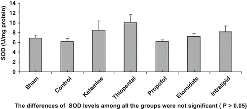
I/R procedure significantly increased MDA levels (p < 0.05), decreased CAT activities (p < 0.05), and did not change superoxide dismutase (SOD) levels (p > 0.05). MDA levels were lower in the ketamine, thiopental, and propofol groups compared to control group (p < 0.05, see ). MDA levels of thiopental and propofol groups were lower compared to etomidate group as well (p < 0.05). Our findings showed that CAT levels increased in propofol, thiopental, and ketamine groups (see ). When CAT levels were evaluated, it was shown that the lowest results were in the propofol group and the highest results were in the sham group. When mean SOD levels were evaluated, the lowest was in the propofol group and the highest was in the thiopental group (see ).
Figure 1. MDA levels (nmol/mg protein).
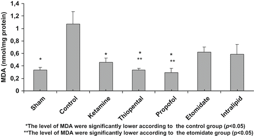
Figure 2. CAT levels.
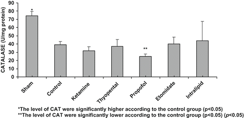
Figure 3. SOD levels.
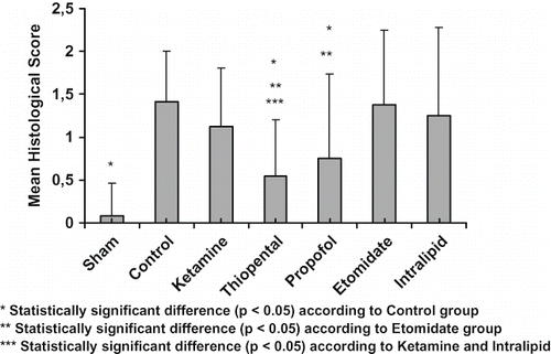
Despite using a light microscope, epithelial necrosis and hyaline cast formation were not observed in kidney tissues that were stained with Hematoxylin-Eosin. In the sham group, no tubular dilatation was observed, although mild and moderate dilatation had been observed in the other groups (see ). In the sham group, no interstitial edema was observed. The most apparent interstitial edema was observed in the control group, in a moderate degree. Tubular dilatation, which was not present in the sham group, was observed in other groups in mild and moderate degrees. In the ischemic group, medullar congestion has been observed in mild and moderate intensity (see ). In the thiopental and propofol groups, the levels of histopathological scores was significantly decreased compared to control and etomidate groups in ischemia-reperfusion (see ).
Figure 4. Mild tubular dilatation.
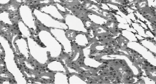
Figure 5. Severe congestion.
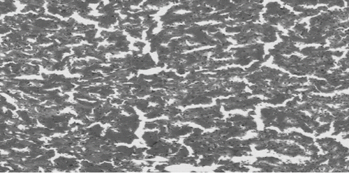
Figure 6. Mean histological scores.
DISCUSSION
Reperfusion damage is the chain of events related to free oxygen radicals produced during tissue ischemia and reperfusion. These chains of events continue with the activation of endothelial cells and the inflammation developed by the circulating leukocyte migration to the area.[Citation10,Citation[11]] The kidney I/R damage, which is a critical clinical problem, has been the subject of many clinically and experimental studies in the development of transplantation surgery. Regarding kidney I/R damage, there are some comparative studies examining propofol and thiopental, which are among the anesthetic agents.[Citation12,Citation[13]] Basu et al. detected that propofol decreased oxidative stress and inflammatory response in patients who had kidney transplantation.[Citation14]
In his study on I/R damage, Darendelioglu stated that ketamine, propofol, and etomidate, when applied in both anesthetic and subanesthetic doses, showed protective effects against I/R damage, though the most significant effects were observed in subanesthetic doses.[Citation15] In our study, we applied ketamine at 20 mg/kg, thiopental at 10 mg/kg, propofol at 25 mg/kg, and etomidate at 10 mg/kg (ip) at subanesthetic doses. We applied 10% of intralipid at a 250 mg/kg dose according to the phosphatide ratio it contained. The early phase of reperfusion—the first hour, when the damage is at its maximum—was expected to benefit from the maximum effects. For this reason, these active materials were applied 15 min before the reperfusion.
As oxygen radicals are short-lived and the kidney tissue has a complex structure, it is hard to detect oxygen radicals. It is executed indirectly with the lipid peroxidation measuring originated from free oxygen radicals. One substance produced by lipid peroxidation is MDA.[3] So, in our research, we studied MDA levels in the kidney tissue in order to show the damage and compare the anesthetics' effects in decreasing the damage. The highest MDA rate was detected in the control group. In addition, MDA levels in thiopental and propofol groups were determined to be lower when compared to control groups (p < 0.05).
In the metabolism, there are enzymatic organisms against the harmful effects of free oxygen radicals. Measuring these enzyme levels gives us indirect information about the free radical mediated damage. We examined SOD and CAT levels in kidney tissue. In examining the SOD levels, we detected that the lowest level was in the propofol group but the highest level was in the thiopental group.
Because it is concordant with the other medications and has a wide confidence interval, ketamine is one of the anesthetic materials with the widest usage range in experimental applications.[Citation16] For this reason, we prefer ketamine as an anesthetic material. Active materials used in this study are intravenously used anesthetic materials. In researching the effects of subanesthetic doses of these anesthetic materials on kidney ischemia reperfusion damage, we found that ketamine was the anesthetic material in this study with the most positive effects. In order to standardize this effect in our study, the anesthesia in all the groups was provided by ketamine. When examining the results of the sham and control groups, in which no active material was used apart from the anesthetic material ketamine, we can see I/R damage occurred in kidney. Therefore, we are convinced that the results we obtained were independent from the effect of ketamine. However, in order to put forward the total effects of ketamine, which we used as anesthetic agent, different studies in which it interacts and is combined with other materials are required.
This study showed that higher doses of ketamine had a better effect than the low doses of ketamine. There is scant information about the effects of ketamine at higher doses with renal failure. However, in a study that investigated the effect of high dose ketamine following incomplete cerebral ischemia in rats, it was observed that high-dose ketamine improved the neurological outcome.
The MDA values obtained in the thiopental group were significantly lower in comparison with the control group (p < 0.05). Considering the histopathology findings, a significant recovery in the thiopental group was detected compared with the control group. This recovery in the thiopental group was better when compared to the other groups, and there were major differences among the ketamine, etomidate, and intralipid groups (p < 0.05). In their studies examining the effects of etomidate, thiopental, and propofol induction on the hypoperfusion reperfusion event, Yagmurdur et al. stated that high lipid resolution material thiopental has the effects of lipid peroxidation inhibition and free radical-related hemolysis, inhibiting antihemolytic activity and antioxidant. Thus, in clinically suitable concentrations, thiopental is stated to repress the neutrophil originated free oxygen radicals significantly.[Citation17] Basu et al. found that propofol had an inflammatory response-decreasing effect compared to thiopental in a study in which oxidative stress and inflammatory response after the renal transplantation were examined.[Citation14] However, we did not detect any significant difference between these two groups in our study. According to this information and based on the data we obtained from our study, it is determined that thiopental decreases the kidney I/R damage via free oxygen radicals in rats, and this effect is significant compared to etomidate.
Compared to control and etomidate groups, the MDA levels of propofol group was significantly low (p < 0.05). In the histopathological analysis of kidney tissue of the propofol group, significant recovery was observed. Considering the increase in average histological scores, it was also found statistically low compared to the control and etomidate groups. The anesthetic material propofol, which has high lipid resolution, is often used in the induction of anesthesia and in the sedation of patients bound to mechanical ventilation in intensive care unita. The positive effects of many organs such as heart, lungs, brain, liver and testis were observed. Propofol may limit the oxidative damage in various tissues, including kidney. In addition to the effect of propofol to the kidney, I/R damage has been stated in very few studies. In the study of Wang et al.,[Citation18] propofol was detected to decrease the renal ischemia reperfusion damage and renal dysfunction in rats significantly. Furthermore they showed that these protective effects are via heme oxygenase-1 induction. Yagmurdur et al. showed in their studies that propofol hypoperfusion reperfusion condition related to lipid peroxidation inhibition and induction doses of propofol may have many advantages in the suspension of metabolites and free oxygen radicals.[Citation17] In examining tissue antioxidant capacities during the propofol anesthesia, Runzer et al.[Citation19] detected that MDA levels decreased significantly when high-dose propofol was mixed with halothane. Nonetheless, in the same study, it was detected that the protective effect of propofol was significant in all tissues and had a significant protective effect primarily in liver and then kidney, heart, and lungs. When adding propofol in high and low doses, more positive results were observed in high doses. They explained the tissues' different responses, as each tissue has different lipid peroxidation sensitivity.
The forms of etomidate and propofol used in clinics are preparations prepared as 10% lipid emulsion. In studying how those components affect the I/R damage, we also used intralipid solution, including 10% lipid contents as another group. In his article, Szekely mentions intralipid explosion reaction redoubling the effect of neutrophils.[Citation20] Mathy-Hartert, on the other hand, compared propofol and intralipid in his study of free oxygen radicals effect, and found that intralipid had an effect in a low level.[Citation21] In Runzer's study, it was confirmed that intralipid has a low antioxidant effect. In the combination of intralipid and propofol, however, they stated that in low concentrations, this effect was more evident.[Citation19] Our diagnoses are supported by the results of those studies. In our study, we confirmed that intralipid in rats may reduce kidney I/R damage at a low level; however, propofol lowers kidney I/R damage at a significant level.
Etomidate is preferred, especially in the induction of cardiac patients. Yagmurdur's research has shown increased MDA levels in etomidate groups. Post-operative examination showed that in etomidate and thiopental groups, AST and ALT levels in 24 hours showed a marked significant increase, but in propofol group no significant change has been marked.[Citation17] Our study showed in that MDA level was higher in etomidate groups than in other groups.
When we compare all of our results, we found a possible reducing effect of ketamine, thiopental, etomidate, and intralipid on kidney I/R damage. Most of this reducing effect was found in the propofol and thiopental groups. While the reducing effect in ketamine and MDA levels was significant, there was no significant improvement in pathological findings. While there was a reduction in MDA levels and pathological findings for etomidate and intralipid, these findings were not significant.
Thiopental and propofol could be chosen in situations where kidney I/R damage was a likely occurrence, such as transplantation surgery in surgery interference and other clinical states to the induction and continuation of anesthesia. However, experimental studies for a wide range of clinical research and the antioxidant effect mechanism of anesthetics are required.
DECLARATION OF INTEREST
The authors report no conflicts of interest. The authors alone are responsible for the content and writing of the paper.
REFERENCES
- Alatas O, Sahin A, Colak O, Inal M, Koken T, Yaşar B, Karahüseyinoglu E. Beneficial effects of allopurinol on glutathione levels and glutathione peroxidase activity in rat ischaemic acute renal failure. J Int Med Res. 1996; 24(1)33–39
- Onal A, Astarcioglu H, Ormen M, Atila K, Sarioglu S. The beneficial effect of L-carnitine in rat renal ischemia-reperfusion injury. Ulus Travma Acil Cerrahi Derg. 2004; 10(3)160–167, [in Turkish]
- Paller MS, Hoidal JR, Ferris TF. Oxygen free radicals in ischemic acute renal failure in the rat. J Clin Invest. 1984; 74(4)1156–1164
- Reiter RJ. Oxidative processes and antioxidative defense mechanisms in the aging brain. FASEB J. 1995; 9(7)526–533
- Rusafa Neto E, Vianna PT, Viero RM, Módolo NS, Ganem EM, Braz RC, et al. Influence of S(+)-ketamine analgesia in renal intraoperative ischemia: Histological study in rats. Acta Cir Bras. 2006; 21(4)242–246
- Fridovich I. Superoxide radical: An endogenous toxicant. Annu Rev Pharmacol Toxicol. 1983; 23: 239–257
- Beutler E. Red cell metabolism2nd. Grune & Stratton, New York 1975
- Ohkawa H, Ohishi N, Tagi K. Assay for lipid peroxides in animal tissues by thiobarbituric acid reaction. Anal Biochem. 1979; 95: 351–358
- Lowry OH, Rosebrough NJ, Farr AL, Randall RJ. Protein measurement with the folin phenol reagent. J Biol Chem. 1951; 193: 265–275
- Singh D, Chopra K. Effect of trimetazidine on renal ischemia/reperfusion injury in rats. Pharmacol Res. 2004; 50(6)623–629
- Hourmant M, Vasse N, le Mauff B, Soulillou JP. The role of adhesion molecules in ischaemia-reperfusion injury of renal transplants. Nephrol Dial Transplant. 1997; 12(12)2485–2487
- Lee HT, Chen SW, Doetschman TC, Deng C, D'Agati VD, Kim M. Sevoflurane protects against renal ischemia and reperfusion injury in mice via the transforming growth factor-beta1 pathway. Am J Physiol Renal Physiol. 2008; 295(1)128–136
- Sánchez-Conde P, Rodríguez-López JM, Nicolás JL, Lozano FS, García-Criado FJ, Cascajo C, et al. The comparative abilities of propofol and sevoflurane to modulate inflammation and oxidative stress in the kidney after aortic cross-clamping. Anesth Analg. 2008; 106(2)371–378
- Basu S, Meisert I, Eggensperger E, Krieger E, Krenn CG. Time course and attenuation of ischaemia-reperfusion induced oxidative injury by propofol in human renal transplantation. Redox Rep. 2007; 12(4)195–202
- Darendelioğlu S. Ketamin, propofol ve etomidatın çizgili kas iskemi-reperfüzyon hasarı üzerindeki etkilerinin karşılaştırılması. Expert thesis, Kahramanmaraş Sütçüimam Üniversitesi, Kahramanmaraş 2008, [in Turkish]
- Hedenquist P, Hellebrekers L. Laboratory animal analgesia, anestesia, and euthanasia. Handbook of laboratory animal science2nd, J Hau, GLV Hoosier. CRC Press LLC, Boca Raton, Fla 2003; 413–431
- Yagmurdur H, Cakan T, Bayrak A, Arslan M, Baltaci B, Inan N, Kilinc K. The effects of etomidate, thiopental, and propofol in induction on hypoperfusion-reperfusion phenomenon during laparoscopic cholecystectomy. Acta Anaesthesiol Scand. 2004; 48(6)772–777
- Wang HH, Zhou HY, Chen CC, Zhang XL, Cheng G. Propofol attenuation of renal ischemia/reperfusion injury involves heme oxygenase-1. Acta Pharmacol Sin. 2007; 28(8)1175–1180
- Runzer TD, Ansley DM, Godin DV, Chambers GK. Tissue antioxidant capacity during anesthesia: Propofol enhances in vivo red cell and tissue antioxidant capacity in a rat model. Anesth Analg. 2002; 94(1)89–93
- Szekely A, Heindl B, Zahler S, Conzen PF, Becker BF. Nonuniform behavior of intravenous anesthetics on postischemic adhesion of neutrophils in the guinea pig heart. Anesth Analg. 2000; 90(6)1293–1300
- Mathy-Hartert M, Deby-Dupont G, Hans P, Deby C, Lamy M. Protective activity of propofol, diprivan and intralipid against active oxygen species. Mediators Inflamm. 1998; 7(5)327–333