
AirwayCentric® is a new school of thought in healthcare, prevention, wellness, and public health. It looks at the airway along with breathing and sleep first. Airway sleep disorders (ASD) impact brain development and neurobehavioral/neurocognitive disorders in children and adults. Airway and breathing are also fundamental in the development of the face: the nasomaxillary complex, mandible, and cervical spine. Sleep bruxism (SB) and temporomandibular disorders (TMD) are also associated with sleep disordered breathing (SDB). AirwayCentric® has important implications for pediatric dentistry, orthodontics, and general and restorative dentistry.
What is it? AirwayCentric® is not a jaw or condylar position. Rather, it is a new method of education and thinking based on the new science of the past 30 years in airway, breathing, and sleep medicine.
The dental profession plays a primary role in ASD because of our involvement everyday with the mandible, maxilla, tongue, soft palate, and palate. A primary site of airway narrowing is behind the jaws at the level of the tongue and soft palate (Fig. 1).
We now understand that airway and breathing problems can start at birth, from the research of Guilleminault,Citation1 with mortal consequences.Citation2 Premature deliveries were found to be associated with high narrow palates, mouth breathing, and narrowed airways at birth.
BonuckCitation3 has identified mouth breathing, snoring, and obstructive sleep apnea at 6, 18, and 30 months of age in a study of 11,000 children over 6 years, which resulted in a significant increase in problematic behavior, hyperactivity, conduct, peer, and emotional difficulties at ages 4 and 7. This neurobehavioral morbidity was 20–60% more likely at age 4 and 40–100% more likely at age 7.
HarvoldCitation4 has shown in primates that nasal disuse or mouth breathing altered facial growth patterns, leading to “adenoid facies” with a long face and downward and backward growth of the maxilla and mandible. Timo Peltomäki demonstrated this effect in childrenCitation5 (Fig. ).
Figure 1 The site of airway narrowing can be seen behind the tongue and soft palate.
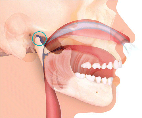
Figure 1A Anatomy of the oropharynx.
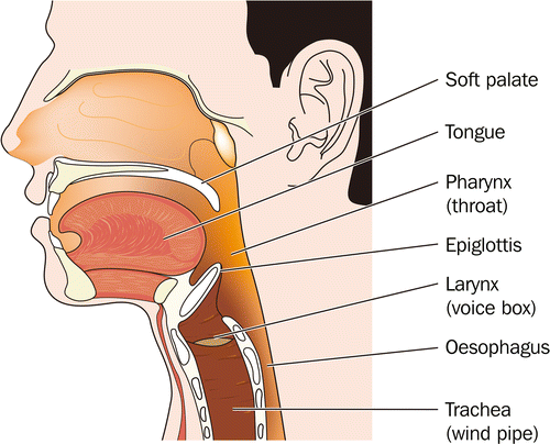
Figure 1B In the supine position, gravity takes the mandible down and back along with the tongue and soft palate.
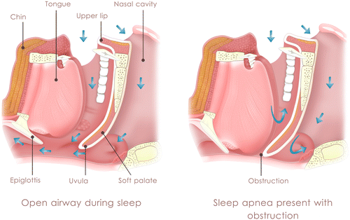
Figure 2 Adenoid Facies or long face syndrome associated with mouth breathing or nasal disuse.

This altered growth and respiration pattern is also associated with an increased incidence of TMD. Reestablishing nasal breathing can reverse this altered growth pattern during a “critical phase” of development.
Mouth breathing or nasal disuse leads to a lack of development of the nasomaxillary complexCitation6 seen as a high, narrow palate, flat cheekbones, and small maxilla. This has major implications on facial esthetics and malocclusion as well as the etiology of TMD.
Bonuck’s workCitation7 links airway obstruction to the development of the brain and prefrontal cortex, which is responsible for executive function, attention, behavioral inhibition, self regulation of affect and arousal, and socio-emotional behaviors. BonuckCitation8 found significant increases in neurobehavioral and neurocognitive disorders in children with SDB, including mouth breathing and snoring. Bonuck and others feel that early childhood is a period of vulnerability, as the critical period of brain development coincides with the greatest need for restorative, oxygenated sleep at a time when adenoids and tonsils are most hypertrophied. Her findings suggest that intervention may be necessary as early as the first year of life. It is, therefore, apparent that tethered oral tissues should be released as early as possible to allow breastfeeding, which helps develop the maxilla and shrink tonsils and adenoids.
A combination of adenotonsillectomy, palatal expansion, and myofunctional therapy is most effective at alleviating mouth breathing, snoring, and Obstructive Sleep Apnea (OSA), and therefore, aiding brain development and function.Citation9
Early intervention may reverse symptoms of attention deficit hyperactivity disorder and oppositional defiant disorder without medication.Citation10 Treatment at any time can improve brain function, cognition, and behavior.
As pediatric ENTs are being trained in sleep medicine, tonsils and adenoids are being removed earlier for obstructing airways rather than for infection. Pediatric dentists and orthodontists who understand the connection with brain development and neurobehavioral/cognitive disorders are now intervening as early as 2½–3 years of age to expand narrow palates and improve nasal breathing. Myofunctional therapists and occupational therapists (OTs) are now highly focused on the airway and intervening earlier in children and adults to establish ideal function in breathing, swallowing, and reestablishing tone through neuromuscular re-education.Citation11
There are numerous orthodontic and pediatric educators who are focused on a variety of techniques for early expansion of the airway through palatal development, both laterally and anteriorly.
AirwayCentric® dentistry
Sleep bruxism
One of the most common chief complaints from dental patients is nighttime clenching and grinding, which results in broken restorations, fractured cusps, worn teeth, and facial pain. SB is the internationally agreed upon Diagnostic Classification from the international classification of sleep disorders.Citation12
SB is defined as a stereotyped movement disorder characterized by rhythmic masticatory movement activity associated with tooth grinding and occasional tooth clenching.Citation13 The prevalence in the general population is somewhere between 5.5 and 12.5%.Citation14
SB can lead to clinical consequences seen by most dentists everyday in their practices:
| (1) | Severe occlusal and incisal wear, chipping, tooth fracture, and attrition | ||||
| (2) | Tooth mobility | ||||
| (3) | Hypersensitivity of teeth | ||||
| (4) | Failure of implant integration | ||||
| (5) | Periodontal concerns, abfraction lesions, recession | ||||
In the past 10 years, it has been hypothesized that there may be a link between SB, SDB, and TMD.Citation15
OhayonCitation16 studied risk factors for SB in the general population and found that individuals with OSA, loud snoring, moderate daytime sleepiness, heavy alcohol consumption, and caffeine intake were at the highest risk.
The current treatment among dentists has progressed from occlusal adjustment and equilibration in the 60s, 70s, and 80s to the use of maxillary occlusal night guards from the 1990s until today. These appliances are often fabricated in a retruded mandibular position, which was taught in dental school according to the dogma of the time, with the bite often taken lying back in the supine position, letting gravity take the mandible back to a retruded, terminal hinge-like position.
Unfortunately, a few studies over the last 10 years show that stabilization appliances may be associated with a risk of aggravating SDB. In one study, the apnea hypopnea index (which is a measure of how many times an hour a patient either stops breathing for 10 s or longer, or partially stops breathing with a 4% drop in oxygen), was increased by 50%. The Respiratory Disturbance Index (RDI) which is a combination of apnea, hypopnea and Resiratory Effort Related Arousals (RERA) per hour of sleep, increased by 30%. RERAs are respiratory effort-related arousals, and a more sensitive measurement of Sleep Disturbed Breathing (SDB). Snoring time increased by 40%.Citation17
A 2013 randomized controlled trial published in the Journal of Orofacial PainCitation18 found that an occlusal stabilization splint is associated with a risk of aggravation of OSA. There is currently not enough evidence to establish a causal relationship between OSA or ASD and SB, although the dentist and dental team must be aware of clinically relevant associations between SB and OSA. Every crown, bridge, implant, partial denture, denture, or occlusal splint or appliance can either open or close the airway. With the popularity of full field Cone Beam Computed Tomography (CBCT), we can now view the airway in three dimensions for every patient in our practice with very low levels of radiation in a 4.8-second scan. Home sleep studies are accessible, inexpensive, and produce accurate results, which can be interpreted by board-certified sleep MDs.
It has been observed that genioglossus and masseter muscle activity increased with an increase in CO2 as the airway collapses during sleep. It is also hypothesized that the masseter fires to enable the genioglossus to work more efficiently to maintain the tongue anteriorly, sparing the airway.Citation19
An interplay exists between the anatomy of the oropharyngeal airway, the tone or collapsibility of the masticatory, pharyngeal and tongue muscles, and gravity.
The oro and hypopharyngeal airway is surrounded by the tongue, soft palate, maxilla, mandible, and the posterior pharyngeal wall. The airway is a flexible tube that facilitates human speech but which leaves it vulnerable to collapse. In the supine position particularly, any loss of tone or protective reflex of the masticatory, tongue and submental muscles combined with a posterior or down and backward shift of the mandible and tongue can result in oro and hypopharyngeal narrowing and increased upper airway resistance.
The morbidity of ASD arises from a combined effect of intermittent hypoxia from lowered oxygen saturations and the brain arousals associated with upper airway obstruction. Arousals have been identified during SB and OSA.
The most common presentation to the primary care physician in the supposed “healthy” population is fatigue, pain, and reflux. Pain complaints include headache, neck and back pain, but it can also involve multiple tendons and muscles. Pain, fatigue, and reflux are all associated with SDB and disturbed sleep. Chronic disease such as diabetes, cardiovascular disease, and obesity also have strong ties to SDB.
TMD and SDB
The Operra cohort in 2013Citation20 found that a high likelihood of OSA was found with greater incidence in the first onset of TMD as well as chronic TMD. In other words, the ASD preceded the expression of TMD symptoms.
This is not surprising, as with the development of OSA, we see the induction of inflammatory cascadesCitation21 and increase in TNF-alpha, Apo-E, and changes in DNA Methylation.Citation22
TMD patients, including those with masticatory myofascial pain who have insomnia, reflux, difficulty concentrating, daytime sleepiness, and snoring, should be evaluated for a sleep study.
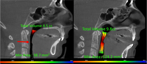
The stabilization splint, which has been recommended for the last 30–40 years, may reduce tongue space as well as the airway volume and smallest area (choke point) of the oro and hypopharyngeal airway18 (Fig. ).
Figure 3 CBCT measures minimal area of the airway and total volume.
It is becoming more common for dentists to recognize a loss of vertical dimension in the lower third of the face. Simply increasing vertical dimension with restorative dentistry or an occlusal splint without mandibular protrusion might aggravate OSA in some patients.Citation23
Practitioners should, therefore, screen their patients for OSA prior to fabricating an oral appliance or a full arch temporary bridge or denture.
In patients with confirmed SB and concomitant SDB after confirming with CBCT or an ENT exam that there is no nasal obstruction, a mandibular advancement device (MAD) or Continuous Positive Airway Pressure may be prescribed. The same is true for TMD patients with SDB.
Dentists must be aware that their current standard maxillary stabilization devices and occlusal splints, which protect teeth from wear and attrition, should be inserted by a dentist who is familiar with the anatomy and function of the temporomandibular joint.
The bite taken for a MAD should be within the patient’s envelope of function, e.g. a phonetic position of the sibilants, or roughly 2/3 of the distance between maximum retrusion and maximum protrusion. Clinical judgment is required. Maxillary and mandibular cants should be correlated with a trauma history and the findings on CBCT of the temporomandibular joints.
In the United States, a dentist is not allowed to diagnose sleep apnea in isolation. This means that a board-certified sleep MD must read and interpret the sleep study with recommendations for treatment. ‘Practice parameters for the treatment of snoring and obstructive sleep apnea with oral appliances: an update for 2005’24 was published in Sleep, and established guidelines, which allowed dentists to treat mild and moderate obstructive sleep apnea with oral appliances, as the primary provider of treatment.
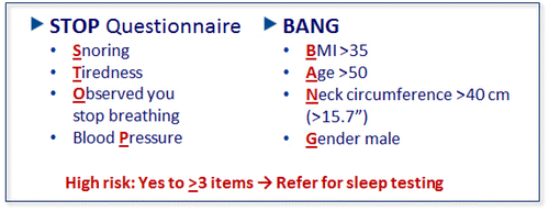
AirwayCentric® Education teaches dentists how to identify the 44% of men and 24% of women who are sitting in their dental chairs everyday, undiagnosed. Those patients with a high likelihood of having an ASDCitation26 (Fig. ) would be referred for a sleep study, either in their home or at a Sleep Center.
Figure 4 STOP BANG – Questionnaire to determine who needs a sleep study.
Depending on the medical history, morbidity, physical characteristics, and patient desire, a team approach may be recommended for the patient. The airway team may include an ENT, pulmonologist, sleep specialist, myofunctional therapist, pediatric dentist, orthodontist, general dentist, physical therapist, chiropractor, osteopath, pediatrician, OT, oral surgeon, and others.
AirwayCentric® Orthodontics and Pediatrics is a subject for another article. The theory is face-focusedCitation25 and built around an airway the size of a garden hose and emphasizing early intervention.
The AAPMD is The Academy of Physiologic Medicine and Dentistry and includes everyone on the airway team and provides an equal seat at the table (www.aapmd.org) The AAPMD is a leader for interdisciplinary collaboration, education, and training as well as advocacy for optimal growth, development, and function of the upper airway. Over 85% of patients with ASD are still undiagnosed and sitting in the chairs of dentists throughout the country. This article strives to improve awareness of dentistry to its primary role in airway management day and night.
The Foundation for Airway Health is a 501(c)(3) dedicated to educating the public about ASD. The Foundation increases awareness of what Airway Breathing Disorders are, their impact on growth and development, on the brain and the heart, and on systemic inflammation. Dentists seeking to learn more about Airway Health are encouraged to contact the Foundation and to form a pod in their community. We will match you up with a like-minded ENT or pediatric ENT in your area as well as a sleep specialist who understands the role of dentistry in Airway Health. www.foundationforairwayhealth.org
For more information about AirwayCentric® Dental Education contact:
References
- Huang Y, Guilleminault C. Pediatric obstructive sleep apnea and the critical role of oral-facial growth: Evidences. Front Neurol. 2012;3:1–7.
- Rambaud C, Guilleminault C. Death, nasomaxillary complex, and sleep in young children. Eur J Pediatr. 2012;171(9):1349–1358. doi:10.1007/s00431-012-1727-3.
- Bonuck K. Sleep disordered breathing in a population based cohort: behavioral outcomes at 4-7 years. Pediatrics. 2012;129(4):e857–865.
- Harvold EP, Chierici G, Vargervik K. Experiments on the development of dental malocclusions. Am J Orthodont. 1972;61:38–44.10.1016/0002-9416(72)90174-1
- Peltomäki, Timo. The effect of mode of breathing on craniofacial growth. Eur J Orthodont. 2007;29(5):426–429.
- Kim JH, Guilleminault C. The nasomaxillary complex, the mandible, and sleep-disordered breathing. Sleep Breath. 2011;15(2):185–193.10.1007/s11325-011-0504-2
- Bonuck K, Grant R. Sleep problems and early developmental delay: implications for early intervention programs. Intell Develop Disabil. 2012;50(1):41–52.10.1352/1934-9556-50.1.41
- Bonuck K, Rao T, Xu L. Pediatric sleep disorders and special educational need at 8 years: a population cohort study. Pediatrics. 2012;130(4):634–642.
- Guilleminault C, Huang YS, Monteyrol PJ, Sato R, Quo S, Lin Ch. Critical role of myofascial reeducation in pediatric sleep disordered breathing. Sleep Med. 2013;14(6):518–525.10.1016/j.sleep.2013.01.013
- Personal Communication, Valerie Deegan. Finding Connor Deegan, Lurie Children’s Hospital, Kevin Boyd, Darius Loghmanee, Steven Sheldon. Available from www.YouTube.com – Finding Connor Deegan.
- Moeller J, Paskay L, Gelb M. Sleep Med Clin. 2014;9:235–243.10.1016/j.jsmc.2014.03.002
- American Academy of Sleep Medicine. International classification of sleep disorders. 2014; 3rd ed. Westchester, IL: AASM, ICSD-3.
- Lobbezoo F, Ahlberg J, Manfredini D, Winocur E. Are bruxism and the bite causally related?. J Oral Rehab. 2012;39(7):489–501.10.1111/jor.2012.39.issue-7
- Maluly M, Anderson ML, Dal-Fabbro C, Garbuio S, Bittencourt L, de Siqueira JT. Polysomnographic study of the prevalence of sleep bruxism in a population sample. J Dent Res. 2013;92(7 supp):975.
- Balasubramaniam R, Klasser G, Cistulli P, LaVigne G. The link between bruxism, sleep disordered breathing and TMD: an evidence based review. Table 1 not published, JDSM 2014;1(1):27-37.
- Ohayon MM, Li KK, Guilleminault C. Risk Factors of sleep bruxism in the general population. Chest. 2011;119:53–61.
- Gagnon Y, Mayer P, Morrison F. Aggravation of respiratory disturbances by the use of an occlusal splint in apneic patients: a pilot study. Intl J Prosthodont. 2004;17(4):447–453.
- Nikolopoulou M, Ahlberg J, Visscher CM, Hamburger HL, Naeije M, Lobbezoo F. Effects of occlusal stabilization splints on obstructive sleep apnea: a randomized controlled trial. J Orofac Pain. 2013;27(3):199–205.
- Remmers JE, DeGroot WJ, Sauerland EK, Anch AM. Pathogenesis of upper airway occlusion during sleep. J Appl Physiol. 1978;44(6):931–938.
- Sanders AE, Essick GK, Fillingim R, Knott C, Ohrbach R, Greenspan JD. Sleep apnea symptoms and risk of temporomandibular disorders: OPPERA cohort. J Dent Res. 2013;92(7 supp):570–577.
- Tan H, Gozal D, Gozal L. Obstructive sleep apnea in children: a critical update. Nat Sci. 2013;5:109–123.
- Kheirandish-Gozal L, Gozal D. Genotype-phenotype interactions in pediatric obstructive sleep apnea. Respir Physiol Neurobiol. 2013;189(2):338–343.10.1016/j.resp.2013.03.016
- Nikolopoulou M, Naeije M, Aarab G, Hamburger HL, Visscher CM, Lobbezoo F. The effect of raising the bite without mandibular protrusion on obstructive sleep apnoea. J Oral Rehab. 2011;38(9):643–647. doi:10.1111/jor.2011.38.issue-9.
- Kushida CA, Morgenthaler TI, Littner MR, Clete A. Kushida, Timothy I. Morgenthaler, Michael R. Littner, Cathy A. Alessi, Dennis Bailey. Practice parameters for the treatment of snoring and obstructive sleep apnea with oral appliances: an update for 2005. Sleep. 2006;29(2):240–243.
- Lecture from Dr. William Hang. Available from www.facefocused.com.
- Fenton ME, Stewart SA, Skomro R, Gjevre J, Reid J. Utility of stop-bang in the prediction of obstructive sleep apnea in primary care. Chest. 2015;4.