
Abstract
Objective: Airway Centric® Dentistry/Orthodontics was defined in a previous article (Gelb M. Airway centric TMJ philosophy. CDA Journal. 2014) that also suggested airway considerations were more important than condylar position issues in determining patient health and welfare. Indeed, that article called for a new paradigm in the profession, but specific treatment techniques to achieve optimal airways and avoid reducing the airway were not discussed. The present article amplifies on that article and identifies specific orthodontic treatment methods, which are or are not, congruent with this new paradigm.
Method: The basis of traditional orthodontic diagnosis is outlined with references from the literature that show the scientific foundation for treatment is weak. A new approach to diagnosis and treatment with the goal of airway optimization is discussed.
Discussion: Six keys for optimal orthodontic outcomes are presented as new goals, and none involve the teeth. Ten specific treatment goals are outlined, and some are the diametric opposite of the current standard of care in the profession.
Conclusion: We recommend that optimizing the airway for every patient and never doing any treatment which will diminish the airway, even minutely, needs to become the standard of care in Airway Centric® Dentistry.
Introduction
A previous article proposed a new Airway Centric® TMJ philosophy.Citation1 That article carefully outlined historical TMJ philosophies and the people who proposed or supported them. Acknowledging the importance of previous emphasis on condylar position and occlusion, it introduced the idea that airway adequacy was an overlooked and more important factor in a person’s comfort and overall health. The article outlined why people in all developed countries have faces with both jaws more retruded than our ancestors from even only a few hundred years ago. It correlated how this lack of forward growth of the entire lower face can reduce the airway size, and therefore, one’s ability to breathe easily both night and day.
The article introduced important concepts in treating patients with sleep problems, which is a rapidly growing and emerging outgrowth of the dental profession. It was stated that it is important to prevent problems by optimizing forward growth in growing individuals and to do nothing to compromise the airway in anyone. The anatomy of the airway and how reductions in airway size anywhere along the path from the nose to the lungs can have a deleterious effect on health was a central point of the article.
The purpose of this article is to build on the previous article by outlining specific interventions that can be done in dentistry, and especially in orthodontics, to optimize the airway. A logical outgrowth of the Airway Centric® TMJ philosophy is a new model: Airway Centric® Orthodontics, featuring techniques which can be done to increase the airway. Perhaps more importantly, we identify approaches that should not be done if we are to avoid reducing the airway/tongue space. Unfortunately, some of the approaches which are not acceptable to the Airway Centric® Orthodontics philosophy are currently standard protocol in much of dentistry and orthodontics.
Discussion
Background of the orthodontic profession and current trends
The orthodontic profession is over 100 years old and is dentistry’s first specialty. The focus of orthodontics from its inception has been the straightening of the teeth and development of methods to do so. Virtually every child grows up hearing the term ‘buck teeth,’ which is an expression describing what orthodontists call a Class II Division 1 malocclusion. The idea that the upper teeth actually protrude in the face has been pretty much an unquestioned ‘fact’ since the start of the profession. Apparently, once this idea was established, it was not questioned enough for anyone to change it. The introduction of the cervical headgear was a solution for that ‘problem.’
An article by McNamara Citation2 described jaw positions in Class II patients in 1981 and found that protrusion of the maxilla was actually not common. This refuted the prevailing thought in the profession. Indeed, he concluded that it was more common to have maxillary retrusion than protrusion in Class II patients. He suggested treatment approaches that might develop the mandible forward were more appropriate than those that would restrict the development of the maxilla and/or retract the maxilla. If one were to really understand his data, it seems logical that treatment approaches should have been innovated to first develop the maxilla forward, followed by developing the mandible forward. MewCitation3 recognized this and has been advancing maxillae for children under age 10 since the 1950s. Mew has even successfully treated adolescents by developing their jaws forward for better facial esthetics when they have been previously diagnosed and given a treatment plan for surgery to advance both jaws. Mew recognized the need for better facial development and developed this approach (Orthotropics®) almost two decades before McNamara’s article.Citation2
The 1980s featured a large movement among general dentists (followed by some orthodontists) to use so-called ‘functional appliances’ to develop the lower face forward. WitzigCitation4 was perhaps the strongest proponent of this approach and had a large following among general dentists. McNamaraCitation5 was an orthodontist who advocated the use of the Frankel appliance, the Bionator, and the Herbst appliance as ways to develop the mandible forward. Many orthodontists tried the appliances and had some success in correcting Class II’s to Class I’s. JohnstonCitation6 compared results of some studies of functional appliances with more traditional orthodontic treatments that focused on retraction, and concluded that there was little difference in outcomes with both groups having ‘moderate mid-facial dentoalveolar retrusions.’
Studies on ‘functional appliances’ and Johnston’s articleCitation6 made no mention of airway reduction that might be associated with such treatments. Evidence today points strongly to the need for such concern. It would appear that a logical conclusion from Johnston’s articleCitation6 would be that research to establish better ways to develop both the maxilla and mandible forward would be of utmost importance if facial balance and airway development (rather than straight teeth) are the primary goals. Public awareness and interest in optimizing facial balance is rising. If one then considers that facial balance (or lack thereof) is in intimate relationship with airway size, this becomes an important public health issue.
MewCitation7 has shown definitively that it is possible to develop both the maxilla and the mandible forward in growing children (generally under age 10) using Orthotropics®. He treats adolescents successfully, but with more limited facial changes. Facial changes in his patients were judged to be superior to that of other treatment approaches.Citation7 He feels that his success comes because he is able to overcome the ‘headgear effect,’ which plagues all functional appliances.Citation3 He is quick to distinguish his approach from ‘functional appliances,’ which do have a headgear effect, by calling his appliance a ‘postural appliance.’ His appliance has a mechanism to prevent the child from allowing the mandible to fall back and retract the maxilla with it. This treatment approach has not achieved acceptance in the mainstream orthodontic community in spite of excellent articles in the refereed literature showing the results. Mew’s appliance also seeks to make a permanent change in the patient’s rest oral posture, which, if corrected, produces very stable results.
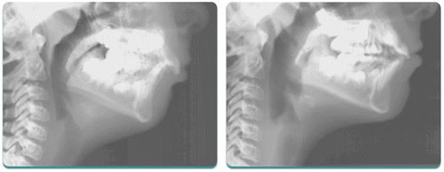
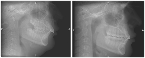
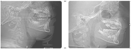
HangCitation8 has shown that the airway can be dramatically improved with Orthotropics® (Figure ). A 31% increase in the airway at the level of the palate, a 23% increase at the angle of the mandible, and a 9% increase at the level of the hyoid bone was achieved on a sample of children.
Figure 1 The patient illustrated above presented with a Class II Division 1 malocclusion and was treated with Orthotropics® to advance the maxillary anterior teeth, followed by advancement of the mandible. The airway improved dramatically in her case as both jaws were developed forward.
There are a number of other appliances that are used to correct Class II malocclusions (Herbst, Jasper Jumper, MARA, Forsus, Twin Force bite corrector, etc.) but none have shown dramatic forward development of the mandible. We are unaware that any involve a protocol to first develop the maxilla forward before attempting to develop the mandible forward (the obvious way to get more forward facial growth). None have shown effective reduction or elimination of the headgear effect.
Other than Orthotropics®, there appears to be no predictable way to develop the entire lower face forward. For children over age 10 with Class II malocclusions, one might conclude that surgery to advance both jaws in the face might be more appropriate than any form of retractive treatment, if optimizing facial balance and airway are goals of treatment. Whereas these recommendations are sometimes made, many orthodontists today still try to avoid surgery and do so with ‘camouflage’ treatment. In an attempt to make Class I cuspids and reduce overjets to achieve incisal and cuspid guidance, many patients have treatment approaches to retract upper teeth. One common approach is to remove the upper first bicuspid teeth in Class II cases when there is no crowding in the lower arch. The upper teeth are retracted into the extraction spaces, and the overjet is reduced. This is still done even years after McNamara’s articleCitation2 found few patients with upper teeth actually too far forward. Until recently, little has been discussed about how this might affect the airway. As noted earlier, Herbst appliances started to find their way into orthodontics in North America in the early 1980s and were purported to ‘grow the mandible.’ PancherzCitation9 has done the most research on the Herbst appliance, and his studies have shown very little forward growth of the mandible. The effects are more dentoalveolar with a pronounced ‘headgear effect’ of retracting the maxillary anterior teeth being a universal finding. BerkmanCitation10 more recently describes the Herbst appliance as an appliance to distalize upper molars (rather than advance the mandible). No one has mentioned what effect such retraction might have on the airway.
Largely in response to the desire of the public to have fewer teeth extracted, there are many treatment approaches to retract the maxillary anterior teeth involving temporary anchorage devices (TADs). They have largely taken the profession by storm with few studies chronicling their safety and effectiveness.Citation11 To our knowledge, no concerns have been expressed about possible airway reduction in such patients, although one can see in the refereed literatureCitation12 that the airway is reduced in some cases treated with them.
Andrews’ ‘Six Keys to Normal Occlusion’Citation13 was a landmark in the orthodontic profession and called for specific goals in how the teeth should fit together. This article is probably required reading in most university graduate orthodontic programs. None of the keys have anything to do with facial balance or airway. AndrewsCitation14 asked rhetorically why there are so many cephalometric analyses. He answered the question with ‘because none of them work!’ Most orthodontists still rely heavily on a cephalometric analysis (usually the one they were taught in their orthodontic training program) to make treatment decisions, even though we are unaware of any cephalometric analysis that includes airway measurements. In subsequent years, Andrews developed his Six Elements Orthodontic Philosophy Course,Citation15 which includes facial balance, but not airway, and describes the forehead as the preferred referent.
More recently, use of cervical headgear has come into question in the profession because of a possible correlation with obstructive sleep apnea (OSA).Citation16 It was concluded that cervical headgear should not be used for patients who exhibit ‘airway inadequacy’ or have recessed mandibles. If one truly understands McNamara’s 1981 article,Citation2 then most Class II patients have recessed mandibles and should not be treated with cervical headgears. If one uses the BoltonCitation17 norm superimposed on glabella and the bridge of the nose, it is clear that all Class II’s have recessed mandibles. Clearly, there is no reason for anyone to wear a cervical headgear.
MewCitation18 has compared a hypothetical Paleolithic profile with two common cephalometric analyses (SteinerCitation19 and McNamaraCitation20), and has shown how both jaws are massively back in both analyses relative to the Paleolithic norm. It is impossible to study airway size from Paleolithic records, but if airway size follows jaw position even a little bit, it is hard not to conclude that we are dealing with much smaller airways than our ancestors’.Citation21 It stands to reason that if the maxilla is retruded, so is the soft palate, which extends from the maxilla. It also stands to reason that if the mandible is retruded, so is the tongue, which is attached to the mandible. We know of no evidence that either the soft palate or the tongue is smaller today than in Paleolithic times. In fact, the tongue is bigger today, due to obesity, and the human face is becoming more like the Bulldog in the Brachycephalic dog model.Citation22
From the Angell Animal Medical Center in Boston, Dr. William Rosenblad, a canine-tooth expert, explains:
We’ve shortened the face of this breed so much,… that there’s just not enough space for everything to fit. The tongue, the palate, it’s all compressed. The teeth often look like they’ve been thrown in there. They have little tiny nostrils. The end result of all the compression is that many bulldogs can barely breathe.Citation23
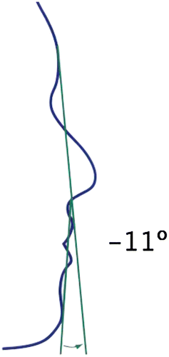
The term ‘bimaxillary protrusion’ (now more commonly referred to as ‘bialveolar protrusion’) has been in the orthodontic profession for decades and generally refers to someone whose face is considered too full. The Esthetic Line (Figure ) is one of a number of measurements used to diagnose this condition. Patients whose lips are ahead of this line are generally considered ‘full in the face.’ Most orthodontists have been taught to remove four bicuspid teeth and retract the teeth back in the face to correct this ‘deformity.’ Rarely is the position of the mandible or the size of the airway taken into account in diagnosing these cases, and yet the use of the Esthetic Line assumes a well-positioned mandible. The Facial Contour Angle (Figure ) has been in the profession for years and describes the position of the mandible. The more the mandible is retruded, the more likely the Esthetic Line is to show the face being too full. Most of the patients in the journals who have been diagnosed with ‘bimaxillary protrusion’ would not be considered to have faces that are too full if their mandibles were properly positioned forward or if their oropharyngeal airway was considered.
Figure 2 Esthetic line.
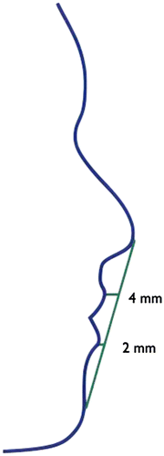
Figure 3 Facial contour angle.
One might also speculate how nose size might also impact the diagnosis of ‘bimaxillary protrusion.’ Superimposing the Bolton norm on faces to assess facial balance for more than 17 years has shown one author that the majority of Asians and African-American individuals have noses that are shorter in the A–P dimension than those of Caucasians by about 3–4 mm. Treatment of ‘bimaxillary protrusion’ in Asians has become very popular, especially in Korea. Until recently, nothing has appeared in the literature to discuss how such treatment might impact airway adequacy. A study on Chinese patients treated for ‘bimaxillary protrusion’Citation24 found that the airway was reduced by such treatment. No mention was made that this reduction in airway might somehow have long-term health consequences, such as producing OSA. It is hard to imagine that such an airway reduction would not, at least in some cases, produce a problem with OSA.
Conclusions of reviewing orthodontic history and current trends in orthodontics
The extraction/non-extraction debate has raged in the orthodontic profession for over 100 years, and the focus has usually been on stability of lower incisor crowding. Despite recognizing over 30 years ago that the maxilla rarely protrudes in Class II cases and is more commonly recessed, treatment is still often focused on trying to reduce so-called ‘protrusion’ of the upper teeth.
American Board of Orthodontics (ABO) requirementsCitation25 for board certification mandate that the orthodontist present at least one extraction case, where four bicuspid teeth are removed and the spaces are closed completely. The ABO website has pictures and guidelines showing how such space reduction must be complete or the orthodontist will be downgraded on the quality of the result. There are no universally accepted criteria for facial balance and no mention of protecting a patient’s airway. Instead, the orthodontic profession is concerned with aligning teeth and making them fit in a way that has never been shown to have any long-term benefit. In fact, Ackerman and ProffitCitation26 have an interesting perspective on occlusion as noted by this quotation:
Although the concept of ideal occlusion has taken precedence as the ultimate goal in clinical orthodontics for some 110 years and serves well as an adopted arbitrary convention and a clinical gold standard, it has no verifiable scientific validity. No one has yet demonstrated that ideal occlusion provides significant benefits in oral or general health, or that it significantly improves oral function.
A new paradigm for all orthodontic treatment and dental treatment in general
Can retraction of the front teeth be done to the degree that the airway might be reduced to the point that OSA is produced? This is a question that must be answered in the profession, and the sooner the better. Is it possible that retraction of teeth has just been ‘grandfathered’ into the profession because it ‘has always been done that way,’ and no one questions it? Since it has now been shownCitation23 that such retraction can reduce the airway, isn’t the obvious next question whether such retraction can produce OSA? One author has been reopening bicuspid extraction spaces for 26 years and has shown that reopening upper bicuspid extraction spaces alone can result in elimination of OSA. The following is taken from a sleep report (signed by an M.D. sleep specialist) of a woman in her mid 40’s who suffered from OSA until one of the authors reopened her maxillary bicuspid extraction spaces:
SL had mild obstructive sleep apnea-hypopnea syndrome with an REM dominant component. Her sleep apnea has completely resolved with orthodontic therapy despite the 10+ pounds of interim body weight gain. It is quite remarkable how much improvement she has had in her apnea severity despite the presence of a large tongue and crowded oropharynx.
With this history and direction of the profession, it seems that a completely new set of goals and treatment protocols be put forward to be considered by the profession. Following are our proposed goals of orthodontic treatment (Figure ).
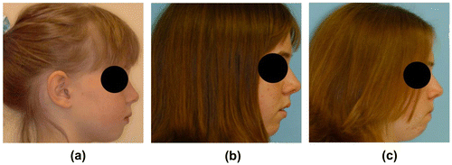
Figure 4 This little girl’s face grew more vertically (less forward) due to poor rest oral posture.
Six specific goals for Airway Centric® orthodontic treatment
| 1. | Face more balanced (not less) with unfavorable vertical growth of the face converted to favorable (horizontal) growth. Much of the orthodontic profession feels that vertical facial growth is genetically determined and cannot be changed without surgery. This is not the case, as has been shown by Mew,Citation3,20 Harvold,Citation26 Hang,Citation27 and others for many years. As the face develops forward, the maxilla and mandible are further forward, along with the soft palate and tongue, which attach to them. Achieving forward movement of the hard tissues of the face opens the airway. This has been shown to be possible.Citation8 | ||||
| 2. | No decrease in (and optimally increasing) airway after treatment. Ideally, pre- and post-PSGs and Cone Beam Computed Tomography (CBCTs) with airway analysis would become the standard of care. | ||||
| 3. | TMJs healthy with condyles not distalized. We can argue about what is considered ‘ideal’ condylar position. Ideal condylar position is not achieved when the anterior teeth hit even a nanosecond before the posterior teeth and distalize the condyles. Distalized condyles are never a good thing. If one were to err in any direction, it is better to leave a slight overjet rather than trying to achieve ideal ‘incisal guidance’ (whatever that is). In one author’s experience of reopening bicuspid extraction spaces, it has been found that the patient’s symptom pattern almost always goes away or is dramatically lessened as soon as a slight overjet is created, allowing the mandible (and condyles) the freedom to go where the muscles want them to go. The GelbCitation28 4/7 position describes a healthy joint relationship. Pre- and post-CBCT TMJ analysis can be helpful in documenting condylar position. | ||||
| 4. | Cheeks fuller, not flatter, after treatment of growing individuals. Patients with high cheeks are deemed almost universally more attractive than those with flat cheeks. High cheeks are an indication of more forward maxillary development and an obvious feature one associates with a larger airway. | ||||
| 5. | A smile ‘complete to the corners of the mouth.’ This isn’t common in our society. Dentists discuss ‘dark triangles’ at the corners of the mouth and know they are unattractive. Smiles are narrow because of low tongue posture. The broader the smile is, the higher the tongue posture and the greater the chance the tongue can be postured firmly to the palate at rest. A broad smile, complete to the corners of the mouth, dramatically increases the patient’s chance of posturing their tongue firmly to the palate and making them more likely to be nose breathers. MewCitation20 recommends a 42 mm intermolar width, but very few have this. It should be our goal, as orthodontists, to make room for the tongue to be positioned firmly to the palate at rest and have all our patients be nasal breathers, as humans were meant to be. With the tongue postured upward and forward into a fully developed maxillary arch, the chances of OSA and/or UARS go down dramatically. | ||||
| 6. | Ideal rest oral posture with nasal breathing should be achieved (or as close as is clinically possible). Ideal rest oral posture involves having the teeth together lightly (or very slightly apart), the tongue positioned firmly to the palate with the tip behind the upper incisors, and the lips together without strain. Patients with mentalis muscle strain are almost universally mouth breathers, and such unfavorable rest oral posture will ultimately cause their faces to fall back over time (Figure ). | ||||
‘Chronic oral breathing is an important clinical marker of orofacial muscle dysfunction, which may be associated with palatal growth restriction, nasal obstruction, and/or a primary disorder of muscular or connective tissue dysfunction,’ according to Guilleminault and Sullivan.Citation29 Continuation of mouth breathing after adenotonsillectomy and palatal expansion may lead to recurrence of sleep disordered breathing in adolescence and adulthood.Citation30
Very rounded cheeks are a symptom of hypertrophic buccinator muscles, and these patients do not have proper rest oral posture either.
Myofunctional therapy should be routine, referring our patients for therapy or providing it in our offices. Our goal should be to produce patients with jaw positions that are as forward as is clinically possible so that their airways are ideally developed and to correct patients’ poor rest oral posture to help them maintain the jaw positions we worked hard to achieve. Indeed, restoration of nasal breathing may be the only valid endpoint of treatment, according to Guilleminault.Citation30
Figure 5 Patient presented with generalized spacing of the upper arch in A. Spacing was closed in the anterior and placed between the upper second bicuspid teeth and first molar teeth without retracting or reducing the arch circumference in B.
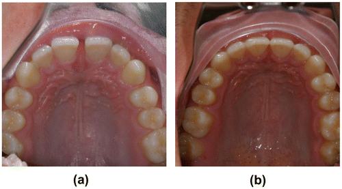
Figure 6 The patient pictured above had his snoring completely resolve after we advanced his lower anterior teeth and opened spaces for extra bicuspid implants. No recession has occurred more than 10 years after the treatment was completed.
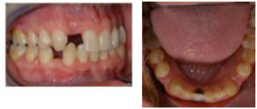
Figure 7 Patient above, in A, was given a removable appliance to advance lower anterior teeth in a Class II case. Spaces were created between lower bicuspid teeth as a consequence of this movement, as shown in B. Temporary anchorage devices were placed bilaterally to allow for all posterior teeth to be brought forward to close the space that had been created. Effectively, the entire lower dentition was advanced over the time in treatment. The airway improved in his case as shown below (Figure ).
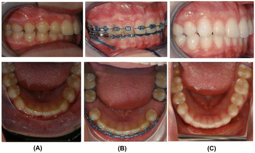
Figure 8 Airway improvement in a patient where lower incisors were advanced, spaces were created between lower bicuspid teeth, and posterior teeth were brought forward with TADS to close the spaces that had been created.
Ten specific treatment protocols that must be adhered to in order to produce Airway Centric®/Airway Focused® orthodontic results:
| 1. | Stop using headgears. If the refereed literature was clear 33 years ago that upper anterior teeth rarely protrude in the face, how can one justify pushing the anterior teeth back or trying to restrain forward development of the maxilla with a headgear? ‘It has always been done this way’ is not an excuse for ignoring the facts. If such retraction can reduce the airway even a little bit and we are not clear on where such airway reduction can produce a problem, there can be no justification for use of headgears. | ||||
| 2. | Stop using appliances with a headgear effect. The decade of the 1980s was the ‘functional appliance’ decade, where orthodontists and dentists doing orthodontics were trying to develop the mandible forward with ‘functional appliances.’ Johnston’s studyCitation6 was pretty clear that patients end up with a ‘moderate dentoalveolar retrusion.’ ‘Headgear effects’ with all these appliances can be expected because there is nothing to prevent the mandible from exerting that effect on the maxilla and retracting the maxilla long term. Perhaps no better evidence for this is the article referencing the Herbst appliance as a molar distalizing appliance.Citation10 Aside from occasions where maxillary molars have drifted mesially secondary to primary molar loss, maxillary molars should never be distalized in trying to correct a Class II or any other malocclusion. Perhaps the most common thing used in treating Class II patients is Class II elastics from the lower molars to the upper cuspid teeth. Class II elastics retract the upper anterior teeth and cannot be justified in Airway Centric® orthodontics. Leaving patients with an overjet must become acceptable and the standard of care! | ||||
| 3. | Accept the need for orthognathic surgery in correcting overjets. Many orthodontists spend massive amounts of time and energy trying to camouflage Class II patients to avoid surgery. In virtually all such cases, they are using mechanics that will retract the anterior teeth, essentially ‘trying to make teeth fit on jaws that do not fit.’ Orthognathic surgery must be considered in such cases in order to optimize facial balance by advancing both the maxilla and mandible. If the patient rejects surgery, a compromise treatment may be agreed upon mutually by the patient and doctor. This could involve leaving an overjet (but aligning the teeth) or repositioning the mandible and accepting a dual or open-bite. | ||||
| 4. | Stop extraction of bicuspids and retraction of anterior teeth. The orthodontic profession has had more than a century-long argument about extraction vs. non-extraction treatments. Indeed, Dr Robert Little states, ‘The idea of balance and stability is more a hope and myth than a reality.’Citation31 Arguments for stability have been cited, but no study has shown extraction cases to be more stable. Most orthodontists are recommending lifetime retention of some sort if perfect alignment of the teeth is desired. Virtually all orthodontic residents have been warned that advancing teeth forward on either the maxilla or mandible will result in recession and possible loss of teeth. ArtunCitation32 and MelsenCitation33 have laid the recession concern to rest and removed it as a reason for extraction. Wang et al.Citation23 have shown narrowing of the airways in bimaxillary protrusive patients treated with four bicuspid extraction. In light of the current epidemic of OSA and OSA related morbidities, such reduction in airway cannot be justified. Instead of arguing about whether or not a proposed orthodontic treatment protocol involves extraction of teeth or not, we must substitute the question about how it affects the airway. Clearly, extraction/retraction must stop. | ||||
| 5. | Stop closing generalized spacing by retraction. Many patients have teeth that are too small for the arches. This includes the apparently simple case of a maxillary midline diastema. Many cases of anterior spacing are that way because of small or misshapen (peg laterals) lateral incisors. Any closure of diastemas done orthodontically should not include any retraction whatsoever. Such seemingly minor retraction can cause reduction in tongue space/airway to produce problems. Spacing of teeth for veneering or bonding in the case of small teeth (particularly small lateral incisors) is to be preferred to any form of retraction. Alternatively, anterior spacing can be placed distal to second bicuspid teeth and arch circumference protected. Such spacing can be completely cleansable if it is large enough and not subject to food impaction. If the spaces are too small, it is easy, and not invasive, to bond the adjacent teeth with composite resin to provide tight contacts (Figure ). | ||||
| 6. | Stop using TADs (temporary anchorage devices) for retraction. The eternal extraction/retraction debate has more recently given way to the argument that retraction to correct an overjet no longer requires extraction of bicuspids when a TAD can be placed and the anteriors retracted. The effect on the airway is the same, however, and cannot be condoned. VigCitation11 has expressed concern about the thousands of articles on TADs without studies confirming their safety, etc. A case report by Tai et al.Citation34 featuring the use of TADS to reduce bimaxillary dentoalveolar protrusion indeed shows an airway reduction. | ||||
| 7. | Create space in lower arch in Class II’s to reduce overjet in non-surgical cases. Virtually all patients with Class II malocclusions have recessed mandibles when one looks critically at the face. McNamara stated this well in 1981.Citation2 Without a predictable way to bring the mandible forward on non-growing patients, surgery is the ideal treatment. Many patients choose not to do surgery or cannot afford it. It is possible and desirable in some such cases to offer the alternative of advancing the lower anterior teeth to increase the tongue space/airway. This can be done, and done safely, without causing recession, despite ‘prevailing wisdom.’ ArtunCitation32 and MelsenCitation33 give evidence in the refereed literature that it can be done without concern for recession. The patient pictured here had his snoring completely disappear after spaces were opened for implants to add an extra bicuspid in each of the lower quadrants (Figure ). | ||||
| 8. | Advance the entire lower dentition in Class II patients to correct the overjet. This can be done by first advancing the lower anterior teeth and creating space in the buccal segments. Once the anterior teeth are in ideal positions relative to the maxillary anterior teeth (which have not been retracted in any way), the spaces created in the lower arch can be closed by placing TADs in the mandibular anterior area. The posterior teeth are then brought forward and all spaces closed with the entire lower dentition effectively moved forward (Figures and ). | ||||
| 9. | Treat early. The controversy over ‘early treatment’ has raged on for decades with the American Association of Orthodontists (AAO) periodically holding conferences to explore whether it is worthwhile or not. The focus of these discussions is almost always the teeth and whether it is better to correct the Class II situation early or later. The corrections that are studied almost always involve retraction. Treating in the primary dentition is possible and ideal when the teeth are viewed as a handle to the face, with improvement in facial balance and airway being the prime goals. BonuckCitation35 demonstrated that mouth breathing, snoring, and apnea at 6, 18, and 30 months of age significantly increased the risk of neurobehavioral and neurocognitive disorders at ages four and seven. She hypothesized that alterations in executive function and behavior were due to changes in the prefrontal cortex, and therefore, early intervention was paramount. Whereas Orthotropics® or any form of orthodontics is not practical at this age, it is essential that these conditions be treated as soon as is feasible. | ||||
The patient illustrated in Figure suffered from OSA and failure to thrive at age five. Development of both jaws forward with Orthotropics® resulted in an elimination of the OSA. GozalCitation36,37 discusses one of the pathophysiologic mechanisms of pediatric sleep disordered breathing (PSDB) as an airway of reduced dimensions with increased collapsibility. Airway Centric® orthodontics, in the form of Orthotropics®, addresses the craniofacial and anatomic factors at the earliest possible age to enlarge the pharyngeal airway space and transform the dentofacial morphology for the largest possible airway. Treating such patients early must be considered a top priority in the profession.
| 10. | Modify ABO requirements to eliminate extraction/retraction treatment for certification, and reward doctors who purposefully leave spaces in the arches rather than blindly close them to adhere to a strict rule with no basis in science. We are indeed primates, and other primates have spaces between the teeth. We simply cannot blindly close spacing in arches and assume it will not have any effect. | ||||
Figure 9 Patient above is a five-year old with Pierre Robin Sequence, suffering from OSA. Orthotropic® treatment to advance both jaws resulted in complete elimination of OSA.
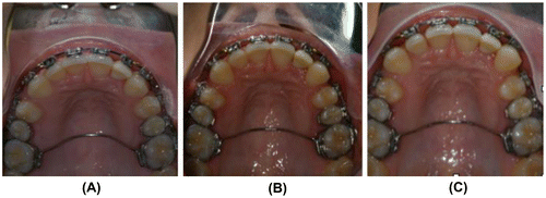
The case in Figure illustrates this. Image A shows the maxillary arches when this patient presented for a second opinion about another matter. She had severe pain to intrameatal palpation, with all the muscles of the face and neck exquisitely tender. She had an ongoing, three week-long headache for which she had been unable to identify a cause. The closing loop, which was being used to close the spaces without regard to the fact that there was no overjet, was cut out of the arch. By merely cutting the closing loop, the pain pattern was eliminated in two days. In just one week, the spaces had opened up some as shown in Image B. By six weeks, the tongue had pushed the six anterior teeth forward to the degree shown in Image C. The patient remained symptom free. There were no other variables operating here, other than cutting the closing loop.
Figure 10 This patient presented with headaches and closing loops to close the spaces remaining distal to the cuspids. After cutting the closing loops, the spaces opened slightly, relieving her headaches. B shows the space three weeks after cutting the closing loops. C shows the even larger space six weeks after cutting the closing loops.
Conclusion
We are very aware that the ideas presented here are controversial, and many will rise quickly to emotionally attack them without even considering that they may have merit. There will be those who will cite the status quo and say that all we do is ‘evidence based.’ On the contrary, much of dentistry is NOT evidence based, but dogma, as BaderCitation38 suggests. Isn’t it time to move on from the meaningless professional benchmark of quality known as lower incisor stability? After about a century of fighting over this, it can pretty much be concluded that LittleCitation31 was correct in stating, ‘stability is a myth.’ It is even more meaningless since most orthodontists are recommending lifetime retention. Isn’t it time for the orthodontic profession to become a part of the health care profession and embrace airway health as the goal of our treatment? Indeed, dentists are the gatekeepers to the airway and have yet to fully realize that role.
Notes on contributors
William M. Hang has an orthodontic practice outside Los Angeles, CA which strives to integrate a beautiful smile with a better functioning body. Treatments include Orthotropics® which has been shown to improve the airways of children, non-retractive orthodontics, and reopening previous extraction spaces. Hang is an international lecturer and serves on a number of professional boards.
Michael Gelb is a dentist in New York City with a practice devoted to Airway, TMJ, and headache disorders and how they effect performance, fatigue, memory and pain. Gelb is a Clinical Professor at NYU College of Dentistry and a Diplomate of the American Board of Orofacial Pain. He has pioneered the concept of Airway Centric® Dentistry and is the co-author of the book ‘Gasp.’ He is also the co-founder of the AAPMD and The Foundation for Airway Health.
Conflict of Interest
Dr Hang owns an orthodontic lab Face Focused® Orthodontic Lab and an educational entity known as AIRWAY-kening™ Academy. He is also the holder of the patent for the ADAPT-LGR®.
Dr Gelb is the co-founder and partial owner of Airway Centric® Dentistry, an educational and device company.
References
- Gelb M. Airway centric TMJ philosophy. CDA J. 2014;42:551–562.
- McNamara JA Jr. Components of class II malocclusion in children 8–10 years of age. Angle Orthod. 1981;51:177–202.
- Mew J. Biobloc therapy. Flo-Print: Langton Green; 1979.
- Witzig J, Spahl T. The clinical management of basic maxillofacial orthopedic appliances: temporomandibular joint. Chicago, IL: Year Book Medical Pub; 1991.
- McNamara JA Jr. Personal communication. Detroit; 1982.
- Johnston LE. Growing jaws for fun and profit. What doesn’t and why. In: McNamara, (ed.) Craniofacial growth series 35. Center for Human Growth and Development. Ann Arbor: University of Michigan; 1999.
- Mew J. Facial changes in identical twins treated by different orthodontic techniques. World J of Orthod. 2007;8:174–188.
- Singh GD, Garcia-Motta AV, Hang WM. Evaluation of the posterior airway space following biobloc therapy: geometric morphometrics. Cranio. 2007;25:84–89.10.1179/crn.2007.014
- Pancherz H, Ruf S. The Herbst appliance: research-based clinical management. Chicago, IL: Quintessence Pub ; 2008.
- Berkman M, Haerian A, McNamara JA Jr. Interarch maxillary molar distalization appliances for class II correction. J Clin Orthod. 2008;62:35–42.
- Vig K. Reliable evidence and clinical practice. PCSO Bulletin; 2010, Winter: 37.
- Jeon J, Yu H, Baik H, Lee J. En-masse distalization with miniscrew anchorage in Class II nonextraction treatment. JCO. 2006;40:472–476.
- Andrews LF. The six keys to normal occlusion. AJO-DO. 1972 Sep; 62:296–309.
- Andrews LF. Personal communication. Foundation for Orthod Research Mtg, Niagara-on-the-Lake; May 1993.
- Andrews LF. The 6E orthodontic philosophy. The Andrews Foundation. San Diego, CA; 2013.
- Pirlia-Parkkinen K, Pirttiniemi P, Nieminen P, Lopponen H, Tolonen U, Uotila R, et al. Cervical headgear therapy as a factor in obstructive sleep apnea syndrome. Ped Dentistry. 1999;21:39–44.
- Steiner CC. Cephalometrics for you and me. AJO. 1953;39:729–755.
- McNamara JA Jr. Cephalometric analysis of untreated adults with ideal facial and occlusal relationships. Int J Adult Orthodon Orthognath Surg. 1988; Apr:221–231.
- Hans M, Broadbent BH Jr, Nelson S. The Broadbent-Bolton growth study – past, present, and future. AJO-DO. 1994;105:589–603.
- Mew J. The cause and cure of malocclusion. Heathfield: John Mew; 2013.
- Liberman D. The evolution of the human head. Cambridge: Belknap Press of Harvard University Press; 2011.
- Can the Bulldog be saved? NY Times.com, 22 November 2011.
- Wang Q, Jia P, Anderson N, Wang L, Lin J. Changes of pharyngeal airway size and hyoid bone position following orthodontic treatment of Class I bimaxillary protrusion. Angle Orthod. 2012;82:115–121.10.2319/011011-13.1
- American Board of Orthodontics. Selecting cases – case record requirements – case criteria. St Louis, MO: ABO Board; 2015.
- Ackerman JL, Proffit WR. Guest editorial – a not-so-tender trap. AJO-DO. 2009;136:619–620.
- Harvold EP, Tomer BS, Vargervik K, Chierici G. Primate experiments of oral respiration. AJO. 1981;79:359–372.
- Singh GD, Medina LE, Hang WM. Soft tissue facial changes using biobloc appliances: geometric morphometrics. Int J of Orthod. 2009;20:29–34.
- Gelb H. Clinical management of head neck and TMJ pain and dysfunction – a multi-disciplinary approach to diagnosis and treatment. Philadelphia, PA: WB Sanders; 1977.
- Guilleminault C, Sullivan S. Towards restoration of continuous nasal breathing as the ultimate treatment goal in pediatric obstructive sleep apnea. Enliven: Pediatr Neonatol Biol. 2014;1(1):1–5.
- Guilleminault C, Huang YS, Monteyrol PJ, Sato R, Quo S, Lin CH. Critical role of myofascial reeducation in pediatric sleep disordered breathing. Sleep Med. 2013 Jun;14:518–525.
- Little R. Clinical implications of the University of Washington post-retention studies. JCO. 2009;63:645–651.
- Artun J, Grobety D. Periodontal status of mandibular incisors after pronounced orthodontic advancement during adolescence: a follow-up evaluation. AJO-DO. 2001;119:2–10.
- Melsen B, Allais D. Factors of importance for the development of dehiscences during labial movement of mandibular incisors: a retrospective study of adult orthodontic patients. AJO-DO. 2005;127:552–561.
- Tai K, Park JH, Tanino M, Ikeda K. Bimaxillary dentoalveolar protrusion treated with lingual appliances and temporary anchorage devices. JCO. 2012;12:739–746.
- Bonuck K. Sleep disordered breathing in a population based cohort: behavioral outcomes at 4–7 years. Pediatrics. April 2012;129(4):1–9.
- Gozal L, Gozal D. Geotype-phenotype interactions in pediatric obstructive sleep apnea. Respir Physiol Neurobiol. 2013 Nov; 189(2):338–343.
- Tan H, Gozal D, Gozal L. Obstructive sleep apnea in children: a critical update. Nat Sci Sleep. 2013 Sep;25(5):109–123.
- Bader JD. Commentary – challenges in quality assessment of dental care. JADA. 2009;140:1459–1464.