ABSTRACT
Objective
Myofascial pain diminishes the stomatognathic function and hinders clinical diagnosis. Therefore, initial pain reduction is crucial before definitive treatment. Here, the clinical validity of non-pharmaceutical therapies, including the Aqualizer® splint, physiotherapy, and dry-needle acupuncture was comparatively assessed.
Methods
Myofascial pain patients (n = 28; 20–65 years old) were examined through a visual analog scale, and intra- and extra-oral muscle palpation. Mandibular maximum opening and neck mobility were also evaluated. Changes in parameters through time were analyzed via the Kruskal-Wallis test, while the Friedman test and dot-plots were used for comparative therapies assessment. General patient improvement was represented via an isometric Principal Component.
Results
The Aqualizer® and physiotherapy resulted in improvement of all parameters except for mouth opening. Acupuncture improved extra-oral muscle pain and neck mobility.
Conclusion
The Aqualizer®, physiotherapy, and oral acupuncture are effective initial pain therapies. Among all, physiotherapy provided the greatest benefits, followed by the Aqualizer®.
1. Introduction
Craniomandibular disorders are complex diseases causing myofascial pain and mechanical limitations, interfering with the critical functions of the orofacial complex, and thus decreasing patients’ quality of life. Protracted pain and functional limitations are known to increase the level of anxiety and worsen depression [Citation1]; thus, relieving patients from pain is one of the main aims of the clinician. Additionally, initial pain treatment facilitates proper clinical assessment, when the presence of pain in combination with the deriving muscle tension or spasm makes it difficult for both patient and clinician to localize the pain or interpret the symptoms. Furthermore, performing functional diagnostic procedures, such as the recordings of the mandibular movements, might become arduous in case of muscle pain and tension.
Several methods are available to practitioners to alleviate patients’ orofacial pain in the early treatment stages. Among these, non-pharmacological interventions [Citation2] present the appreciable advantage to spare the patients from the possible medicine’s side effects. There exist different kinds of non-pharmacological therapies acting mainly on the body (e.g., manual physical therapy and exercise therapy), on the mind (e.g., psychotherapy or support groups), or both (e.g., meditation, acupuncture). They share the same purpose to achieve muscle relaxation and improve local blood flow and analgesia, thus reducing excessive stress and strain on the masticatory structures. The choice of the most suitable technique depends on numerous factors, including pain etiology, patient preferences, and the clinical setting in which the treatment is performed. Various kinds of physical therapies are used in oral medicine for reducing pain and functional limitations, like laser or heat therapy, oral appliances, physiotherapy, and acupuncture. More often, clinicians recommend psychological treatment or cognitive therapy to help patients manage the effects of pain on their daily life.
Therapeutic approaches such as physiotherapy and acupuncture have shown variable degrees of efficacy in the short term [Citation3] although evidence is not definitive. The validity of the different types of oral appliances available has been tested in various works, with contrasting results [Citation4,Citation5]. Based on the clinical experience of the authors (I.S. and E.P.), preliminary tests were performed in a series of pilot studies for promising therapeutic approaches for the initial treatment of pain, evaluating the short-term outcomes of the Aqualizer® occlusal splint [Citation6], physiotherapy consisting in active and passive physical exercises for orofacial muscles relaxation [Citation7] and dry-needle oral acupuncture [Citation8], respectively. In the current study, a comparative assessment of the outcomes of these pilot studies is presented, based on subjective and clinically diagnosed orofacial pain, maximum mouth opening, and neck mobility. Furthermore, the clinical applicability of initial pain therapies tested is discussed.
2. Materials and methods
2.1. Patient selection
For each of the three pilot projects, 10 patients affected by myofascial facial pain were selected. Both males and females between the age of 18 and 80 years were considered. In 2019, the subjects were recruited in order of presentation from the pool of temporomandibular disorder patients attending the Clinical Division of Prosthodontics, University Clinic of Dentistry, Medical University of Vienna, Vienna, Austria. Ethical approval for these projects was granted by the Ethical Commission of the Medical University of Vienna (approval Nr. 1145/2019). Exclusion criteria were: orofacial infections, concomitant treatment with anticoagulants such as heparin, nonsteroidal anti-inflammatory drugs or plasma substitutes, heart failure and/or disorders of the cardiovascular system, respiratory insufficiency, myasthenia gravis, inability to comply with the study requirements and to understand the study measures, and severe mental illness. Patients were excluded also in case of systemic diseases affecting the stomatognathic system, co-occurrence of joint pain, or degenerative diseases such as arthritis or jaw lock. Patients already receiving other craniomandibular therapies or participating in other clinical studies were not considered. A dropout of 10%, namely 1 patient per study group, was tolerated. The patients’ clinical data were pseudonymized and managed according to the ethical requirements and privacy rules of the Medical University of Vienna (https://www.meduniwien.ac.at/web/en/rechtliches/good-scientific-practice/), which are in line with the Declaration of Helsinki. Hence, informed consent was obtained from each of the patients before the commencement of the study.
The three therapeutic approaches tested in this study were applied and assessed separately, and the corresponding clinical outcomes were then compared. Since the patients were recruited and clinically evaluated independently for every pilot study, each of these projects is to be considered a prospective, randomized, single-center study with a longitudinal design over three survey dates (see section 2.3. Initial pain therapies). The pilot study using the Aqualizer® splint was conducted by N.O.; the pilot study applying physiotherapy was conducted by M.B.; the pilot study based on dry-needle oral acupuncture was conducted by N.A. These works were closely supervised by E.P. and I.S.-K.
2.2. Diagnosis and clinical assessment
The diagnosis of myalgia was carried out based on the Research Diagnostic Criteria for Temporomandibular Disorders (RDC/TMD) [Citation9]. The clinical evaluation included pain assessment through the visual-analog scale (VAS) and intra- and extra-oral palpation of muscles, along with measurement of maximum mouth opening and scoring of neck mobility. Thus, the patients compiled the VAS form in which the perceived orofacial pain level is represented by the patient on a continuous scale from 0 (i.e., no pain) to 100 (i.e., worst pain ever experienced). The values were rounded to intervals of 5. Muscle palpation was performed bilaterally at the atlanto-occipital articulation, anterior, medial, and posterior temporal muscles, muscles of the craniomandibular junction, lateral and medial pterygoid, masseter, digastric, supra- and infra-hyoid muscles, and sternocleidomastoids. Additionally, palpation was carried out intraorally at the vestibular and retromolar maxillary and mandibular areas on both sides. Palpation pressure was calibrated on each patient on the first dorsal muscle (the spot between the first and the second fingers). Pain at palpation was rated as: (0) no pain [Citation1]; light pain [Citation2]; moderate pain [Citation3]; severe pain. The sum of the scores obtained for each palpated muscle was analyzed separately from the sum of the scores obtained intraorally.
To assess maximum jaw opening the distance between the incisal edge of the upper and lower central incisors was measured (in mm). An interincisal mouth opening of at least 40 mm was considered sufficient based on the indications in Meyer [Citation10]. Moreover, mobility of the head along its three axes of rotation was assessed by evaluating right and left rotation and tilting, and forward and backward movements, and was classified into four groups: (0) no limitation; (1) minor limitation; (2) moderate limitation; (3) severe to total limitation. The time needed for the clinical assessment of each patient was approximately 45 minutes. To evaluate the effect of the initial pain therapies, the clinical assessment was performed during the first session, before therapy (T1), and then after 2 weeks (T2) and 4 weeks (T3) from the beginning of the therapy. Thus, the overall therapy duration for each patient from T1 to T3 was 4 weeks. A follow-up session was not considered necessary since this study aimed at assessing the short-term effects of the various initial pain therapeutic approaches.
2.3. Initial pain therapies
2.3.1. The Aqualizer® dental splint
At the first appointment, the patients were instructed about the use and cleansing of the Aqualizer® according to the manufacturer’s instructions. The Aqualizer® is a soft splint with water-filled pads that, according to Pascal´s law stating that “the pressure applied to any part of the enclosed liquid will be transmitted equally in all directions through the liquid”, permits occlusal forces to be distributed evenly on the dental arches, thereby compensating for possible effects of malocclusion [Citation11]. Its validity as initial pain therapy has been shown in about 80% of the cases, an outcome that is comparable to that obtained with hard splints [Citation12,Citation13]. Patients were instructed to wear the splint overnight, and, if possible, also during the day time, for no less than 8 hours in the 24 hours. For hygienic reasons, the product should not be used for longer than 2 weeks; thus, the patients were provided with a new splint during the second appointment. Additionally, the appliance was replaced in case of damage. The Aqualizer® model ultra with medium volume and thickness of approximately 2 mm was chosen because suitable for most patients, thus allowing standardization of the method.
2.3.2. Physical therapy
In this pilot study, patients performed both active and passive physical exercises aimed mostly at relaxing and stretching the upper quarter of the body with a particular focus on the orofacial muscles, neck, and shoulders. Patients were instructed to perform the exercises every day in the evening, after work. A morning session was considered optional, however, skipping the evening session for more than 1 day per week was considered a reason for exclusion. Each session began with a progressive, general relaxation of the body and continued with more specific exercises for the areas of interest. At T1, the exercises were demonstrated by the therapist, and more complex movements were repeatedly trained by the patient before they were performed. This training was repeated at T2. In particular, the following protocol was applied combining exercises from different sources [Citation14–18]. Figures for the exercises illustrated below can be found in the open-access master thesis describing this pilot study (https://repositorium.meduniwien.ac.at/obvumwhs/content/titleinfo/4941965); references to the relevant figures are reported for the various exercises, as “A.” for “Abbildung” meaning “figure” in the German language, followed by the respective figure and page numbers.
1 – Progressive muscle relaxation in a seated position. Illustration A.1, p. 20. The intensity exercise is to be performed while seating in an armchair with a headrest. The body muscles are contracted as much as possible for a few seconds with hands clenched into tight fists. Afterward, both body and hands are relaxed while quietly exhaling. The exercise is repeated at approximately 50% of the maximum contraction, and then again at approximately 25% of the maximum contraction.
2 – Further progressive muscle relaxation. Illustration A.2, p. 20. This exercise consists of three phases:
(a) While lying supine on a mat, the arms are slightly flexed at the elbow and are pressed into the sides of the body, while the hands are clenched into fists. The shoulders are pulled down towards the mat; the back, the abdominal muscles, and the legs are tightened; the feet are flexed bringing the toes forward and downward. After achieving maximum contraction, the patient tries to fully relax the muscles.
(b) To activate the facial muscles, frowning, squinting of the eyes, wrinkling of the nose, clenching of the teeth, and forced closing of the lips are performed simultaneously. For this exercise, the patients are invited to think of the facial expression they would make if they were to bite a lemon. After a few seconds, the facial muscles are relaxed.
(c) To stretch the neck muscles, the head is tilted backward and forward with slow, smooth, and pain-free movements, while sitting. In the forward movement, the chin is pointed towards the breastbone. In the rest phase, the head is balanced on the shoulder without stretching or bending the neck.
3 – Stretching exercises for cervical and thoracic muscles. This set of exercises is meant to stretch the upper quarter of the body:
(a) Illustration A.3, p. 21. The levator scapulae muscles are stretched using a towel. While sitting straight, one extremity of the towel is positioned under one buttock and the other extremity is slung over the ipsilateral shoulder. The towel is pulled downward with the ipsilateral hand while the contralateral cheek is turned towards its ipsilateral shoulder. The position is held for about 30 seconds before returning to the rest position. The exercise is repeated three times in a row for each side.
(b) Illustration A.4, p. 22. To stretch the trapezius muscles the patient assumes the position as in 3a but this time tilts the head towards the ipsilateral shoulder to move the ear as close as possible to it. The movement is repeated three times for each side.
(c) Illustration A.5, p. 22. The stretching of the pectoral muscle is performed while standing next to a wall, the arm is stretched back along the wall at a right angle to the vertical axis of the body and the palm is pressed against the wall. The ipsilateral leg is placed forward and diagonally. This position is held for 30 seconds and is repeated three times for each side.
4 – Stretching of the mandible using a rope. Illustration A.6, p. 23. The rope passes behind the head at the occiput while its extremities are held with the contralateral hands. One extremity runs over the forehead and the other runs along the body of the mandible. The strings are pulled gently while the mouth is slightly open to permit the mandible to shift to the side. The mandibular shift is repeated 10 times on one side, then the position of the rope strings is inverted and the mandible is pulled to the other side.
5 – Relaxation of the tongue retraction muscles. Illustration A.7, p. 23. The tongue is rolled up and far to the back. The tip of the tongue should rest on the back of the palate. In this position, slow opening and closing jaw movements are performed 10 times. The suprahyoid muscle, temporalis posterior, and digastricus posterior are the targets of this exercise.
6 – Clenching of the mandible. Illustration A.8, p. 24. This exercise is performed while sitting at a table, having the head supported by the forearm by positioning a closed fist below the chin with the elbow resting on the tabletop. In this position, the teeth are clenched 5 times with the fist counterpressure and 5 times without counter pressure.
7 – Protrusion-retrusion movements of the mandible. Illustration A.9, p. 24. The mandible is displaced forward of about 7-10 mm, if possible, in a straight line. Meanwhile, the tongue should rest on the palate and, ideally, no joint noises should be heard. The mandible is then retracted and the teeth are brought back into maximum intercuspation. These movements should be repeated 10 times.
8 – Distraction of the mandibular condyle. Illustration A.10, p. 25. This exercise is performed while sitting at a table with both elbows positioned on the tabletop and the chin resting on the palm of the hands which are joint medially at their heels to create a two-armed mandibular lever. A 2-3-mm-thick wine cork disc is placed between upper and lower molars, as posteriorly as possible, on the affected side. If both sides are affected, cork discs should be placed bilaterally. The tongue should rest on the palate and the pressure on the mandible should be exerted for about 30 seconds three times. This exercise relieves the temporomandibular joint because the condyle is displaced caudally.
9 – Intra- and extra-oral massage of the masseter. Illustration A.11, p. 26. This exercise is performed while sitting at a table with the arm performing the massage while resting on the tabletop with its elbow. The thumb is placed intra-orally and the index and middle fingers extra-orally. The masseter muscle is rubbed by moving all fingers in circles for about 2 minutes for each side. For hygienic reasons, it is recommended to wear sterile gloves (latex-free in case of allergies), otherwise, the hands should be washed thoroughly.
10 – Massage of the temporal muscle. Illustration A.12, p. 27. Circular movements are performed on the temples with the heels of the hands for 2 minutes. The fingertips can be used to identify tender points onto which the massage should be performed. This exercise is carried out while sitting at a table with the elbows on the tabletop.
11 – Massage of the lateral pole of the condyle. Illustration A.13, p. 27. The lateral poles of the condyles are massaged with circular movements of the index fingers, 2 minutes per side. The condyle lateral pole can be found in front of the ear canal and feels like a lump below the fingertip. Opening and closing the mandible repeatedly allows easy localization of the condyle head, but the massage should be performed in maximum intercuspation.
12 – Massage of the masseter and medial pterygoid muscles. Illustration A.14, p. 28. The thumb is placed medially to the jawline and the index and middle finger laterally on the body of the mandible at the level of the masseter (thus posteriorly, close to the jaw angle). The thumb is pushed cranially while the index and middle fingers are moved in a circle. The massage should last about 2 minutes per side and can be performed simultaneously on the right and left sides.
13 – Isometric mandibular exercise. Illustration A.15, p. 28. The elbow is placed on the wall and the palm rests against the side of the face, on the mandible. The mandible is shifted laterally towards the hand to resist the pressure exerted by the palm for 10 seconds. This exercise is repeated 5 to 10 times per side.
14 – Tuning of the chewing muscles. Illustration A.16, p. 29. The sublingual surface of the tongue tip is placed in contact with the palatine surface of the upper incisors with the mouth slightly open and is moved back inside the mouth after a few seconds. This movement can be performed up to a maximum of 20 times.
15 – Localization of the tongue tip. Illustration A.17, p. 29. In this exercise, the tongue is moved to reach the upper canine tooth tip on one side. Five attempts to open the mouth without losing contact between the tongue and the tip of the canine should be performed. The position should be maintained for a few seconds. Afterward, the exercise is carried out for the other side.
2.3.3. Dry-needle oral acupuncture
In this pilot study, patients received dry-needle oral acupuncture based on the “very point” technique [Citation19] over three sessions of the duration of about 10 minutes each, administered every 2 weeks (at T1, T2, and T3). The therapy consisted of an increasing pressure exerted on the “very points”, namely the most sensitive point of a certain body area, with the empty cannula of a disposable syringe (BD Micro-Fine® Insulin syringe 0.3 × 8 mm). Painful zones often manifest through swelling due to accumulated lymphatic fluid or overloaded muscle areas. Within these areas of higher sensitivity, the “very point” was identified by the therapist by triggering the patient’s reaction with a light pressure of the needle tip. Once the “very point” was identified, the patient was punctured and the needle was held in place for 5 seconds before removal.
The “very point” acupuncture technique is usually performed by injecting small quantities (1–3 drops) of low concentration (0.5%) anesthetic fluid (such as Procaine) although in the current study only the prick effect of the injection cannula was exploited since the ethical committee did not allow the injection of the anesthetic fluid nor of saline (NaCl) solution. The points considered in this study were selected based on Gleditsch´s Micro Acupuncture Point System [Citation20,Citation21], and thus included also extra oral points that provide beneficial, therapeutic effects to the oral cavity, even if not anatomically belonging to it. In particular, the sternal midline, the area of the ear internal rim, the parasagittal lines in the middle of the forehead, the retromolar and vestibular regions of both upper and lower jaws, and the acupuncture points on the hands located between the first and second finger and at the base of the fifth finger, laterally, were included. However, the number of points needing treatment varied between patients. Acupuncture has been long applied for the treatment of orofacial pain and its efficacy in reducing pain has been shown [Citation22,Citation23].
2.4. Statistical analyses
Because of the limited sample size (9 or 10 patients per subsample), a power analysis was performed to determine the power to detect a therapeutic effect (from T1 to T3) for each of the 5 relevant clinical endpoints at a global significance level of 5%. Assuming a correlation between time points of 0.7, a clinically relevant effect size expressed as a partial eta-square of 0.15 (i.e., 15% of the total variance explained by the therapeutic effect) had a power of 79% to be detected with the given sample size.
Descriptive statistic was performed for the samples, both by pilot study and cumulatively, including mean age and standard deviation, and distribution of males and females in the three subsamples. Statistical differences in age and sex distribution between the three samples were tested using the Kruskal-Wallis test. The same test was applied also to verify whether there were statistical differences between the various clinical parameters between the different subsamples at different evaluation times. In case of significant differences revealed by the Kruskal-Wallis test, a pairwise comparison between the three therapeutic approaches was carried out via Dunn’s test.
The comparative analysis of the efficacy of the three different therapeutic methods was carried out by considering the changes in parameters between T1 and T3 so that an improvement, such as the increase in mouth opening and neck mobility and decrease in VAS score and muscle and intraoral pain, would always result in a positive value. Accordingly, a worsening of the patient’s parameters would result in a negative value. These treatment outcomes were represented using dot plots: positive x-axis values signified improvement of condition; negative x-axis values resulted from a worsening, and 0 meant no changes in the parameter scores. A cumulative treatment outcome score was computed by summing the outcomes of the five clinical parameters. Given the fact that the score range of the clinical parameters differed, before summing them up a normalization by the root mean square around zero was needed. The cumulative scores were analyzed via an isometric Principal Component analysis. The Friedman test for one-way repeated measures analysis of variance by ranks, followed by Nemenyi’s test for all-pair pairwise comparison, was performed to test the differences in treatment outcomes between the three therapeutic approaches for each of the five clinical parameters. Furthermore, correlation tests were carried out to investigate a possible relationship between clinical presentation at T1 and improvement at T3 based on age and sex. Statistical significance for all analyses was set at 0.5. Analyses and plots were generated in the R software environment [Citation24].
3. Results
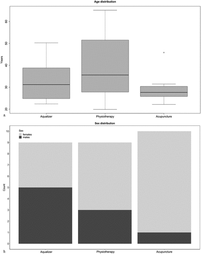
Patients’ age ranged from 19.92 and 65.12 years (average: 33.41 years; standard deviation: 11.26 years). In , average age and standard deviation are reported by subsample. Age and sex distribution are also represented in , respectively. One of the patients enrolled in the Aqualizer® pilot study dropped out because was unable to hold the splint in his mouth and felt uncomfortable wearing it. Similarly, one patient of the physiotherapy subsample dropped out, not showing up for the second appointment and without providing any explanations. The samples did not differ by their median age (Kruskal-Wallis rank sum test, p = .4638), but were not comparable in terms of distribution of the sexes (Kruskal-Wallis rank sum test, p = .03691) as is evident by observing .
Figure 1. a) age distribution in years, and b) distribution of males and females are shown for each therapeutic approach, i.e., Aqualizer®, physiotherapy, and dry-needle oral acupuncture.

Table 1. Descriptive table reporting the number, sex (m = male; f = female), and age in years of the patients considered, by therapeutic approach. SD = standard deviation.
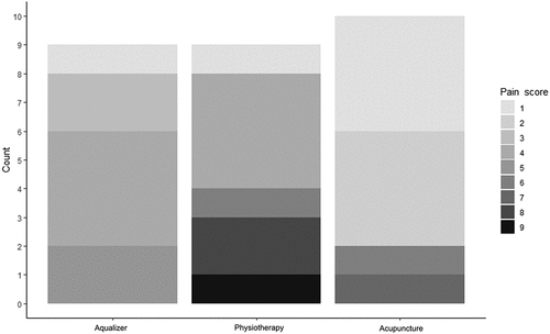
The values for the clinical parameters collected during sessions T1, T2, and T3 are reported in . The scores for each of the parameters did not differ significantly between subsamples at any of the sampling times, except for intraoral pain at T1 (p = .03215). The post hoc Dunn’s test showed that among the pairwise comparisons, intraoral pain at T1 differed significantly only between the physiotherapy and oral acupuncture samples (uncorrected p = .00906; adjusted p = .02717). The distribution in recorded pain scores, ranging from 1 to 9, is shown in . The clinical parameters at T1 did not correlate with either sex or age ().
Figure 2. Distribution of the intraoral pain scores at baseline (i.e., T1) for the Aqualizer®, physiotherapy, and oral acupuncture subsamples.

Table 2. Descriptive table reporting the parameters for mouth opening, neck mobility, muscle, and intraoral pain, and VAS by treatment sessions (T1 = baseline; T2 = 2 weeks from first treatment; T3 = 4 weeks from first treatment).
Table 3. Clinical parameters at baseline (T1) did not correlate to sex or age.
reports the outcomes of the Friedman test, which was used to show whether there were significant changes from T1 to T3 in the various clinical parameters scored. The use of the Aqualizer® and the physiotherapy yielded significant changes in all clinical parameters (Aqualizer®: muscle pain, p = .00012; intraoral pain, p = .000912; VAS, p = .02075; neck mobility, p = .00461. Physiotherapy: muscle pain, p = .00012; intraoral pain, p = .00056; VAS, p = .00139; neck mobility, p = .00239) except for mouth opening, which did not show significant differences in the oral acupuncture group, either. In the latter, instead, only muscle pain and neck mobility were significantly improved (p = .00745 and 0.00509, respectively). The Nemenyi’s all-pairs comparisons test showed that the differences observed within the treatment parameters for both Aqualizer® and physiotherapy owed to a significant change between T1 and T3 (Aqualizer®: muscle pain, p = .00007; intraoral pain, p = .00280; VAS, p = .02600; neck mobility, p = .01300. Physiotherapy: muscle pain, p = .00007; intraoral pain, p = .00180; VAS, p = .00180; neck mobility, p = .00620). The significant differences between the clinical parameters in the oral acupuncture sample, owed to both differences between T2 and T3 (muscle pain = 0.03700; neck mobility, p = .03700) and between T1 and T3 (muscle pain, p = .01000; neck mobility, p = .02700).
Table 4. Results of the statistical analyses testing the effect of the treatments at different times. The difference between baseline (T1), 2 weeks, and 4 weeks from the first treatment (T2 and T3, respectively) were tested using the Friedman test. Pairwise comparison between T1 and T2, T2 and T3, and T1 and T3 was conducted using Nemenyi’s all-pairs comparisons test. Significant p-values (<0.05) are in bold font.
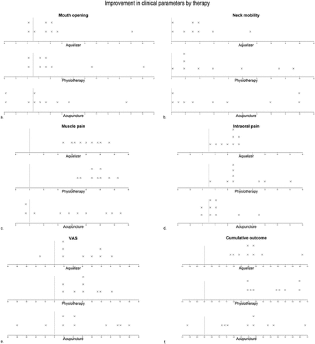
The changes in clinical parameters are graphically represented in dot plots in . Markers on the positive side of the horizontal axis represent an improvement (i.e., a decrease in pain or an increase in mouth opening and neck mobility). On the contrary, the markers on the negative side of the axis represent a worsening (i.e., increase of pain or decrease in mouth opening and neck mobility). The patients wearing the Aqualizer® showed a general improvement of all parameters with the following exceptions: two patients exhibited slightly decreased mouth opening after treatment and one showed no changes; one patient showed a slight decrease in mouth opening and no changes in neck mobility; one patient showed no improvement in neck mobility, and one reported worsening of pain through the VAS. Similarly, the physiotherapy patients mostly showed an improvement except for one showing a worsening in mouth opening, one showing no changes in intraoral pain, and one with a slight decrease in mouth opening and no changes in neck mobility. Patients receiving oral acupuncture showed an improvement except in the following instances: one patient presented a worsening in three parameters (i.e., mouth opening, muscle pain, and VAS), one patient showed no changes in mouth opening and worsening of intraoral pain and VAS, five patients slight worsening or no improvement of two parameters and other two patients of only one parameter. shows the cumulative outcome of the three different therapies obtained by summing the scores of the 5 clinical parameters, after normalization, and demonstrating a general improvement in the patients except for one subject in the acupuncture group who experienced a worsening. The improvement observed at T3 did not correlate to either sex or age ().
Figure 3. Changes in a. muscle pain, b. intraoral pain, c. VAS, d. mouth opening, and e. neck mobility. In f. the cumulative changes derived by the sums of the normalized parameters are shown. Markers on the positive side of the horizontal axis represent an improvement (i.e., a decrease in pain or an increase in mouth opening and neck mobility). On the contrary, the markers on the negative side of the axis represent a worsening (an increase of pain or decrease in mouth opening and neck mobility).

Table 5. Correlation between the improvement of the clinical parameters at 4 weeks from first treatment (T3), age, sex. The improvement observed did not correlate with these demographic variables.
4. Discussion
For their complex nature, orofacial pain and dysfunction require specialized medical attention. Careful diagnosis for an accurate evaluation of the patient’s condition is necessary to identify the most suitable treatment. An initial therapy providing immediate pain relief is recommended because it brings at ease the distressed patient presenting muscle pain or spasms in the face and neck. Additionally, it allows for conducting a more reliable clinical assessment, thus facilitating a correct diagnosis. Several approaches exist for the initial treatment of orofacial pain aimed at reducing discomfort and improving function. Among the non-pharmacological options, various techniques are known in the field of oral medicine to provide initial treatment to the orofacial pain patient.
In this work, the effectiveness of the Aqualizer®, a fluid-based dental splint, physical therapy mostly focused on the upper quarter of the body, and dry-needle oral acupuncture were assessed comparatively. These approaches have been widely used in clinical settings and evidence for their efficacy has been provided in the literature [Citation13,Citation25–27]. The pilot studies presented in this work contributed convincing results about the validity of these therapies, which could improve the general condition of the patients altogether, in a relatively short amount of time. The physical therapy sessions and the use of the Aqualizer® resulted in more positive outcomes than acupuncture. Moreover, physiotherapy, as the Aqualizer®, could reduce intraoral pain significantly although the initial condition of the physiotherapy patients was more severe than in the other two samples. These results are in agreement with previous findings as reported in recent reviews considering, however, physiotherapy in comparison to occlusal splits other than the Aqualizer® [Citation25,Citation28].
Although acupuncture was not conducive to as great improvement as the other two approaches, the results achieved are surprising considering that they were obtained with just 5-second-long dry-needle pricks, thus using a simpler technique that does not exploit the full potential of conventional oral acupuncture [Citation29,Citation30]. Usually, a few drops of low-concentration anesthetic fluids are injected, providing both mechanical and anti-inflammatory benefits on the “very point”. The anesthetic can also be substituted by saline solution, in this case forsaking the anti-inflammatory effects, and keeping the needle in place for longer could also be more effective. Dry-needle acupuncture was found helpful in reducing orofacial pain and increasing mouth opening in combination with upper cervical spinal manipulation [Citation27], but in this case, the needles were left in place for 20 minutes, thus compensating for the lack of injection fluid.
The outcomes for the patient receiving acupuncture and experiencing a general worsening of the condition need detailed discussion. This patient did not show a particularly worrying baseline situation. However, since the ethical commission recommended to include also patients with initial, light symptoms, she was recruited because still met the inclusion criteria of the performed randomized trial. Taking into account the generally positive outcome for the rest of the patients and the muscle relaxing effect of acupuncture, it can be assumed that, by releasing muscle tension, the patient could perceive the pain more distinctively, thus increasing her awareness of pain.
Despite the generally positive results, the use of the Aqualizer® presented also some drawbacks. Breakage of one appliance occurred with consequent leaking of the contained distilled water, which, although it is a rare occurrence based on the experience of the clinicians in this group, might be accounted as a possible disadvantage of this therapy. The dropout patient in the Aqualizer® cohort, disliking the feeling of the splint in his mouth, is a reminder that individuals possess different preferences and that no treatment can be considered universal. Thus, while treating patients, alternative types of splints should be considered, as well. Similarly, the patient dropping out of the physiotherapy group, although initially motivated, did not show up to the second appointment. For the physiotherapy sessions, intense communication between the therapist and the patient occurred to ensure the correct execution of the exercises. As this patient was not familiar with either German or English – the idioms spoken by the therapist – it is reasonable to think that he was discouraged by the language barrier. Since these three different initial therapies showed beneficial effects, it can be concluded that in the clinical, non-experimental, settings, patients can be recommended for the approach most suitable to their condition and preferences.
Through a combination of stretching, isometric contractions, and relaxation exercises for the upper quarter of the body, physiotherapy presents the advantage to improve posture, body awareness, mobility of the spine and temporomandibular joints, thus also positively affecting the position of the mandible, which partly depends on the condition of the head and neck muscle [Citation31]. Tongue exercises are especially relevant in case the tongue does not rest properly on the palate in a habitual position, thus exerting excessive pressure on its anterior part causing tension of the pterygoid muscle as well as supra- and infrahyoid muscles [Citation32]. It requires the active participation of the patients and must be performed regularly to be most beneficial, thus enough time has to be dedicated to its practice, which can represent a drawback for some.
The Aqualizer® acts by decoupling the occlusion, allowing a relaxed position of the mandible, dictated by the temporomandibular joint rather than by occlusion. It provides an immediate change of muscle pattern, deprogramming the muscle, by raising the bite and distracting the temporomandibular joints; bruxism occurs on a softer surface, thus strains are reduced. Since it is not specifically designed for the patient, it should not be used for a long time. Additionally, some do not find it easy to wear, especially for many hours per day, because it is loose in the mouth. However, it can be considered a useful tool for acute therapy, interrupting the vicious circle of pain [Citation12,Citation13].
Oral acupuncture presents several advantages including providing immediate improvement without requiring patient cooperation. It can be considered a causal therapy in that it is an effective analgesic and spasmolytic, through muscle relaxation, endorphin release, and improved lymphatic flow [Citation33]. Acupuncture is known for activating the body’s regulation and healing capacities. It is also economical and allows for a certain degree of flexibility in the modality of application, so that, in case patients fear needles, acupressure can be practiced as an alternative. However, it requires high clinical proficiency, deriving from long-term training and practice.
The evidence stemming from the pilot studies conducted for this work should be considered as preliminary results, encouraging further research on the effectiveness of initial therapies for myofascial pain patients. To overcome the limitations of the present study, future works should include larger patient cohorts, possibly assessed by the same, blinded, operator, to explore the medium to long-term effects of these therapeutic approaches. The efficacy of various oral acupuncture techniques, using anesthetic fluid versus dry needles, or with variable duration of the sessions, should also be thoroughly investigated.
5. Conclusions
Initial pain treatment is beneficial in the management of patients presenting with orofacial myalgia. Non-pharmacological therapeutic approaches such as physiotherapy and the Aqualizer® splint and dry-needle oral acupuncture represent useful ways to reduce orofacial pain and improve neck mobility, thus contributing to patients’ well-being and treatment success. Since these methods present different advantages and drawbacks, they can be applied depending on the patient’s preferences and needs.
Acknowledgments
We thank the personnel of the Clinical Division of Prosthodontics, University Clinic of Dentistry, Medical University of Vienna, Vienna, Austria for managing the patients’ appointments. We are grateful to Tássio Drieu Bellezia de Sales for insightful comments on the manuscript. Michael Kundi performed the analysis to determine the power to detect a therapeutic effect.
Disclosure statement
The authors declare no conflicts of interest.
Additional information
Funding
References
- Barroso J, Branco P, Apkarian AV. Brain mechanisms of chronic pain: critical role of translational approach. Transl Res. 2021;238:76–89.
- Fleming PS, Strydom H, Katsaros C, et al. Non-pharmacological interventions for alleviating pain during orthodontic treatment. Cochrane Database Syst Rev. 2016;12(12):Cd010263. Epub 20161223 DOI:10.1002/14651858.CD010263.pub2.
- Argueta-Figueroa L, Flores-Mejía LA, Ávila-Curiel BX, et al. Nonpharmacological interventions for pain in patients with temporomandibular joint disorders: a systematic review. Eur J Dent. 2022;16(3):500–513. Epub 20220308. DOI:10.1055/s-0041-1740220.
- Riley P, Glenny AM, Worthington HV, et al. Oral splints for temporomandibular disorder or bruxism: a systematic review. Br Dent J. 2020;228(3):191–197.
- Amin A, Meshramkar R, Lekha K. Comparative evaluation of clinical performance of different kind of occlusal splint in management of myofascial pain. J Indian Prosthodont Soc. 2016;16(2):176–181.
- Pilot Study ÖN. Aqualizer as acute therapy for craniomandibular dysfunction” [pilotstudie: „aqualizer als akuttherapie bei craniomandibulärer dysfunktion“] [doctoral thesis]. Vienna, Austria: Medical University of Vienna; 2020.
- Balla M. Pilot study: relaxation techniques as acute therapy for craniomandibular dysfunction [pilotstudie: entspannungstechniken als akuttherapie bei craniomandibulärer dysfunktion] [doctoral thesis]. Vienna, Austria: Medical University of Vienna; 2020.
- Artacker N. Pilot study: intraoral insertion of an injection needle without injection as acute CMD therapy [pilotstudie: intraoraler einstich einer injektionsnadel ohne injektion als CMD-Akuttherapie] [doctoral thesis]. Vienna, Austria: Medical University of Vienna; 2020.
- Dworkin SF, LeResche L. Research diagnostic criteria for temporomandibular disorders: review, criteria, examinations and specifications, critique. J Craniomandibular Disord: Facial Oral Pain. 1992;6(4): 301–355. Epub 1992/01/01.
- Meyer G. Short clinical screening procedure for initial diagnosis of temporomandibular disorders. J Aligner Orthodontics. 2008;2(2):91–98.
- Hobson J, Esser B. Utilizing an Aqualizer® appliance to address back pain through a dental/physical therapy approach. CRANIO®. 2022;40(2):93–94.
- Buchbender M, Keplinger L, Kesting MR, et al. A clinical trial: aqualizer ™ therapy and its effects on myopathies or temporomandibular dysfunctions. Part II: subjective parameters. CRANIO®. 2021;1–7. DOI:10.1080/08869634.2021.1885887.
- Buchbender M, Keplinger L, Kesting MR, et al. A clinical trial: aqualizer ™ therapy and its effects on myopathies or temporomandibular dysfunctions. part I: objective parameters. CRANIO®. 2021;1–9. Epub 20210216. DOI:10.1080/08869634.2021.1885886.
- Aigner B, Klose C. Physiotherapy techniques from a to Z [Physiotherapietechniken von a – Z]. Stuttgart: Georg Thieme Verlag; 2018.
- Stelzenmüller W, Wiesner J, Ricken C, et al. Therapy for jaw joint pain - a treatment concept for dentists, orthodontists and physiotherapists [Therapie von Kiefergelenkschmerzen - Ein Behandlungskonzept für Zahnärzte, Kieferorthopäden und Physiotherapeuten]. Stuttgart: Georg Thieme; 2004.
- Bartrow K. The culprit of the jaw joint – finally relaxed and pain-free again: 60 exercises with an immediate effect [übeltäter kiefergelenk – endlich wieder entspannt und schmerzfrei: 60 übungen mit soforteffekt]. Stuttgart: TRIAS Verlag; 2012.
- Chaitow L. Muscle energy techniques [muskel-energie-techniken]. 2nd ed. München: Urban & Fischer Verlag; 2008.
- Schomacher J. Diagnosis and therapy of the musculoskeletal system in physiotherapy [diagnostik und therapie des bewegungsapparates in der physiotherapie]. Stuttgart: Georg Thieme; 2001.
- Gleditsch J. The “very point” technique: a needle based point detection method. Acupunct Med. 1995;13(1):20–21.
- Gleditsch J, Markert IU. Initial therapy of CMD using acupuncture [Initialtherapie der CMD mittels Akupunktur]. ZWR - Das Deutsche Zahnärzteblatt. 2017;126(12):622–627.
- Gleditsch JM. Textbook and atlas of micro acupuncture point system (MAPS) [lehrbuch und atlas der mikro-aku-punkt-systeme (MAPS)]. 2nd ed. Berlin: Quintessenz (KVM); 2007.
- Fang CY, Yu JH, Chang CC, et al. Effects of short-term acupuncture treatment on occlusal force and mandibular movement in patients with deep-bite malocclusion. J Dent Sci. 2019;141:81–86. Epub 20181204. DOI:10.1016/j.jds.2018.11.003.
- Zotelli VL, Grillo CM, Gil ML, et al. Acupuncture effect on pain, mouth opening limitation and on the energy meridians in patients with temporomandibular dysfunction: a randomized controlled trial. J Acupunct Meridian Stud. 2017;105:351–359. Epub 20170922. DOI:10.1016/j.jams.2017.08.005.
- R Core Team. R: a language and environment for statistical computing. Vienna, Austria: R Foundation for Statistical Computing; 2022.
- Paço M, Peleteiro B, Duarte J, et al. The effectiveness of physiotherapy in the management of temporomandibular disorders: a systematic review and meta-analysis. J Oral Facial Pain Headache. 2016;30(3):210–220.
- Simma I, Simma L, Fleckenstein J. Muscular diagnostics and the feasibility of microsystem acupuncture as a potential adjunct in the treatment of painful temporomandibular disorders: results of a retrospective cohort study. Acupunct Med. 2018;36(6):415–421.
- Dunning J, Butts R, Bliton P, et al. Dry needling and upper cervical spinal manipulation in patients with temporomandibular disorder: a multi-center randomized clinical trial. CRANIO®. 2022;1–14. Epub 20220412. DOI:10.1080/08869634.2022.2062137.
- Zhang L, Xu L, Wu D, et al. Effectiveness of exercise therapy versus occlusal splint therapy for the treatment of painful temporomandibular disorders: a systematic review and meta-analysis. Ann Palliat Med. 2021;106:6122–6132. Epub 20210510. DOI:10.21037/apm-21-451.
- Irnich D, Behrens N, Gleditsch JM, et al. Immediate effects of dry needling and acupuncture at distant points in chronic neck pain: results of a randomized, double-blind, sham-controlled crossover trial. Pain. 2002;99(1–2):83–89.
- Simma I, Gleditsch JM, Simma L, et al. Immediate effects of microsystem acupuncture in patients with oromyofacial pain and craniomandibular disorders (CMD): a double-blind, placebo-controlled trial. Br Dent J. 2009;20712:E26. Epub 20091030. DOI:10.1038/sj.bdj.2009.959.
- Sodhi A, Nair P, Hegde S. Physiotherapy: key to the kinetics of orofacial musculature. JIAOMR. 2014;26(4):419–424.
- Nascimento ALO, Reis F, Bérzin F, et al. When tongue strength exercises reflect in the cervical region. Codas. 2020;324:e20180285. Epub 20200731. DOI:10.1590/2317-1782/20202018285.
- National Institutes of Health. Acupuncture. 1997 Nov 3-5. Report No.: 1080-1707 (Print) Contract No.: 5.
