
We have compiled the results in three tables, the first contains the eyes of emmetropes, the second those of the myopic, the third those of the hypermetropic. This division was made to determine the influence of the length of visual axis. It is been proven that, ordinarily, hypermetropia and myopia depend almost exclusively on the length of visual axis. In the cornea their cause is not to be found (1). Nor has a difference in the focal length of the lens been found. Accordingly one could decide in favor of a difference in length of the visual axis per exclusionen that is, in addition, confirmed by direct observation. It was therefore to be expected that the point of rotation would not be located at the same depth in ametropia as in normal emmetropic eyes. To determine the location, in relation to the length of the visual axis, we have calculated this length for each case, according to the degree of the ametropia, thereby assuming that cornea and crystal lens and, consequently, the cardinal points, would conform to those of the schematic eye of Helmholtz (2).
Figure 1. See text for full explanation.
The first 15 eyes, listed in this table, are free from any emmetropic refractive anomaly, the last four suffered from astigmatism (3), with almost emmetropia existing in at least one of the meridians.
The 15 emmetropic eyes are ranked according to age. However, a relation between age and location of the pivot point, or between age and angle between corneal axis and visual axis does not emerge.
With regard to the location of the pivot point, its distance from the base of the cornea segment is maximally 11.44mm, minimally 10.43mm; we found 10.94mm, on average.
In emmetropic eyes the cornea is always cut on the inner (medial) side of its center corresponding to its top (4), by the visual axis. The angle between corneal axis and visual axis was maximally 7°, minimally 3.5°; on average 5.082°.
Note that this angle is larger in the four astigmatic eyes, and rises to 7° or 8°. In simple hypermetropic astigmatism (emmetropia in one, hypermetropia in the other principal meridian) this seems to be the rule. Regarding the distance between the pivot point and the base of the cornea segment, these eyes hardly deviate from the purely emmetropic eyes.
Finally it should be noted that movement was only limited in one of all eyes listed in . Accordingly, with one exception they were able to deviate the corneal axis to at least 28º right and left of the mid position.
Table 1. Emmetropia
, containing the results obtained in myopic eyes, shows that the pivot point is located here further behind the basis of the corneal segment, that the angle between the corneal axis and the visual axis is smaller and that eye movements are not rarely limited.
Table 2. Myopia
The myopic eyes are ranked according to the degree of myopia, ranging from 1/16 to 1/2.25. The distance between the pivot point and the base of the cornea segment ranges from 10.58mm to 13.37mm, 11.96mm on average, and thus much larger than that in emmetropic eyes (10.94mm). In addition, that distance increases, in general, with the degree of myopia. In the first seven eyes with myopia between 1/16 to 1/6.25, it averaged 11.42mm; in the last ten eyes with myopia of between 1/5.25 and 1/2.25, the average rises to 12.27mm. Comparison only between the lowest and the highest degrees of myopia in the table shows this influence even more strongly.
Evidently, its location is related to the length of the axis of the eye. The longer the axis of the eye, the further the pivot point lies behind the base of the cornea segment, but the farther it nevertheless remains removed from the rear end of the axis of the eye. The length of the axis of the eye has been calculated under the assumption that the dioptric system was the same as in emmetropic eyes, and this result has been added in the last column.
Secondly, the relation between the corneal axis and the visual axis is very remarkable. The chart clearly shows that the angle between these two is much smaller in myopes than in emmetropes. We find a maximum of 5.25º, a minimum of -1.5º (the negative sign means that the angle lies lateral from the corneal axis) and an average of slightly less than 2º. We come back later to discuss the cause of this.
Finally, we should remark that in 9 out of 17 cases eye movement was limited to such an extent that, starting from the visual axis with normal state of the head, an eye movement to each side of almost 28° was impossible. Among the ten cases with the highest degree of myopia, eye movement was free only in three cases.
The number of hypermetropic eyes that was examined, presented in , is 12.
Table 3. Hypermetropia
These eyes have a shorter axis of the eye (last column of ), which was also calculated here from the degree of the ametropia, on the premise that the shape and orientation of the refracting surfaces of the cornea and crystalline lens were similar to those of the emmetropic eye. In relation to the shortness of the axis of the eye, the pivot point also appears to lie nearer to the cornea. The maximum of the distance between pivot point and the base of the corneal segment is 12.07mm and – if we disregard the first two weakly hypermetropic eyes – the average is only 11.21mm; the minimum is 9.72mm. However, a clear relation between the distance and the degree of hypermetropia does not emerge from these few cases.
Regarding the angle between corneal axis and visual axis, the hypermetropic eyes have results opposite to those of myopic eyes. First, here the angle is always positive; moreover the smallest angle (6°) found is larger than the largest (5.25°) found in myopia; the largest angle found in hypermetropia is 9° and we found almost 7.3°, on average. We found limited eye movement only in two eyes.
We will now compare the eyes with different refraction more closely. To this end, above was prepared with the outcomes for the average emmetropic eye, the average myopic eye, the average hypermetropic eye, the most myopic eye and the most hypermetropic eye.
TABLE 4. Summary
Regarding the column “Length of the axis of the eye” it is to be noted that to the axis of the eye, calculated in , and , has been added: the thickness of the rear layers of the eye, in emmetropic and hypermetropic eyes 1.2 mm, in myopic eyes, with much thinner rear layers of the eye, 0.6 mm, and for the largest myopia only 0.18 mm.
Regarding the column “Behind the top of the cornea” 2.6mm is added in order to obtain, from the detected location of the pivot point behind the base of the corneal segment, its location behind the anterior surface of the cornea.
From this t appears:
that the pivot point of the emmetropic eye is located a significantly (1.77mm) behind the midst of axis of the eye.
that in the myopic eye the pivot point is located deeper in the eye, but also further away from the rear layers of the eye, and that in myopic eyes in general, as well as in the most myopic eye, the ratio between the parts of axis of the eye located before and behind the pivot point, is substantially the same as in the emmetropic eye. In the most myopic eye the pivot point seems to lie somewhat relatively forward, in the mean myopic eye somewhat backward.
that in hypermetropic eyes the pivot point is located a somewhat less deeply, but much closer to the rear layers of the eye, so that in the ratio, especially in strongly hypermetropic eyes, the part behind the pivot point is much smaller.
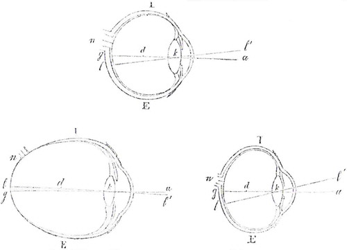
To illustrate the relationship between corneal axis and axis of the eye, we have added the figure above. The upper drawing represents an emmetropic eye, the lower left a myopic and the lower right a hypermetropic eye. All are seen in horizontal cross-section, through the optic nerve: I is, accordingly, the inner (translator: medial) side, E is the outer (translator: lateral) side of the eye; d is the pivot point of the eye, ll’ is the axis of the eye. Please note that these eyes differ from one another, in particular regarding the relative lengths of the axis of the eye. The myopic eye pictured here (lover left) is the largest that has occurred to us so far. The angle l’ka, between the visual axis and the corneal axis, is larger in the hypermetropic eye (lower right) than in the emmetropic eye. In the myopic eye it is smaller when the myopia is larger and it ultimately becomes negative, as shown in the lower left drawing.
That the corneal axis and the visual axis do not coincide was evident from the studies by Senff, Helmholtz, Knapp and others. That the angle is smaller in myopic eyes than in emmetropic eyes was proved by Donders: he found sufficiently equal radii of curvature of the cornea, at equal distances from the visual axis. He had then already applied this to the position and movements of eyes with myopia. That, however, the opposite is true of hypermetropia first follows from the investigations performed by us, reported here, while, in addition, that what was found previously for myopes was confirmed with the method used here for a larger number of eyes.
The influence of ametropia on the relation between corneal axis and visual axis is important in more than one respect. First, it is related to the shape of ametropic eyes and the change of shape that an eye with progressive myopia undergoes. Regarding the latter, the matter is simple. The direction of the visual axis is determined by two points of intersection: 1. the common nodal point k; 2. the macula lutea l. When the myopia increases, k distances itself from the retina; and if the distance lg remains the same, the angle between corneal axis and visual axis, consequently, becomes smaller. This, however, is not so much an option here. That a more important factor is at work is shown by the fact that the angle not only becomes smaller, but may even become negative. That important factor is the elongation which, albeit, takes place in the rear portion of the eye (staphyloma posticum) but, nevertheless, more so on the outer (translator: lateral) side E in particular. As a result, the optic nerve which lies on the inside will be located more on the inside, and the same is true of the yellow spot (macula lutea), in spite of the facts that the distance between n and l becomes considerably greater and that atrophy of the choroid tends to start just here. That uneven stretching of the inside and the outside makes the yellow spot shift more and more to the point at which the corneal axis is directed and, finally, even passes this point.
As for the hypermetropic eye, it seems to us that this is an imperfectly developed eye, similar to a slight degree of microphthalmos. Not infrequently, the function of the retina is not perfect. In addition, the hypermetropic eye is more subject to asymmetry. Research on the course of its development must still clarify this. At the same time one sees easily that, when the distance between the nodal point and the macula is smaller, the latter only needs to be located at the usual distance on the outside of the extended corneal axis, for the angle between this axis and the visual axis to increase.
Related to the different angle between the visual axis and the corneal axis is the peculiar look of myopic and hypermetropic eyes. When the direction of the visual axes of the eyes is parallel, the corneal axes of emmetropic eyes diverge at an angle of 2 x 5° = 10°. However, this mode of looking does not convey the impression of divergence to us, but rather of parallelism. Based on this starting point, in myopes, whose corneal axis and visual axis coincide, an apparent convergence of the cornea axes exists of approximately 2 x 5° = 10° and, vice versa, in hypermetropes, in whom the divergence can amount to 2 x 9° = 18º, the divergence will only seem to be 8º. At correctly directed visual axes of the eye, the extremes of ametropia cause a difference in the direction of the corneal axes of 8º + 10º = 18º, which may even rise to 20º. It is not surprising that the look gets something peculiar and often even suggests strabismus. Certainly this is one of the additional reasons why, in myopia, the convergence and, in hypermetropia, the divergence of axes of the eye is often inadequate for binocular vision, and why myopia is much more common in divergent strabismus, and hypermetropia more common in convergent strabismus (5). Besides, one finds the movements of the eyes rarely to be limited in hypermetropes. The opposite is true, as we saw, in myopes, and this comment brings us back to the pivot point of the eye.
We know that Johann Mueller was of the opinion a long time that the pivot point could be found at the back of the eye. He derived the main argument for this view from comparative anatomic dissection; it certainly did occur to him, that the more the pivot point shifted forward, the more the increased movement of the back of the eyeball, with the optic nerve connected to it, would restrict motility.
For motility, the distance from the pivot point to the rear of the eye, and in particular to the insertion of the optic nerve, is one of the most important factors.
When that distance is large, the visual axis cannot describe a wide arc. The range of eye movement becomes much more understandable now that we found that, in the emmetropic eye, the pivot point is located not in the midst of axis of the eye, but only 10 mm from the rear area of the eye and from the insertion of the optic nerve. But when we now further see that in myopic eyes, as we investigated, that distance averages 10.08 mm and may rise even up to 12.24 mm, the main reason for the restriction of movement found in these eyes has already been identified. Indeed, the increased size of the eye in all its dimensions by itself hampers the free movement while, furthermore, shortening of a muscle by displacement of its point of insertion on the surface of the greater eyeball results in less excursion in degrees. Overall, the motility of the eyes in relation to the ordinary and to the abnormal shapes merits a separate investigation. The insertion of the optic nerve, the shape of the orbit in relation to the exlarged myopic eye, the relation of the muscles and many other points should thereby be investigated more closely. However, this is for the moment beyond our concern. We wished only to determine the location of the pivot point in the eyes with different refraction, and underline, in general, the importance of a correct understanding of this point. Moreover, we do not deny that even with regard to the determination of the pivot point much remains to be done. In the first place, we did not examine to what extent the pivot point can be regarded as an immovable point. Our investigations extend only to horizontal rotation and that, almost always, with an equal range. In addition, we have derived the length of the axis of the eye only from the degree of the ametropia by calculation, and this cannot be very precise. The dioptric system of the myopic eye differs from the emmetropic firstly by a deeper position of the crystalline lens and, concerning the hypermetropic eye, the mere fact that the pivot point was found relatively deeply in the eye, advocates the opinion that the crystalline lens of these eyes have a greater focal length and that, therefore, the calculated length of axis of the eye was too small. So we can only vouch for the exactness of what was assessed directly: the distance between the base of the cornea segment and the pivot point of the eye, at a range of eye movement in the horizontal plane, for eyes of different refraction.
Notes
1. Donders F.C. Royal Netherlands Academy of Science, Verslagen en Mededelingen, Afd. Natuurkunde, Eerste Reeks, p159.
2. The calculation is done with the formula for the conjugate focal lengths. The degree of ametropia allows determination of the location of the anterior focal length (in the air), from which the posterior focal length that coincides with the retina is determined.
3. Compare: Donders FC: Astigmatisme en cylindrische glazen (Astigmatism and cylindrical glasses). Utrecht, 1862.
4. From the investigations of Helmholtz and Knapp it has been found that the top of the ellipsoid almost fully corresponds to the center of the cornea. We have assumed, therefore, that the axis of the cornea coincides with its center. Some measurements of the corneal radius on both sides at equal distances from the center, also provided us with almost entirely the same outcomes.
5. Compare de Haas JH: Geschiedkundig onderzoek omtrent de hypermetropie en hare gevolgen (Historical research on the hypermetropia and its consequences). Diss. Inaug. Utrecht 1862.