To the Editor
Platelets can be observed in a direct blood smear for approximate quantity and shape or detected by hematology analyzer using voltage-pulse counting or electro-optical counting. In both systems, the collected blood is diluted and counted by passing the blood through an electronic counter. The primary functions of a platelet count are to assist in the diagnosis of bleeding disorders and to monitor certain patients (marrow failure, patients with solid tumors and therapy-induced thrombocytopenia, massive hemorrhage and in thrombocytopenic patients undergoing major surgery). Besides, rapid laboratory results would allow thrombolysis to be discontinued promptly even after initiation in patients subsequently found to have thrombocytopenia in acute ischemic stroke of thrombolytic agents, which could be avoided in patient with an acute ischemic stroke and thrombocytopenia Citation[1].
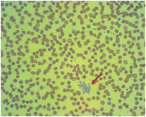
All of the hematology instruments use software-generated flagging algorithms for identifying abnormal platelet distributions that may indicate the presence of platelet-specific interference or the presence of platelet clumping. In our stat laboratory, we must review the blood smear before sending out the complete blood cell count (CBC) reports when the flag of platelet clumping occurred. In this study, we selected 100 blood specimens with the platelet level ranging from 14,000 to 249,000 µL. Those patients included 57 females ranging in age from 12 to 90 years (median: 62 years) and 43 males ranging from 1 to 77 years (median: 53 years). All peripheral blood smears were reviewed by a senior medical technologist. According to our findings, we found two true positive of platelet clumping using Coulter LH 750 hematology analyzer (Beckman Coulter Inc., Fullerton, CA, USA). The first true positive is due to the clotting of blood specimen. The second true positive is due to EDTA-induced thrombocytopenia. A 73-year-old woman was admitted to our emergency department (ED) and laboratory showed platelet level of 51,000 µL with standard EDTA tube. Because she was a new patient we then asked the on-duty nurse to resend her paired blood samples (EDTA and sodium-citrate tubes). After reanalysing, the platelet level was 53,000 µL and 206,000 µL, respectively. The platelet count was erroneously low due to platelet clumping with standard EDTA tube and the presence of platelet clumping on peripheral blood smear ().
Figure 1. The presence of platelet clumping on peripheral blood smear (red arrow).
To interpret the flag of platelet clumping, the three simple steps could be followed: (1) To ensure evidence of clots whether clot and/or agglutinin appears on the surface of tube by visual observation; (2) To compare the platelet results to the recent platelet results using the Laboratory Information System (LIS); (3) It would be advisable to draw paired samples-tubes (EDTA and sodium-citrate tubes) for rechecking platelet count if he or she is new patient. If the flag of platelet clumping continues to appear, it is unavoidable to carry out the blood smear. So, if the flag of platelet clumping occurred, the result of platelet count can be sent out rapidly if the technologist follows the rules mentioned above step by step.
Platelet clumping in EDTA anticoagulants continues to pose procedural problems for the clinical hematology laboratory Citation[2]. The presence of platelet clumps on blood smear, whether platelet count is normal or low, has no clinical significance and does not represent a disease. The most common cause of platelet clumping in blood is ethylenediaminetetra-acetic acid (EDTA), the anticoagulant used in test tubes for complete blood counts. This phenomenon is sometimes called “EDTA-induced thrombocytopenia or pseudothrombocytopenia” Citation[3]. Occasionally, it is due to cold-reacting antibodies that aggregate platelets at room temperature. Nevertheless, it only occurs with an incidence of approximately 0.1% in the general population Citation[4]. In addition, the risks for a platelet count test are minimal in normal individuals.
In fact, determination of platelet count is a frequent cause of treatment delay. In contrast to glucose, which can be measured by using widely available point-of-care testing devices, platelet-count measurement requires venipuncture and blood collection, careful specimen-labeling, transportation to a central laboratory where automated equipment is available, sample analysis and communication of results to the treating physician Citation[1]. Delay can be introduced at numerous points along this pathway. A previous study Citation[5] reported that the Coulter LH 750 hematology analyzer incorporates new algorithms for platelet enumeration that significantly reduce the number of samples requiring further review. This feature represents a major advantage over previous impedance hematology counters. However, another study Citation[6] showed that the Coulter LH 750 demonstrated the poorest sensitivity for platelet clumps and they supposed that failure to flag platelet clumps could lead to erroneous reporting of low platelet counts if laboratory algorithms for confirming low platelet counts are inadequate or not followed carefully.
In summary, if all smears must be reviewed for platelet count verification when a platelet clumps flag is generated, it will influence the turnaround time for complete blood cell count (CBC) counts. Based on our findings, our recommended steps may allow the improvement in the turnaround time (TAT), avoiding delays that are crucial, particularly in the outpatient setting. Besides, improvement in the ability to overcome EDTA-induced platelet clumping would contribute greatly to rapid and accurate platelet count reporting by clinical laboratories.
Declaration of interest: The authors report no conflicts of interest. The authors alone are responsible for the content and writing of the paper.
References
- Cucchiara BL, Jackson B, Weiner M, Messe SR. Usefulness of checking platelet count before thrombolysis in acute ischemic stroke. Stroke 2007; 38: 1639–1640
- Bartels PCM, Schoorl M, Lombarts AJ. Screening for EDTA dependent deviations in platelet counts and abnormalities in platelet distribution histograms in pseudothrombocytopenia. Scand J Clin Lab Invest 1997; 57: 629–636
- Allerheiligen D, Houston R, Vermedahl B. EDTA-Induced Pseudothrombocytopenia. J Am Board Fam Prac 1996; 9: 212–214
- Mori M, Kudo H, Yoshitake S, Ito K, Shinguu C, Noguchi T. Transient EDTA-dependent pseudothrombocytopenia in a patient with sepsis. Intens Care Med 2000; 26: 218–220
- Keeney M, Brown W, Yee IC. Platelet counts and flagging rates from the LH 750 hematology analyzer compared with the ICSH/ISLH platelet reference method and the Gen. S hematology analyzer. Lab Hematol 2001; 7: 204–210
- Sandhaus LM, Osei ES, Agrawal NN, Dillman CA, Meyerson HJ. Platelet counting by coulter LH 750, sysmex XE 2100 and advia 120: A comparative analysis using the RBC/platelet ratio reference method. Am J Clin Pathol 2002; 118: 235–241