ABSTRACT
We report the artificial synthesis from T-2 toxin of a type A trichothecenes complete antigen. First, 3-Ac-T-2 was made from T-2 following acetylation. Then 3-Ac-NEOS as a T-2 skeleton structure was synthesized by enzymolysis and identified by liquid chromatography-tandem mass spectrometry and nuclear magnetic resonance. Next, a hapten (3-Ac-NEOS-HS) was formed by modifying the C8 position of 3-Ac-NEOS using succinic anhydride method. 3-Ac-NEOS-HS-BSA/-OVA were prepared by conjugating hapten with bovine serum albumin (BSA) or ovalbumin (OVA) by carbodiimide method and identified by UV spectroscopy and sodium dodecyl sulphate-polyacrylamide gel electrophoresis. Results showed that conjugation ratio of 3-Ac-NEOS-HS to BSA was 8.76: 1 and OVA 7.24: 1, indicating 3-Ac-NEOS-HS-BSA as a complete antigen was better. Next we used it to immunize rabbits and obtained a 1:64,000 antibody titre. In conclusion, a type A trichothecenes complete antigen was successfully synthesized, which was the foundation for antibody preparation and enzyme-linked immunosorbent assay kit development for all type A trichothecenes (parent + modified/masked type A trichothecenes).
1. Introduction
Trichothecenes are a class of fungal toxins produced by the Fusarium species. They are classified according to their chemical structure into four types: A, B, C and D (Ueno, Citation1977). Toxins belonging to the A and B types may compromise food safety because they can contaminate edible crops. Type A trichothecences have been shown to be immunotoxic and cytotoxic to animals (Sklan et al., Citation2003; Sokolović, Garaj-Vrhovac, & Šimpraga, Citation2008). They have also been shown to inhibit protein and DNA synthesis both in vitro and in vivo (Mylona & Magan, Citation2011).
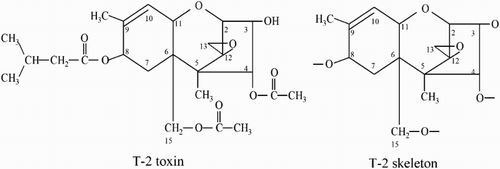
Type A trichothecenes have a four-ring structure and are characterized by the presence of either a hydrogen atom or a hydroxyl group (or the esters of the hydroxyl group) at the C8 position in the trichothecene nucleus (), while type B trichothecenes are characterized by a carbonyl group at the C8 position. They both have a reactive group at the C3 position.
T-2 toxin is the most common and also the most hazardous of the type A trichothecenes in food and feeds (Ueno, Citation1984). In the past few years, a high incidence of T-2 has been reported in raw oats from several northern European countries (Edwards, Citation2009; Pettersson et al., Citation2011). It is mutagenic and carcinogenic even at low concentrations. Skin irritation experiments in rats have shown that opening the epoxy ring in the skeletal structure of the T-2 toxin (de-epoxidation) can lower toxicity to 1/400 of that of the unaltered T-2 toxin (Zhou et al., Citation2010). This confirms that the epoxy group in type A trichothecenes is responsible for most, if not all of their toxicity. Therefore, identification of the specific skeleton structure of type A trichothecenes is key to minimizing their harm to human and animal health (Eriksen & Alexander, Citation1988; Ueno, Citation1983).
Techniques to detect type A trichothecene structures have included chromatographic technologies such as gas chromatography (GC) (Valle-Algarra, Mateo, Mateo, Gimeno-Adelantado, & Jimenez, Citation2011), gas chromatography-mass spectrometry (GC-MS) (Eskola & Rizzo, Citation2001), gas chromatography-triple quadrupole mass spectrometry (GC-MS/MS) (Jekel & Van Egmond, Citation2014; Nielsen & Thrane, Citation2000; Rodríguez-Carrasco, Berrada, Font, & Manes, Citation2012), liquid chromatography-mass spectrometry (LC-MS) (Dall’Astaa et al., Citation2004; Fuchs, Rabus, Handl, & Binder, Citation2003; Nakagawa, Sakamoto, Sago, Kushiro, & Nagashima, Citation2013; Razzazi-Fazeli, Rabus, Cecon, & Bohm, Citation2002) and liquid chromatography-tandem mass spectrometry (LC-MS/MS) (Tamura, Nakagawa, Uyama, & Mochizuki, Citation2014; Yang et al., Citation2013). Although these detection technologies characteristically have a high-flux and low detection limit, not all type A trichothecenes and their metabolites are recognized especially if the concentration of the parent compound is extremely low and the metabolites are unknown. Therefore an immunological detection technique such as enzyme-linked immunosorbent assay (ELISA), using the principle of specific binding of antigen to antibody, has been attempted by producing antibodies to the skeleton structure of type A trichothecenes to specifically identify type A trichothecenes and their metabolites. However, due to the lack of a complete antigen to type A trichothecene skeletons, the development of a high-throughput specific identification method has been hindered. Therefore, there is a need to build an antigen for the complete type A trichothecene skeleton, so that specific immunological monitoring techniques can be developed to detect even extremely low concentrations of type A trichothecenes and their metabolites.
Because T-2 toxin has a skeleton structure similar to those characteristic of type A trichothecenes, by replacing the groups at C4, 8 and 15 with H, T-2 can be converted to a type A trichothecene skeleton (). In a previous study, we used Fusarium poae to prepare T-2 toxin (Dai et al., Citation2011). By coupling a type A trichothecene skeleton to a protein, an antigen with immunogenicity can be synthesized. Wei and Chu (Citation1987) and Fan, Schubring, Wei, and Chu (Citation1988) developed a method to successfully synthesize 3-Ac-NEOS by subjecting T-2 toxin to enzymatic hydrolysis of pig liver extract, and confirmed that the resulting 3-Ac-NEOS-HS can be used as the hapten for the type A trichothecene skeleton structure, by coupling it to bovine serum albumin (BSA) to produce an immunogenic compound. So, 3-Ac-NEOS can be formed by modifying the C8 group of the T-2 toxin to expose the type A trichothecene skeleton. However, this methodology is complex and a wide variety of by-products are formed, making it difficult to separate and purify the hapten. Therefore, in this study, the preparation of the 3-Ac-NEOS hapten (3-Ac-NEOS-HS) was optimized by using the liquid from enzymatic hydrolysis of rat red blood cells (RBC), instead of pig liver extract, to activate the C8 group (Johnsen, Odden, Johnsen, & Fonnum, Citation1988). Several column chromatographic methods were used to separate and purify the hapten, which was confirmed by nuclear magnetic resonance (NMR) and LC-MS/MS methods. The hapten was then coupled with BSA and OVA proteins and subjected to UV-Vis spectrometry and sodium dodecyl sulphate-polyacrylamide gel electrophoresis (SDS-PAGE) to calculate the coupling ratios using Equation (2). By using the T-2 toxin in vitro to prepare an antigen of the complete type A trichothecene skeleton, a reliable immunogen and a coating antigen for polyclonal antibody preparation were developed. 3-Ac-NEOS-HS-BSA can be used to immunize animals and thereby prepare a complete polyclonal antibody. This lays the foundation for antibody preparation and ELISA kit development for all type A trichothecenes (parent + modified/masked type A trichothecenes).
2. Materials and methods
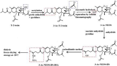
Because the C8 group in the skeleton of T-2 toxin is the recognition site of type A trichothecenes, in order to connect the macromolecular protein to the recognition site, the C8 group of the T-2 toxin was first hydroxylated and then replaced by an active side chain. Since competitive connections often occur between C3 and C8 side chains, we first acetylated the modified C3 hydroxyl groups so that a complete antigen to type A trichothecenes can be constructed in vitro. The synthetic pathway for preparing the complete antigen to type A trichothecenes is shown in the mother nucleus structure of the T-2 toxin in .
2.1. Acetylation of T-2 toxin
First, 10 mg of T-2 toxin (21259-20-1, purity > 98%, ENZO Co., New York, USA) was added to 1 mL pyrimidine and 2 mL acetic anhydride in a test tube, with oscillation reaction at room temperature (RT) for 2 h. Next, it was flushed with nitrogen (N2) gas for enrichment, and 3 mL alcohol was added to the concentrate followed by mixing and flushing with N2 at 70°C to concentration it further. This was followed by separation and purification of acetylated T-2 toxin concentrate using a silica gel column (1 × 19 cm) packed with silica gel G-60 (mesh 70-230, Merck AG, Darmstadt, Germany). The column was washed with 100 mL hexane followed by elution from the silica gel column with hexane: ethyl acetate (1:1). The effluent 3-Ac-T-2 was further concentrated, and crystalized by passing through N2 gas using a pressure blowing concentrator (DC-12, ANPEL, Shanghai, China), and stored at −20°C.
2.2. Detection of acetylated T-2 toxin (3-Ac-T-2) by mass spectrometry (MS)
A Hypersil GOLD analytical column (5 μm, 100 × 2.1 mm; Thermo Scientific Co., Waltham, USA) was used. The electrospray ionization (ESI+) mode was used in the MS settings. Detection conditions were as reported by Lu et al. (Citation2016) for the T-2 toxin.
2.3. Synthesis of type A trichothecene skeleton, 3-Ac-NEOS
2.3.1. Preparation of 3-Ac-NEOS
A solution of rat RBC enzyme was prepared as described by Johnsen et al. (Citation1988). 3-Ac-T-2 toxin was mixed with 1 mL of dimethyl sulfoxide and 1 mL of ethanol and then with 3 mL RBC enzyme solution. pH was adjusted to between 7.4 and 7.5, and the mixture emulsified in an incubator at 37°C for 2 h.
2.3.2. Purification of 3-Ac-NEOS
The emulsion from above was concentrated by flushing with N2 gas. The concentrate was separated by column chromatography (XAD-2, 1.9 × 19 cm, Sigma, San Francisco, USA), with macroporous resin in the column used as absorbent and acetone as the eluting agent. The ion exchange column was eluted with 150 mL acetone, and the eluent was concentrated (approximately 5 mL) in a vacuum rotary evaporator (Shimadzu Corporation, Kyoto, Japan). The concentrate was then transferred to a plastic centrifuge tube and flushed with N2 gas to a viscous concentrate, dissolved in distilled water and extracted with 20 mL trichloromethane four times. The concentrated extract liquor was purified by column chromatography (packed with 6 g Florisil; 1 × 14 cm) with 30 mL chloroform and equilibrated with 25 mL chloroform/methanol mixture at 99:1 (v/v) followed by 98:2 ratio, and finally by 97:3 ratio. The effluent (approximately 5 mL) was concentrated with N2 gas and crystallized.
2.3.3. Identification of 3-Ac-NEOS
The structure of the synthetic 3-Ac-NEOS was confirmed as matching that of the type A trichothecene skeleton by NMR spectroscopy (Varian Mercury AS400, Varian Co., Salt Lake City, USA) and MS analysis (TSQ Quantum Access, Thermo Scientific).
2.4. Synthesis of 3-Ac-NEOS-HS by succinic anhydride method
The C8 position in the synthetic 3-Ac-NEOS was connected to the hapten by a connecting arm using the succinic anhydride method. This was achieved by mixing 4 mg of 3-Ac-NEOS and 80 mg of succinic anhydride and dissolving in 2 mL of pyridine as described by Xu et al. (Citation2010) to build the connection arm – HS.
2.5. 3-Ac-NEOS-HS-BSA or OVA prepared by carbodiimide (EDC) method
2.5.1. Preparation of the immunogen 3-Ac-NEOS-HS-BSA
First, 3 mg of 3-Ac-NEOS-HS was dissolved in 0.2 mL dimethyl formamide (DMF) (A), and 6 mg BSA and 4 mg EDC in 25 mL of distilled water (B). After mixing solution A with B, 0.8 mg of EDC was added to the mixture and pH adjusted to 5.5. The solution was continuously agitated for 24 h at RT, and the mixture was dialysed for 5 d with 0.01 moL/L phosphate buffer saline, pH 7.4 at 4°C. After completion of dialysis, the solution was freeze-dried. The methodology for preparing the coating antigen 3-Ac-NEOS-HS-OVA was the same as for 3-Ac-NEOS-HS-BAS described above except that OVA was used instead of BSA.
2.5.2. Detection of complete antigen
3-Ac-NEOS, 3-Ac-NEOS-HS, BSA, OVA and the complete antigen of the T-2 toxin skeleton were identified by wavelength scans and SDS-PAGE after appropriate dilution. Comparing and contrasting the locations of specific absorption peaks and the electrophoretic bands were used to determine whether the coupling was successful.
2.5.3. Calculation of coupling ratio
Ultraviolet spectroscopic scans were used to determine the maximal absorbance wavelength, and the molar absorption coefficient was measured according to the following equation:(1)
where ε is the molar absorption coefficient [L/(moL, cm)]; A is the sample absorbance; C is the sample concentration (moL/L) and l is the thickness of the cuvette (1 cm).
The conjugation ratio of 3-Ac-NEOS to BSA and OVA can be calculated from the following equation (Xu et al., Citation2010):(2)
2.6. Immunization of animals
New Zealand rabbits were immunized with 3-Ac-NEOS-HS-BSA conjugates synthesized by the carbodiimide method. The rabbits were injected hypodermically at a dose of 400 μg conjugate with Freund’s complete adjuvant in the first instance and at a dose of 200 μg conjugate with Freund’s incomplete adjuvant three other times at a 2-week interval. Serum samples were collected from the marginal ear blood vessels on day 7 after the fourth immunization, and antibody titres determined by indirect ELISA (Cai et al., Citation2014) using 3-Ac-NEOS-HS-BSA as the coating antigen.
3. Results
3.1. Acetylation of T-2 toxin
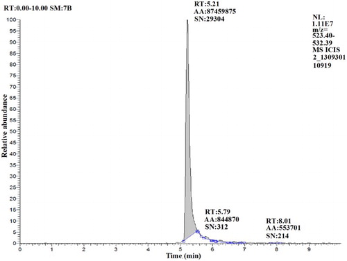
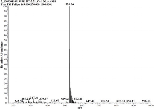
After acetylation of the T-2 toxin, high-resolution MS showed the resultant synthetic product to have a mass spectrum typical of 3-Ac-T-2. Exact mass results as measured by Fourier Transform Mass Spectrometry (FTMS) are as follows. The retention time of the 3-Ac-T-2 toxin was 5.21 min (). For 3-Ac-T-2, m/z = 508, calculated for [M+18]+ 526.66 ().
3.2. Identification of type A trichothecene skeleton, 3-Ac-NEOS
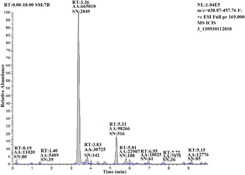
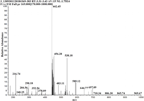
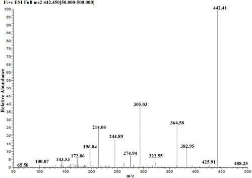
The retention time for 3-Ac-NEOS was 3.36 min as detected by MS (). shows the strongest spectral peak at 442.45 [M+18]+, according to the theoretical molecular weight (MW) of 3-Ac-NEOS (MW = 424). In the secondary mass spectrogram (), characteristic product ion peaks were observed, and structural information was obtained from these product ions.
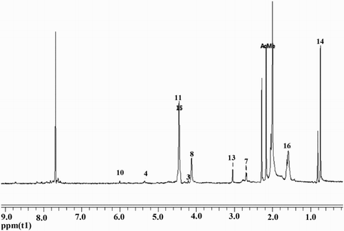
From 1H-NMR (), three strong peaks at 2.024, 2.182 and 2.246 were detected. The target 3-Ac-NEOS’s unique three acetyl characteristic peaks are shown as C3, C4 and C1. These three strong peaks are the three acetyls of the target product’s (3-Ac-NEOS) characteristic peak and the 4.122 peak is the C8 hydroxyl-generated peak. These are consistent with earlier reports (Wei & Chu, Citation1987).
These results confirmed successful synthesis of the target skeleton 3-Ac-NEOS.
3.3. Synthesis of 3-Ac-NEOS-HS
UV absorption peaks of 3-Ac-NEOS and 3-Ac-NEOS-HS were at 194 and 196 nm, respectively. This meant that the connecting arm was successfully introduced into the 3-Ac-NEOS skeleton to synthesize the hapten 3-Ac-NEOS-HS by the succinic anhydride method.
3.4. Synthesis of 3-Ac-NEOS-HS-BSA or OVA by the carbodiimide (EDC) method
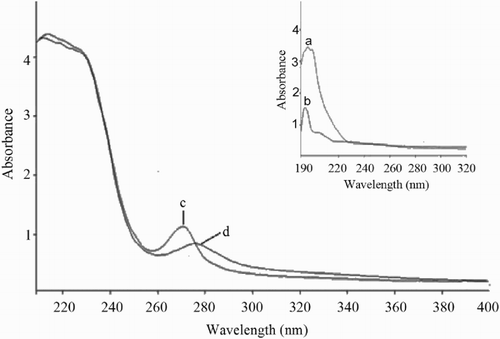
The UV absorption spectra of 3-Ac-NEOS, 3-Ac-NEOS-HS and BSA are shown in with corresponding peaks at 194, 196 and 273 nm. On BSA coupling to 3-Ac-NEOS-HS, the UV peak shifted to 277 nm. This illustrates that the hapten 3-Ac-NEOS-HS was connected to the carrier protein and that the artificial antigen was successfully synthesized. Thus the maximum absorption peaks of 3-Ac-NEOS-HS-BSA and BSA are at wavelengths 277 and 273 nm, respectively. Based on Equation (2), the 3-Ac-NEOS: BSA coupling ratio was determined as 8.76:1.
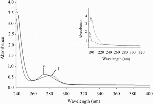
The UV absorption spectra of 3-Ac-NEOS, 3-Ac-NEOS-HS and OVA peak at 194, 196 and 274 nm are shown in . The maximum absorption peak of 3-Ac-NEOS-HS-OVA at 282 nm is offset to the right. This indicates that the hapten 3-Ac-NEOS-HS was connected to the carrier protein and that the artificial antigen was successfully synthesized. Based on Equation (2), the 3-Ac-NEOS and OVA coupling ratio was determined as 7.24:1.
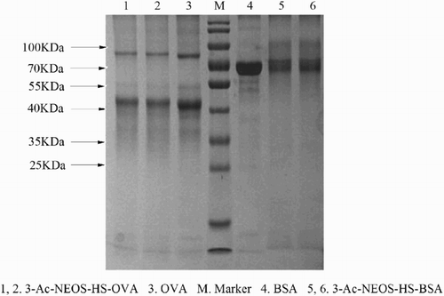
The coupling of 3-Ac-NEOS-HS was shown by protein analysis using SDS-PAGE (). The protein band of 3-Ac-NEOS-HS-BSA is slightly higher than that for BSA (MW = 66.446 KDa). This confirms that coupling of 3-Ac-NEOS-HS-BSA was successful. Similarly, the 3-Ac-NEOS-HS-OVA band was slightly higher than the MW of OVA, showing further evidence for successful coupling of 3-Ac-NEOS-HS-OVA.
3.5. Antibody titre with immunization of animals
The results of indirect ELISA showed that immunization of rabbits with 3-Ac-NEOS-HS-BSA could elicit significant immune response. The antibody titre induced by 3-Ac-NEOS-HS-BSA was 1:64,000 (by the carbodiimide (EDC) method).
4. Discussion
Immunochemical methods are now commonly used to detect fungal toxins in food and feed. Examples include those used to detect deoxynivalenol (DON) and T-2/HT-2 in grains and grain products (Baumgartner, Führer, & Krska, Citation2010), zearalenone (ZEA), T-2/HT-2, DON, and fumonisins (FB) in grain (Lattanzio, Von Holst, & Visconti, Citation2013), and ELISA to detect DON, nivalenol (NIV), and T-2/HT-2 in wheat kernels (Lattanzio et al., Citation2013). Peters, Bienenmann-Ploum, De Rijk, and Haasnoot (Citation2011) used the multiplex cytometric microsphere immunoassay to detect several mycotoxins including DON, T-2 toxin, FB and ZEA simultaneously. Wang et al. (Citation2012) used the immune chip effectively to quantify DON, ochratoxins (OTA), T-2 toxin and ZEA. Methods that can simultaneously detect all type A trichothecenes are rare. Wei and Chu (Citation1987), Fan et al. (Citation1988) and Lee, Wei, and Chu (Citation1989) reported immunochemical methods to detect type A trichothecenes, but none of these methods can detect all type A trichothecenes. Our research has extended these findings by further optimizing the preparation of an antigen to type A trichothecene skeletons. T-2 toxin was first acetylated, and enzymolysis liquid of rat RBC was used to produce 3-AC-NEOS. In our study, the 3-AC-NEOS synthesis was a two-step process instead of the three steps used by others (Fan et al., Citation1988; Wei & Chu, Citation1987). The process of preparing T-2 toxin antigen is by linking T-2 toxin to succinic anhydride and then coupling it to protein BAS, N-hydroxysuccinimide (NHS), keyhole limpet haemocyanin (KLH), OVA, histidine-rich protein (HRP) and poly lysine (PLL) (Wang, Duan, Zhang, & Wang, Citation2010). In our study 3-AC-NEOS was linked only to BAS and OVA after connecting with succinic anhydride. This way, both the number of processes and the preparation times were reduced. According to Chu, Grossman, Wei, and Mirocha (Citation1979), succinic anhydride was used to link T-2 toxin with carrier protein to produce the hapten. In our study, succinic anhydride was used as the link arm to couple 3-AC-NEOS with carrier proteins (BAS and OVA). Succinic anhydride, with fewer carbon atoms, can reveal the hapten better. The products were identified by LC-MS/MS and NMR.
T-2 toxin belongs to the type A trichothecenes class of compounds, which have a common characteristic skeleton structure, 3-Ac-NEOS; because 3-Ac-NEOS is a small molecule, it does not possess immunogenicity and therefore cannot act like a natural antigen that can be used directly in animal immunity studies to stimulate and elicit specific antibodies (Song, Wang, Wang, Tang, & Deng, Citation2010). Therefore, the 3-Ac-NEOS hapten was coupled with a protein to produce a complete antigen. The antigen was prepared using EDC and succinic anhydride methods. The specificity of the antibody and antigen depends not only on the structure and properties of the hapten, but also on the number of haptens that connect with carrier protein. Therefore, it was essential to calculate the number of antigen couplings with a protein carrier that would allow calculation of a coupling ratio. In general, to achieve immunogenicity in an antigen, a good coupling ratio (3–45:1) is required. A high titre of the antibody would appear only if a coupling ratio (of antigen to protein carrier) of 8–25:1 could be achieved (Erlanger, Citation1980). The experimentally prepared artificial antigens 3-Ac-NEOS-HS-BSA and 3-Ac-NEOS-HS-OVA with coupling ratios of 8.76:1 and 7.24:1, respectively, met the requirement of an immunogen that could produce high-titre antibodies. The coupling ratios showed that 3-Ac-NEOS-HS-BSA was better. When 3-Ac-NEOS-HS-BSA was used to immunize rabbits, the antibody titre was 1:64,000. This confirmed that the synthesis of a type A trichothecenes complete antigen was successful.
Many mycotoxins and or their metabolites may bind to macromolecules and thereby alter their solubility and other characteristics, making them difficult to detect using conventional methods. These mycotoxins are now referred to as masked/modified mycotoxins (Berthiller et al., Citation2013). Masked/modified mycotoxins have been identified only in recent years and some are as toxic as the parent mycotoxin (Berthiller et al., Citation2013). Therefore, it is important to know the total concentrations of mycotoxins and their metabolites in food/feed, to be certain that total toxicity does not exceed safe limits.
Published immunological detection methods have reported higher mycotoxin concentrations in samples than have been reported using classical traditional techniques such as GC and LC, which usually measure only the parent compound. This is probably because the crosslinking rate is higher with immunological techniques. Zachariasova et al. (Citation2008) analysed 20 beer samples from the European market and showed that apparent DON levels obtained by all immunoassays were significantly higher than the LC-MS/MS values. This may be because of the unknown metabolites (including masked/modified DON) that exist in the samples. There are reports that DON can conjugate with sugars such as glucose (Berthiller, Schuhmacher, Adam, & Krska, Citation2009; Zachariasova et al., Citation2008) or with sulphate (Warth et al., Citation2015). Dorokhin, Haasnoot, Franssen, Zuilhof, and Nielen (Citation2011) found that DON antibodies and masked DON can lead to specific interactions, with coupling rates of 71%, 66% and 36% for 3-ADON, 15-ADON and DON-3G, respectively. There are a variety of masked/modified forms of a mycotoxin and it is difficult to detect all these forms individually. Although LC and MS detection methods give high sensitivity, they do not detect all the different mycotoxin metabolites and therefore the values obtained from these techniques are an underestimation. In addition, these instruments are expensive and require skilled professional operators. Immunological techniques provide a new approach to detecting mycotoxins of similar structure, including masked/modified mycotoxins; the only requirement is that an antigen with a common skeleton structure is required to be synthesized as reported in this study. The new technologies (including immunochemical, MS and associated chromatographic techniques) are evolving as a new direction for analysis of various masked/modified mycotoxins. A combination of these technologies will in future evolve and provide even more possibilities and a greater accuracy for detection of total mycotoxins (including masked/modified mycotoxins) in food/feed and this would enable more accurate risk assessment.
5. Conclusion
In our study, firstly, we acetylated the modified C3 hydroxyl groups of T-2 toxin to synthesize acetylated T-2 toxin, using the MS method to detect of 3-Ac-T-2. Next, by enzymatic hydrolysis, the type A trichothecene skeleton 3-Ac-NEOS was obtained. Synthesis of 3-Ac-NEOS-HS was achieved by the succinic anhydride method after the type A trichothecenes complete antigen was prepared by conjugation of hapten with carrier BSA or OVA (by the carbodiimide method) and identified by UV spectroscopy and SDS-PAGE. The 3-Ac-NEOS:BSA coupling ratio was determined as 8.76:1. The high antibody titre when injected into rabbits confirmed the successful synthesis of a complete antigen, which could be used as the foundation for antibody synthesis and ELISA kit development for all type A trichothecenes (parent + modified/masked type A trichothecenes).
Acknowledgement
This research was conducted at the Guangdong provincial key laboratory of aquatic product processing and safety.
Disclosure statement
No potential conflict of interest was reported by authors.
Notes on contributors
Pengli Lu was studied at the quality and safety of aquatic products, Guangdong Ocean University, Zhanjiang.
Yaling Wang was studied at the quality and safety of aquatic products, Guangdong Ocean University, Zhanjiang.
Yapei Wang was studied at the quality and safety of aquatic products, Guangdong Ocean University, Zhanjiang.
Chaojin Wu was studied at the quality and safety of aquatic products, Guangdong Ocean University, Zhanjiang.
Lijun Sun was studied at the quality and safety of aquatic products, Guangdong Ocean University, Zhanjiang.
Defeng Xu was studied at the quality and safety of aquatic products, Guangdong Ocean University, Zhanjiang.
Xiaodong Sun was studied at the environment and resources of Dalian Nationalities University.
Jianrong Li, professor, mainly studied in aquatic products and fruits and vegetables storage processing and the quality and safety control research in Bohai University.
Ravi Gooneratne, professor, mainly studied at the centre for food research and innovation of Lincoln University.
Additional information
Funding
References
- Baumgartner, S., Führer, M., & Krska, R. (2010). Comparison of monoclonal antibody performance characteristics for the detection of two representatives of A- and B-trichothecenes: T-2 toxin and deoxynivalenol. World Mycotoxin Journal, 3, 233–238. doi:10.3920/WMJ2010.1224
- Berthiller, F., Crews, C., Dall’Asta, C., Saeger, S. D., Haesaert, G., Karlovsky, P., … Stroka, J. (2013). Masked mycotoxins: A review. Molecular Nutrition and Food Research, 57, 165–186. doi:10.1002/mnfr.201100764
- Berthiller, F., Schuhmacher, R., Adam, G., & Krska, R. (2009). Formation, determination and significance of masked and other conjugated mycotoxins. Analytical and Bioanalytical Chemistry, 395, 1243–1252. doi:10.1007/s00216-009-2874-x
- Cai, Q. C., Hou, Y. Z., Deng, R. G., Hu, X. F., Wang, Y., Zhang, X. F., & Wang, F. Y. (2014). Aflatoxin M1 preparation and identification of artificial antigens. Chinese Journal of Immunology, 30, 789–793. doi:10.3969/j.issn.1000-484X.2014.06.015
- Chu, F. S., Grossman, S., Wei, R. D., & Mirocha, C. J. (1979). Production of antibody against T-2 toxin. Applied and Environmental Microbiology, 37, 104–108. Article ID: 0099-2240/79/02-0104/05$02.00/0
- Dai, Z., Wang, Y. L., Sun, L. J., Shi, Q., Xu, D. F., Wang, Y. Z., … Lu, G. Z. (2011). Cultivation condition and extraction method to prepare T-2 toxin using Fusarium poae. Journal of Microbiology, 31(5), 40–44.
- Dall’Astaa, C., Galavernaa, G., Biancardib, A., Gasparinib, M., Sforzaa, S., Dossenaa, A., & Marchellia, R. (2004). Simultaneous liquid chromatography-fluorescence analysis of type A and type B trichothecenes as fluorescent derivatives via reaction with coumarin-3-carbonyl chloride. Journal of Chromatography A, 1047, 241–247. doi:10.1016/j.chroma.2004.07.002
- Dorokhin, D., Haasnoot, W., Franssen, M. C. R., Zuilhof, H., & Nielen, M. W. F. (2011). Imaging surface plasmon resonance for multiplex microassay sensing of mycotoxins. Analytical and Bioanalytical Chemistry, 400, 3005–3011. doi:10.1007/s00216-011-4973-8
- Edwards, S. G. (2009). Fusarium mycotoxin content of UK organic and conventional oats. Food Additives and Contaminants Part A, 26, 1063–1069. doi:10.1080/02652030902788953
- Eriksen, G. S., & Alexander, J. (Eds.). (1988). Fusarium toxins in cereals: A risk assessment. Copenhagen: Nordic council of ministers. TemaNord, 502, 7–27, 45–58.
- Erlanger, B. F. (1980). The preparation of antigenic hapten-carrier conjugates: A survey. Methods in Enzymology, 70(A), 70–74. doi:10.1016/S0076-6879(80)70043-5
- Eskola, M., & Rizzo, A. (2001). Sources of variation in the analysis of trichothecenes in cereals by gas chromatography-mass spectrometry. Mycotoxin Research, 17, 68–87. doi:10.1007/BF02946130
- Fan, T. S., Schubring, S. L., Wei, R. D., & Chu, F. S. (1988). Production and characterization of a monoclonal antibody cross-reactive with most group A trichothecenes. Applied and Environmental Microbiology, 54, 2959–2963. Retrieved from http://aem.asm.org/content/54/12/2959
- Fuchs, E., Rabus, B., Handl, J., & Binder, E. M. (2003). Type A-trichothecenes - quantitative analysis using LC-MS and occurrence in Austrian maize and oats. Mycotoxin Research, 19, 56–59. doi:10.1007/BF02940094
- Jekel, A. A., & Van Egmond, H. P. (2014). Determination of T-2/HT-2 toxins in duplicate diets in the Netherlands by GC-MS/MS: Method development and estimation of human exposure. World Journal, 7, 267–276. doi:10.3920/WMJ2014.1697
- Johnsen, H., Odden, E., Johnsen, B. A., & Fonnum, F. (1988). Metabolism of T-2 toxin by blood cell carboxylesterases. Biochemical Pharmacology, 37, 3193–3197. doi:10.1016/0006-2952(88)90320-6
- Lattanzio, V. M. T., Von Holst, C., & Visconti, A. (2013). The case of a multiplex dipstick for Fusarium mycotoxins in cereals. Analytical Bioanalytical Chemistry, 405, 7773–7782. doi:10.1007/s00216-013-6922-1
- Lee, R. C., Wei, R. D., & Chu, F. S. (1989). Enzyme linked immunosorbent assay for T-2 toxin metabolites in urine. Journal of the Association of Official Analytical Chemists, 72, 345–348.
- Lu, P. L., Wu, C. J., Shi, Q., Wang, Y. L., Sun, L. J., Liao, J. M., … Gooneratne, R. (2016). A sensitive and validated immunomagnetic-bead-based enzyme-linked immunosorbent assay for analyzing total T-2 (free and modified) toxins in shrimp tissues. Food Analytical Methods. Advance online publication. doi:10.1007/s12161-015-0336-y
- Mylona, K., & Magan, N. (2011). Fusarium langsethiae: Storage environment influences dry matter losses and T2 and HT-2 toxin contamination of oats. Journal of Stored Products Research, 47, 321–327. doi:10.1016/j.jspr.2011.05.002
- Nakagawa, H., Sakamoto, S., Sago, Y., Kushiro, M., & Nagashima, H. (2013). Detection of masked mycotoxins derived from type A trichothecenes in corn by high-resolution LC-Orbitrap mass spectrometer. Food Additives and Contaminants A, 30, 1407–1414. doi:10.1080/19440049.2013.790087
- Nielsen, K. F., & Thrane, U. (2000). Fast methods for screening of trichothecenes in fungal cultures using GC-MS/MS. Mycotoxin Research, 16, 252–256. doi:10.1007/BF02940051
- Peters, J., Bienenmann-Ploum, M., De Rijk, T., & Haasnoot, W. (2011). Development of a multiplex flow cytometric microsphere immunoassay for mycotoxins and evaluation of its application in feed. Mycotoxin Research, 27, 63–72. doi:10.1007/s12550-010-0077-0
- Pettersson, H., Brown, C., Hauk, J., Hoth, S., Meyer, J., & Wessels, D. (2011). Survey of T-2 and HT-2 toxins by LC–MS/MS in oats and oat products from European oat mills in 2005–2009. Food Additives and Contaminants Part B, 4, 110–115. doi:10.1080/19393210.2011.561933
- Razzazi-Fazeli, E., Rabus, B., Cecon, B., & Bohm, J. (2002). Simultaneous quantification of A-trichothecenes mycotoxins in grains using liquid chromatography-atmospheric pressure chemical ionisation mass spectrometry. Journal of Chromatography A, 968, 129–142. doi:10.1016/S0021-9673(02)00957-3
- Rodríguez-Carrasco, Y., Berrada, H., Font, G., & Manes, J. (2012). Multi-mycotoxins analysis in wheat semolina using an acetonitrile-based extraction procedure and gas chromatography-tandem mass spectrometry. Journal of Chromatography A, 1270, 28–40. doi:10.1016/j.chroma.2012.10.061
- Sklan, D., Shelly, M., Makovsky, B., Geyra, A., Klipper, E., & Friedman, A. (2003). The effect of chronic feeding of diacetoxyscirpenol and T-2 toxin on performance, health, small intestinal physiology and antibody production in turkey poults. British Poultry Science, 44, 46–52. doi:10.1080/0007166031000085373
- Sokolović, M., Garaj-Vrhovac, V., & Šimpraga, B. (2008). T-2 toxin: Incidence and toxicity in poultry. Archives of Industrial Hygiene and Toxicology, 59, 43–52. doi:10.2478/10004-1254-59-2008-1843
- Song, J., Wang, R. M., Wang, Y. Q., Tang, Y. R., & Deng, A. P. (2010). Hapten design, modification and preparation of artificial antigens. Chinese Journal of Analytical Chemistry, 38, 1211–1218. doi:10.1016/S1872-2040(09)60063-3
- Tamura, M., Nakagawa, H., Uyama, A., & Mochizuki, N. (2014). Simultaneous trichothecenes analysis by LC-MS/MS with a pentafluorophenyl column. Food Hygiene and Safety Science, 55, 19–24. doi: 10.3358/shokueishi.55.19
- Ueno, Y. (1977). Trichothecenes: Overview address. In J. V. Rodricks, C. W. Hesseltine, & M. A. Mehlman (Eds.), Mycotoxins in human and animal health (pp. 189–207). Park Forest South, IL: Phathotox.
- Ueno, Y. (1983). General toxicology. In Y. Ueno (Ed.), Trichothecenes – Chemical, biological and toxicological aspects (pp. 135–146). Tokyo: Kodansha Ltd./Amsterdam: Elsevier Science.
- Ueno, Y. (1984). Toxicological features of T-2 toxin and related trichothecenes. Fundamental and Applied Toxicology, 4, 124–132. doi:10.1016/0272-0590(84)90144-1
- Valle-Algarra, F. M., Mateo, E. M., Mateo, R., Gimeno-Adelantado, J. V., & Jimenez, M. (2011). Determination of type A and type B trichothecenes in paprika and chili pepper using LC-triple quadrupole-MS and GC-ECD. Talanta, 84, 1112–1117. doi:10.1016/j.talanta.2011.03.017
- Wang, J. P., Duan, S. X., Zhang, Y., & Wang, S. (2010). Enzyme-linked immunosorbent assay for the determination of T-2 toxin in cereals and feedstuff. Microchimica Acta, 169, 137–144. doi:10.1007/s00604-010-0318-0
- Wang, Y., Liu, N., Ning, B. A., Liu, M., Lu, Z. Q., Sun, Z. Y., … Gao, Z. X. (2012). Simultaneous and rapid detection of six different mycotoxins using an immunochip. Biosensors and Bioelectronics, 34, 44–50. doi:10.1016/j.bios.2011.12.057
- Warth, B., Fruhmann, P., Wiesenberger, G., Kluger, B., Sarkanj, B., Lemmens, M., … Schuhmacher, R. (2015). Deoxynivalenol-sulfates: Identification and quantification of novel conjugated (masked) mycotoxins in wheat. Analytical and Bioanalytical Chemistry, 407, 1033–1039. doi:10.1007/s00216-014-8340-4
- Wei, R. D., & Chu, F. S. (1987). Production and characterization of a generic antibody against group A trichothecenes. Analytical Biochemistry, 160, 399–408. doi:10.1016/0003-2697(87)90067-4
- Xu, J., Hong, S. J., Fu, J. Y., Cao, J., He, J., Yu, Z. N., & Zhang, J. B. (2010). Preparation of T-2 toxin antigen. Chemistry and Bioengineering, 27, 66–68. (in Chinese)
- Yang, L. C., Zhao, Z. Y., Wu, A. B., Deng, Y. F., Zhou, Z. L., Zhang, J. P., & Hou, J. F. (2013). Determination of trichothecenes A (T-2 toxin, HT-2 toxin, and diacetoxyscirpenol) in the tissues of broilers using liquid chromatography coupled to tandem mass spectrometry. Journal of Chromatography B, 942–943, 88–97. doi:10.1016/j.jchromb.2013.10.034
- Zachariasova, M., Hajslova, J., Kostelanska, M., Poustka, J., Krplova, A., Cuhra, P., & Hochel, I. (2008). Deoxynivalenol and its conjugates in beer: A critical assessment of data obtained by enzyme-linked immunosorbent assay and liquid chromatography coupled to tandem mass spectrometry. Analytica Chimica Acta, 625, 77–86. doi:10.1016/j.aca.2008.07.014
- Zhou, Z. Y., He, Z. F., Li, H. J., Zou, C. H., Han, P. F., & Yang, J. Y. (2010). Research progress in transformation and degradation of trichothecenes. Food Science, 31, 443–448. Article ID: 1002-6630(2010)19-0443-06

