
 ?Mathematical formulae have been encoded as MathML and are displayed in this HTML version using MathJax in order to improve their display. Uncheck the box to turn MathJax off. This feature requires Javascript. Click on a formula to zoom.
ABSTRACT
An indirect competitive enzyme-linked immunosorbent assay (ic-ELISA) based on immunomagnetic beads' (IMBs) clean-up was developed for detection of the residues of maduramicin (MD) in different chicken tissues. IMBs coated with a specific monoclonal antibody (MAb) against MD named MAb 2D6 were applied to enrich the residues of MD in chicken tissues. The specificities of these IMBs were well maintained and the reversibility remained at more than 73% of the original capability after being used for three times. After elution, enriched MD was detected by a conventional ic-ELISA. The limits of detection of MD were 72, 74 and 173 μg/kg in chicken muscle, skin and fat, and liver, respectively. Recoveries ranged from 80.0% to 115.8% with coefficients of variation being less than 11.3%. These results indicated that a rapid, robust clean-up of IMBs combining ELISA provides a simple, time-saving and environmentally friendly method to detect MD in chicken tissues.
Introduction
Polyether antibiotics are widely used as feed additives for the prevention of coccidiosis in chickens (Stanker, Elissalde, Rowe, Beier, & El-Aziz Nasr, Citation1994; Tian et al., Citation2017). Maduramicin (MD), a polyether ionophore antibiotic, is extensively used to prevent coccidiosis in poultry due to its board anticoccidial activity. Furthermore, MD has been widely used as a growth promoter to improve feed conversion rate in ruminants (Le-Minh, Khan, Drewes, & Stuetz, Citation2010). However, the extensive or illegal use of MD can lead to intoxication of animals and residues in chicken tissues (Wang, Zhang, Murtazina, Eremin, & Shen, Citation2008), which constitutes a potential hazard for both human and environment’s health (Sharma, Bhalla, Varma, Jain, & Singh, Citation2005). For example, MD could increase coronary blood flow and induce coronary dilation (Pereira et al., Citation2016), and made the situation even worse on the patients with coronary-artery-related diseases (Elliott, Kennedy, & McCaughey, Citation1998). To minimize its risk, many countries, such as the US, Japan and China, have set the maximum residue limits (MRLs) of MD in various tissues (The Ministry of Agriculture published Announcement Citation235, China; Tolerances for residues of new animal drugs in food, United States Food and Drug Administration; Positive list system for agricultural chemical residues – provisional MRLs list, Japan) (Table S1). For instance, the MRLs are 240 μg/kg for muscles, 480 μg/kg for skin and fat, and 720 μg/kg for liver in China (The Ministry of Agriculture published Announcement Citation235, China).
Numerous analytical methods have been developed for detecting the residues of MD in chickens. Most of them are based on instrumental analysis such as high-performance liquid chromatography (HPLC) with either UV or fluorescent detector and liquid chromatography-electrospray ionization tandem mass spectrometry (LC-MS/MS) (De Jong et al., Citation2004). Due to the unique structure of MD and hydrophobicity, sophisticated pre-treatment and derivative protocols are needed prior to instrumental analysis to decrease matrix effect (Wang et al., Citation2008). Additionally, these instrumental methods are time-consuming, costly and professional, which are not suitable for screening a large number of samples though they are sensitive and accurate (Ha, Song, Ai, & Li, Citation2016). Furthermore, though extraction and purification of MD by immunoaffinity chromatography and solid-phase extraction columns are facilitated to selectively adsorb residual components, they are disposable and expensive. It is necessary to develop a rapid, highly efficient and environmentally friendly method to detect MD in chicken tissues.
Magnetic bead (MB) is a versatile material with great biocompatibility, high specific surface area, robust magnetism and good dispersibility (Holschuh & Schwämmle, Citation2005; Wang et al., Citation2014). It has been widely used for cell separation (Šafařík & Šafaříková, Citation1999), protein or gene analysis (Liao et al., Citation2014; Zhu, Zhou, & Xing, Citation2012) and pathogen detection (Diler, Obst, Schmitz, & Schwartz, Citation2011). MBs with a large surface area have good biocompatibility and fast reactability to couple with antibodies or small molecules (Zhang et al., Citation2017). Immunomagnetic beads (IMBs), which were MBs that functionalized with antibodies on its surface, enable high capability and fast reaction to capture targets of interest to enhance its sensitivity (Teste et al., Citation2010). In addition, IMBs can be easily separated through magnetic separation and remain homogeneously distributed after removal of the magnet (Wang et al., Citation2014; Wei et al., Citation2012). However, few studies based on IMBs were published to analyse the residues of veterinary drugs in food animals (Vanoosthuyze, Van Peteghem, Courtheyn, & Vercammen, Citation1994).
To the best of our knowledge, only few reports focused on the detection of MD using immunoassay methods (Chen, Tang, Ding, He, & Xiao, Citation2009; Guo, Xu, Song, Liu, & Kuang, Citation2017; Kennedy, Blanchflower, & O’Dornan, Citation1997; Shen, Qian, Jiang, & Yang, Citation2001; Wang et al., Citation2008). Enzyme-linked immunosorbent assay (ELISA) is low cost, high-throughput and ready-to-use, which have been widely used for screening large samples for food safety (Liu et al., Citation2014; Zhu, Song, Liu, Kuang, & Xu, Citation2016). In the current study, an IMB-ELISA was developed for the detection of MD residues in chicken tissues based on our specific anti-MD monoclonal antibodies (MAbs), as shown in Scheme 1.
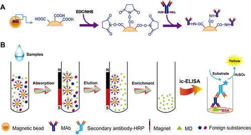
Scheme 1. The#protocol of preparation of IMBs and clean-up based on IMBs.
Note: The production of IMBs (A) and the protocol of IMBs-based ic-ELISA (B).
Materials and methods
Chemicals and materials
Maduramicin ammonium, salinomycin sodium, monensin sodium, lasalocid sodium and narasin were purchased from Dr Ehrenstorfer GmbH (Augsburg, Germany). Bovine serum albumin (BSA), ovalbumin and tetramethylbenzidine (TMB) were obtained from Sigma-Aldrich (St. Louis, MO, USA). Peroxidase-conjugated goat anti-mouse IgG was purchased from Jackson ImmunoResearch Laboratories, Inc. (West Grove, PA, USA). 1-ethyl-3-(3-dimethylaminopropy) carbodiimide (EDC), N-hydroxysuccinimide (NHS) and morpholineethanesulfonic acid (MES) were purchased from Aladdin Chemistry Co. Ltd (Shanghai, China). Other chemicals were supplied by Beijing Reagent Corporation (Beijing, P.R. China). Ninety-six-well microplates were purchased from Corning Life Sciences (New York, USA). Carboxylic-acid-functionalized Dynabeads® magnetic beads (10 mg beads/mL) were acquired from Invitrogen (Carlsbad, CA, USA). Water was obtained from Milli-Q synthesis water purification system (Bedford, MA, USA).
Production of MAbs
The MAbs used in the study were produced as mentioned below. Briefly, eight female BALB/c mice (8 weeks old) were immunized with the MD-BSA antigen prepared by the active ester coupling method, as previously described (Chen et al., Citation2009). After immunization for five times, the titres of antibody in mice were detected by direct competitive ELISA (dc-ELISA) and indirect competitive ELISA (ic-ELISA). The mice exhibiting antibodies with high titre were sacrificed for cell fusion with Sp2/0 myeloma cells. Positive hybridomas were cloned three times and used for MAb production. MAbs were purified from ascites using the saturated ammonium sulphate precipitation method and was stored at −20°C before further use.
Preparation of IMBs
IMBs were synthesized by coupling carboxyl acid groups on the surface of MBs and the amino groups of MAbs (Song et al., Citation2014). 1.5 mg MBs were washed with MES buffer (500 μL, 0.05 mol/L, pH 6.0) for three times, discarding the supernatant through magnetic separation. Then MBs were incubated with 1.25 mg EDC and 1.25 mg NHS in 1000 μL 0.05 mol/L MES buffer (pH 6.0) at room temperature for 30 min. Subsequently, activated MBs were washed with 0.01 mol/L PBS (pH 7.4) containing 0.05% Tween 20 for three times and collected by the external magnetic fields. Activated MBs and 80 μg purified MAbs were mixed in 2000 μL of 0.05 mol/L MES (pH 6.0) at room temperature for 4 h. Later, synthesized IMBs were washed with 0.01 mol/L PBS (pH 7.4) containing 0.05% Tween 20 for three times and 0.01 mol/L PBS (pH 7.4) containing 2% BSA was added to block the free sites on the surface of MBs at room temperature for 30 min. Lately, IMBs were stored in 0.01 mol/L PBS (pH 7.4) containing 1% BSA with the final volume of 1 mL at 4°C for further use.
Clean-up based on IMBs
IMBs were mixed with 500 μL of sample extracts or MD serial standard solution of different concentrations for 10 min with shaking, discarding the supernatant through magnetic separation. After washing, 250 μL of methanol/PBS (60:40, v:v) was added to elute the antigens at room temperature for 5 min. Then the supernatant contained MD was collected after magnet separation and 1–10 diluted with 0.01 mol/L PBS (pH 7.4) before detected by ic-ELISA. IMBs were washed with 0.01 mol/L PBS (pH 7.4) containing 0.05% Tween 20 for four times and re-dispersed in 0.01 mol/L PBS (pH 7.4) containing 1% BSA with the final volume of 1 mL at 4°C for further analysis.
ELISA analysis
For dc-ELISA, 96-well microplates were coated with 100 μL/well of coating antigen in 0.05 mol/L carbonate–bicarbonate buffer (pH 9.6) at 37°C for 2 h and then blocked with 200 μL of 2% casein at 37°C for 2 h. Then 100 μL of the MAbs were added for 30 min at 37°C. Later, 100 μL/well Horseradish Peroxidase (HRP) labelled goat anti-mouse IgG solution (1:5000) was added to incubate at 37°C for another 30 min. After washing with 0.01 mol/L PBS (pH 7.4) containing 0.05% Tween 20, 100 μL/well of TMB substrate solution was added and incubated at 37°C for 15 min. Lastly, 2 mol/L H2SO4 was added to stop the reaction and the optical density was measured at 450 and 630 nm by a Thermo Scientific Microplate Reader MK3. The same procedures were used for ic-ELISA measurement, except that 50 μL/well of the standard solution or sample extracts and 50 μL/well of MAbs were simultaneously added and incubated at 37°C for 30 min.
Derivatives of coccidiostat, including salinomycin sodium, monensin sodium, lasalocid sodium and narasin, were tested for the specificity of the method. The cross-reactivity (CR) was calculated using the following formula (Wang et al., Citation2014):
Sample pre-treatment
MD-free samples obtained from local supermarkets were confirmed by ultra performance liquid chromatography-tandem mass spectrometery (UPLC-MS/MS). 1.0 g of chicken tissues (muscle, skin and fat, or liver) and 1 mL methanol (2 ml methanol for liver) were added into a centrifuge tube to be vortexed for 1 min and shaken for 10 min. The mixture was centrifuged at 4000 rpm for 10 min. Subsequently, 1–10 dilution of the supernatants for the tissues of muscle, skin and fat and 1–15 dilution of liver with 0.01 mol/L PBS (pH 7.4) was employed for IMBs-based clean-up. The stability and feasibility were tested for muscle, skin and fat spiked at the level of 120, 240 and 360 μg/kg and for liver at 360, 480 and 720 μg/kg.
Validation of IMBs-based ic-ELISA
The IMBs-based ic-ELISA method was compared with the UPLC-MS/MS analysis in terms of accuracy and precision. Three tissues spiked with different concentrations of MD were compared. For muscle, skin and fat samples, standard curves were established by spiked levels of MD at 120, 180, 240, 300, 360 and 420 μg/kg. For liver samples, the curve was established at five spiked levels of 270, 360, 450, 540, 630 and 720 μg/kg.
Results and discussion
Preparation of IMBs
In order to prepare IMBs, hydrophilic magnetic beads with carboxyl groups on the surface were used because carboxyl acid groups on the surface of MBs could be coupled with the amino groups in MAbs by EDC and NHS (Jang & Keng, Citation2008; Song et al., Citation2014). To prepare IMBs, several crucial components and parameters were optimized. First, two MAbs candidates (MAb 2D6 and MAb 3B4) with good specificities to MD were chosen in the study. The CR of MAbs 2D6 and 3B4 against MD and other derivatives were calculated based on ic-ELISA measurement. Both MAbs showed a good specificity to MD with CR < 0.1% in Table S2. The affinity constant of MAb 3B4 is 5.35 × 10⁻⁷ mol/L, which is much higher than MAb 2D6 (2.46 × 10⁻⁸ mol/L). IMBs specific for MD used for sample purification were of vital importance. Noncovalent bonds between antibodies with low affinity and antigens could be easily destroyed by physical and chemical parameters such as organic solvent, pH and so on. Except for the specificity of the IMBs, the residues of MD should be easy to elute from the IMBs to enrich the MD for further detection by ic-ELISA. Therefore, MAb 2D6 with lower affinity constant was selected to couple with MBs, and MAb 3B4 with higher affinity constant was chosen for ELISA analysis. Second, to obtain the best efficiency of IMBs, the coupling conditions between MBs and MAbs were optimized, including the amounts of EDC, NHS and MAbs, coupling time and pH. EDC and NHS can active the carboxyl acid groups on the surface of MBs. Insufficient activation will decrease the loading efficiency of antibodies, whereas extensive amounts of EDC and NHS will result in low hydrophilicity of MBs. As shown in Figure S1(A), 1.25 mg EDC and 1.25 mg NHS could achieve the best activation of the carboxyl groups on MBs. The pH of buffer system also played a great role because it could affect the activity and stability of MAbs. Meanwhile, pH could dramatically affect the conjugation efficiency between MBs and MAbs. As shown in Figure S1(B), the reaction reached its best performance at pH 6.0. Proper concentration of protons made the carboxyl groups of beads electrically neutral, which assisted antibody absorption (Tian et al., Citation2017). Additionally, the amount of MAbs and time used were analysed as well. To achieve the best capacity of IMBs, 80 μg MAbs (Figure S1(C)) and reaction time for 4 h (Figure S1(D)) were selected for further study.
Characterization of IMBs
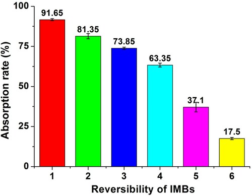
Extracting residues directly from animal tissues using IMBs may not be efficient for drug residues generally occuring in free or bound forms. So IMBs were applied during the clean-up step after extraction to get a better performance in the detection. Based on the prepared IMBs, the ability of IMBs to capture maximum amounts of MD was detected. It showed that 1 mg IMBs could approximately capture 25 ng MD in 10 min (Figure S2(A,B)). High recoveries from IMBs were important for detection of trace-amount of drug residues in animal tissues. To elute MD from IMBs, the proportion of methanol was chosen from 40% to 100% (methanol/PBS, v:v) based on ic-ELISA. Low absorbance at 450 nm indicated a high concentration of MD eluted from IMBs. Sixty per cent methanol showed the best efficiency for elution in 5 min (Figure S2(C,D)). The low concentration of methanol for elution led to a decrease in dilution before ic-ELISA so as to maintain the activity of MAbs and improve the sensitivity. Furthermore, the reversibility of IMBs was evaluated as well. After elution, the elutriate was detected by ic-ELISA. The same process was repeated six times. The specificities of IMBs were well maintained and the reversibility remained at more than 73% of the original capability after three cycles, as shown in . The results indicated a good performance of the developed IMBs for assaying the residue of MD in various chicken tissues.
Figure 1. Reversibility of IMBs for sample pre-treatment.
Notes: The absorption capability of IMBs was tested for six cycles. Data were represented as mean ± standard deviation (SD) of three replicates.
Development of IMBs-based ic-ELISA
To increase the sensitivity of ic-ELISA, the critical parameters (including buffer systems, incubation time and temperatures) were tested. In addition, the concentrations of secondary antibody (Ab2) and substrate/enzyme were optimized to get the best performance (Chen et al., Citation2009). Results indicated that dilutions of 1:10,000 for MAb 3B4 and 1:5000 for the Ab2 were optimal. Using these dilutions, MAb 3B4 could get good performance and work in an adequate working range. Coating buffer, blocking buffer and MAb dilution buffer were also optimized. Moreover, incubation times of 30 min at 37°C were enough for MAb and antigen or Ab2 reaction. All the optimal parameters were shown in Table S3.
Our developed method was challenged with authentic samples collected from local markets in Beijing, China. For the sample pre-treatment, methanol was selected as extracting agent to extract MD from chicken tissues, due to its high extraction efficiency of MD with less influence on immunoassay (Wang et al., Citation2008). The extracts were 1–10-fold diluted with 0.01 mol/L PBS (pH 7.4) for muscle, skin and fat, and 1–15-fold diluted for liver before IMBs-based clean-up. Sixty percent methanol was used to elute MD from IMBs. The supernatant after elution containing methanol was 1–10-fold diluted with 0.01 mol/L PBS (pH 7.4) because it could significantly decrease the activity of MAbs 3B4 in ic-ELISA tests (Figure S3). The standard curves of MD extracts from muscle, skin and fat under 10-fold dilution were comparable to the blank control without matrix effect (Figure S4(A,B)). A similar trend was observed for the standard curve of MD residue in liver (Figure S4(C)).
Standard curves of IMBs-based ic-ELISA were shown in Figure S5. The linear dynamic range was from 118.5 to 435.6 μg/kg, 117 to 513.5 μg/kg and 196.5 to 735.5 μg/kg for muscle, skin and fat, and liver, respectively. Recoveries of MD in three tissues with all spiking levels were higher than 80.0%, with the inter-day coefficients of variation (CVs) less than 11.3% and intra-day CVs less than 10.6% (). The limits of detection (LODs) were calculated as 72, 74 and 173 μg/kg in muscle, skin and fat, and in liver, respectively. These data indicated that our IMB-based ic-ELISA is sufficient enough to detect MD residue in three chick tissues.
Table 1. Recoveries and CVs of MD in different chicken tissues (n = 3).
Comparison of IMBs-based ic-ELISA and UPLC-MS/MS analysis
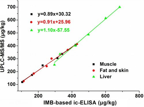
To evaluate the performance of the IMBs-based ic-ELISA method, UPLC-MS/MS was used to confirm the results obtained from same samples. As shown in , good correlations with R2 more than 0.99 were observed for muscle, skin and fat, and liver between two methods. The liner relationships ranged from 120 to 420 μg/kg in chicken muscle, skin and fat, and 270–720 μg/kg in liver. The results from the authentic sample test by IMBs-based ic-ELISA were consistent with the data obtained by the UPLC-MS/MS analysis.
Figure 2. Comparison of IMB-based ic-ELISA and UPLC-MS/MS assay.
Notes: Good linear relationships were observed for all tested samples (R2 ≥ 0.99). Data were represented as mean ± SD of three replicates.
During the IMBs clean-up process, a strong magnetic field should be used to ensure the accurate analysis. In addition, the extraction procedure need to be optimized to get better sensitivity. However, from a long-term perspective, the ic-ELISA method based on IMBs clean-up would greatly save cost and make for an efficient, convenient and environmentally friendly method for analysing MD residues in chicken tissues.
Conclusion
In this study, antibody-functionalized MB (IMB) was synthesized and characterized based on the production of a specific anti-MD MAb. IMB showed good performance and could be repeatedly used for sample pre-treatments. IMBs-based ic-ELISA was subsequently employed to detect MD in three chicken tissues. The LODs of MD in chicken muscle, skin and fat, and liver achieved in this study stratified with the set MRLs of MD in many countries. The results from authentic sample test by IMBs-based ic-ELISA were consistent with the data obtained by UPLC-MS/MS analysis. Altogether, it indicated that a rapid, robust clean-up of IMBs combining ELISA provides a simple, time-saving and environmentally friendly method to detect MD in chicken tissues.
supplemental_20171130docx_Clean.docx
Download MS Word (9.8 MB)Disclosure statement
No potential conflict of interest was reported by the authors.
Additional information
Funding
Related Research Data
References
- Chen, Y., Tang, S., Ding, S., He, F., & Xiao, X. (2009). Monoclonal antibody-based immunoassay for the detection of maduramicin in chicken tissues. Analytical Letters, 42, 2793–2806. doi: https://doi.org/10.1080/00032710903201966
- De Jong, J., Stoisser, B., Wagner, K., Tomassen, M., Driessen, J., Hofman, P., & Putzka, H. A. (2004). Determination of maduramicin in feedingstuffs and premixtures by liquid chromatography development, validation, and interlaboratory study. Journal of AOAC International, 87, 1033–1041.
- Diler, E., Obst, U., Schmitz, K., & Schwartz, T. (2011). A lysozyme and magnetic bead based method of separating intact bacteria. Analytical and Bioanalytical Chemistry, 401, 253–265. doi: https://doi.org/10.1007/s00216-011-5065-5
- Elliott, C. T., Kennedy, D. G., & McCaughey, W. J. (1998). Critical review methods for the detection of polyether ionophore residues in poultry. The Analyst, 123, 45–56. doi: https://doi.org/10.1039/a708698i
- Guo, L., Xu, L., Song, S., Liu, L., & Kuang, H. (2017). Development of an immunochromatographic strip for the rapid detection of maduramicin in chicken and egg samples. Food and Agricultural Immunology, 87, 1–12.
- Ha, J., Song, G., Ai, L. F., & Li, J. C. (2016). Determination of six polyether antibiotic residues in foods of animal origin by solid phase extraction combined with liquid chromatography-tandem mass spectrometry. Journal of Chromatography B, 1017–1018, 187–194. doi: https://doi.org/10.1016/j.jchromb.2016.01.057
- Holschuh, K., & Schwämmle, A. (2005). Preparative purification of antibodies with protein A – an alternative to conventional chromatography. Journal of Magnetism and Magnetic Materials, 293, 345–348. doi: https://doi.org/10.1016/j.jmmm.2005.02.050
- Jang, L., & Keng, H. (2008). Modified fabrication process of protein chips using a short-chain self-assembled monolayer. Biomedical Microdevices, 10, 203–211. doi: https://doi.org/10.1007/s10544-007-9126-7
- Kennedy, D. G., Blanchflower, W. J., & O’Dornan, B. C. (1997) Development of an ELISA for maduramicin and determination of the depletion kinetics of maduramicin residues in poultry. Food Additives and Contamimants, 14, 27–33. doi: https://doi.org/10.1080/02652039709374494
- Le-Minh, N., Khan, S. J., Drewes, J. E., & Stuetz, R. M. (2010). Fate of antibiotics during municipal water recycling treatment processes. Water Research, 44, 4295–4323. doi: https://doi.org/10.1016/j.watres.2010.06.020
- Liao, Y., Huang, R., Ma, Z., Wu, Y., Zhou, X., & Xing, D. (2014). Target-triggered enzyme-free amplification strategy for sensitive detection of microRNA in tumor cells and tissues. Analytical Chemistry, 86, 4596–4604. doi: https://doi.org/10.1021/ac5007427
- Liu, N., Song, S., Lu, L., Nie, D., Han, Z., Yang, X., … Zheng, X. (2014). A rabbit monoclonal antibody-based sensitive competitive indirect enzyme-linked immunoassay for rapid detection of chloramphenicol residue. Food and Agricultural Immunology, 25(4), 523–534. doi: https://doi.org/10.1080/09540105.2013.847065
- MHLW. (2007). Positive list system for agricultural chemical residues – provisional MRLs list. Ministry of Health, Labour and Welfare (Japan), Department of Food Safety, Standards and Evaluation Division.
- Ministry of Agriculture of China. (2002). Agricultural standards [Internet]. No. 235 Bulletin of the Ministry of Agriculture of the People’s Republic of China. Retrieved November 20, 2011, from http://www.moa.gov.cn/zwllm/nybz/index_2.htm
- Pereira, M. U., Spisso, B. F., Jacob, S. C., Monteiro, M. A., Ferreira, R. G., Carlos Bde, S., & da Nobrega, A. W. (2016) Validation of a liquid chromatography-electrospray ionization tandem mass spectrometric method to determine six polyether ionophores in raw, UHT, pasteurized and powdered milk. Food Chemistry, 196, 130–137. doi: https://doi.org/10.1016/j.foodchem.2015.09.011
- Šafařík, I., & Šafaříková, M. (1999). Use of magnetic techniques for the isolation of cells. Journal of Chromatography B: Biomedical Sciences and Applications, 722, 33–53. doi: https://doi.org/10.1016/S0378-4347(98)00338-7
- Sharma, N., Bhalla, A., Varma, S., Jain, S., & Singh, S. (2005). Toxicity of maduramicin. Emergency Medicine Journal, 22(12), 880–882. doi: https://doi.org/10.1136/emj.2004.020883
- Shen, J., Qian, C., Jiang, H., & Yang, H. (2001). Development of an enzyme-linked immunosorbent assay for the determination of maduramicin in broiler chicken tissues. Journal of Agricultural and Food Chemistry, 49, 2697–2701. doi: https://doi.org/10.1021/jf001520g
- Song, F., Zhou, Y., Li, Y., Meng, X., Meng, X., Liu, J., … Zhang, J. (2014). A rapid immunomagnetic beads-based immunoassay for the detection of β-casein in bovine milk. Food Chemistry, 158, 445–448. doi: https://doi.org/10.1016/j.foodchem.2014.02.150
- Stanker, L. H., Elissalde, M. H., Rowe, L. D., Beier, R. C., & El-Aziz Nasr, M. I. A. (1994). Detection of coccidiostats by immunoassay. Food and Agricultural Immunology, 6(1), 45–54. doi: https://doi.org/10.1080/09540109409354812
- Teste, B., Vial, J., Descroix, S., Georgelin, T., Siaugue, J. M., Petr, J., … Hennion, M. C. (2010). A chemometric approach for optimizing protein covalent immobilization on magnetic core-shell nanoparticles in view of an alternative immunoassay. Talanta,, 81, 1703–1710. doi: https://doi.org/10.1016/j.talanta.2010.03.027
- Tian, W., Zhang, X., Song, M., Jiang, H., Ding, S., Shen, J., & Li, J. (2017). An enzyme-linked immunosorbent assay to detect salinomycin residues based on immunomagnetic bead clean-up. Food Analytical Methods, 10(9), 3042–3051. doi: https://doi.org/10.1007/s12161-017-0873-7
- US FDA. (2017). Tolerances for residues of new animal drugs in food. United States Food and Drug Administration, Department of Health and Human Services, Code of Federal Regulations. Title 21 – Food and Drugs, Chapter 1, Part 556.
- Vanoosthuyze, K., Van Peteghem, C., Courtheyn, D., & Vercammen, J. (1994). Use of an immunomagnetic separation-ELISA technique for the fast detection of growth promoters in cattle urine. Food and Agricultural Immunology, 6(3), 241–249. doi: https://doi.org/10.1080/09540109409354835
- Wang, Y., Wang, Y., Wang, H., Ji, W., Sun, J., & Yan, Y. (2014). An immunomagnetic-bead-based enzyme-linked immunosorbent assay for sensitive quantification of fumonisin B1. Food Control, 40, 41–45. doi: https://doi.org/10.1016/j.foodcont.2013.11.025
- Wang, Z., Zhang, S., Murtazina, N. R., Eremin, S. A., & Shen, J. (2008). Determination of the veterinary drug maduramicin in food by fluorescence polarisation immunoassay. International Journal of Food Science & Technology, 43, 114–122. doi: https://doi.org/10.1111/j.1365-2621.2006.01400.x
- Wei, B., Li, F., Yang, H., Yu, L., Zhao, K., Zhou, R., & Hu, Y. (2012). Magnetic beads-based enzymatic spectrofluorometric assay for rapid and sensitive detection of antibody against ApxIVA of Actinobacillus pleuropneumoniae. Biosensors and Bioelectronics, 35, 390–393. doi: https://doi.org/10.1016/j.bios.2012.03.027
- Zhang, X., Song, M., Yu, X., Wang, Z., Ke, Y., Jiang, H., … Wen, K. (2017). Development of a new broad-specific monoclonal antibody with uniform affinity for aflatoxins and magnetic beads-based enzymatic immunoassay. Food Control, 79, 309–316. doi: https://doi.org/10.1016/j.foodcont.2017.02.049
- Zhu, Y., Song, S., Liu, L., Kuang, H., & Xu, C. (2016). An indirect competitive enzyme-linked immunosorbent assay for acrylamide detection based on a monoclonal antibody. Food and Agricultural Immunology, 27, 796–805. doi: https://doi.org/10.1080/09540105.2016.1160369
- Zhu, X., Zhou, X., & Xing, D. (2012). Nano-magnetic primer based electrochemiluminescence-polymerase chain reaction (NMPE-PCR) assay. Biosensors and Bioelectronics, 31, 463–468. doi: https://doi.org/10.1016/j.bios.2011.11.016