
ABSTRACT
Rathayibacter toxicus is Gram-positive bacterium causing gummosis of forage grasses, and it produces a corynetoxin responsible for livestock poisonings in Australia. R. toxicus is a USDA APHIS-listed Select Agent, threatening seed and livestock production areas in the U.S. We therefore are developing diagnostic assays to prepare for the introduction of the pathogen into the U.S. Using proteomic data, we identified proteins expressed in R. toxicus but not in other Rathayibacter spp. Recombinant proteins corresponding to extracellular domains of candidate proteins were used to raise polyclonal and monoclonal antibodies. Antibodies were evaluated for sensitivity and specificity in immunoassays against a panel of Rathayibacter spp. and near-neighbours. Polyclonal and monoclonal antibodies exhibited high sensitivity and specificity for R. toxicus in assays with cultured bacteria. The highest-performing immunoreagents were identified for further testing with field samples collected in Australia and for eventual development of an immunoassay for bacterial detection in grass samples.
Introduction
Rapid, sensitive diagnostic assays are needed for high-consequence plant pathogens for deployment at diagnostic laboratories and ports of entry. Ongoing national and state surveys are conducted annually for selected high-consequence plant pathogens, but development, validation and deployment of diagnostic assays for plant pathogens lag behind these efforts. As a result, surge capacity at national, regional and state labs in the U.S. National Plant Diagnostic Network (https://www.npdn.org/home) is strained during introduction and transportation of high-consequence pathogens such as Ralstonia solanacearum R3b2 (Norman et al., Citation2009; Swanson, Yao, Tans-Kersten, & Allen, Citation2005) and Phytophthora ramorum (Stokstad, Citation2004; Tjosvold, Chambers, & Blomquist, Citation2008).
Rathayibacter toxicus is Gram-positive, nematode-vectored bacterium causing gummosis of forage grasses, and produces a corynetoxin responsible for recurring livestock poisonings in Australia (Murray et al., Citation2017). R. toxicus is a USDA-APHIS-listed Plant Pathogen Select Agent under the Agricultural Bioterrorism Protection Act of 2002 (USDA, Citation2002), and is not present in the U.S. In Australia, the exportation of hay containing R. toxicus seed galls has been a pathway for movement of the pathogen, resulting in trade embargoes on hay exports (Allen, Citation2012). Although no pathways are known to exist for movement of R. toxicus into the U.S, the inadvertent introduction of the pathogen as a contaminant in seed-based commodities would threaten grass seed and livestock production areas in North America. The Australian government has developed and effectively deployed sampling technology and immunoassays (Masters, Gregory, Evans, Speijers, & Sutherland, Citation2006, Citation2011, Citation2017) for detection of R. toxicus in hay destined for export, in a robust programme to protect their industry in Western Australia and South Australia (Allen, Citation2012).
R. toxicus is one of several Select Agent plant pathogens for which no diagnostic assays are currently available. The USDA National Plant Disease Recovery System (NPDRS) identified the need for diagnostic assays in the Recovery Plan for R. toxicus (USDA NPDRS, Citation2015).
We are developing reagents for immunodiagnostic assays (Miller & Martin, Citation1988) to prepare for the possible introduction of the pathogen into the U.S. Applying proteomic, transcriptomic and comparative genomics data, we first identified proteins expressed in R. toxicus but not present in other Rathayibacter spp. Candidate antigens were then selected based upon predicted extracellular localization, antigenicity, and solubility. One protein, designated accession AYW_02955 in the R. toxicus genome (Sechler et al., Citation2017; NCBI Bioproject PRJNA312185), was selected for recombinant protein production and antibody generation. A recombinant protein corresponding to the extracellular domain of AYW_02955 gene was used as the immunogen to raise rabbit polyclonal (pAbs) and mouse monoclonal (mAbs) antibodies. Antibodies were evaluated for sensitivity and specificity in western blot, slot blot and ELISA format immunoassays against a panel of Rathayibacter spp. and near-neighbour genera. This approach has yielded antibodies with high selectivity and sensitivity to cultured R. toxicus cells and secreted protein fractions, with potential for the development and validation of robust immunoassays to detect the pathogen in plant parts, imported commodities and other sample matrices.
Materials and methods
Bacterial cultures
Cultures of R. toxicus (), and Rathayibacter, Clavibacter and Pseudomonas spp. (Table 1S) from our culture collection were maintained on a modified YGM medium (mYGM) (De Boer & Copeman, Citation1980). Liquid cultures were inoculated with a single colony into 7–10 mL liquid mYGM from single colonies and incubated with shaking at 200 rpm at 28°C and grown for 48–72 h until the stationary phase was reached as determined by OD600 above 0.6. Cell concentrations were determined by serial dilution and plating in triplicate on mYGM plates. Colony-forming units (CFU) were counted after incubation at 28°C for 7 days. For limit of detection (LOD) ELISA and slot blot assays with R. toxicus FH 79, 10 mL cultures were grown as described above, pelleted at 6000 g and resuspended in 1 mL PBS. For the time-course study, 10 mL of culture were sampled every 24 h for 11 days. Aliquots were centrifuged at 5000 g for 10 min and supernatant was collected. The pellets were resuspended in 1 mL of 1X PBS. The supernatants were filtered through a 0.45µ filter unit, followed by a 0.22µ filter unit (Millipore Sigma). For generation of the concentrated cells for counter-screening hybridomas, each culture (Table 1S) was centrifuged, resuspended in an equal volume of PBS, then 1 mL of each culture was pooled and heated at boiling temperature for 10 min to inactivate the cells. The pooled, heat-killed sample was sent to the contract provider (Genscript) for counter-screening the hybridoma cell cultures. All experiments utilized at least three individual biological replicates.
Table 1. List of R. toxicus strains from Australia used in this study, with specificity–inclusivity testing of antibodies against strains on chemiluminescent western blots.
Secreted protein fraction
For detection of the secreted protein in R. toxicus FH79 cultures, a 72-h culture of R. toxicus cells was pelleted and the supernatant was filtered through a 0.22µ filter unit. The secreted protein fraction was concentrated 25-fold in centrifuge concentration tubes (Millipore Sigma) and the concentrated secreted fraction was loaded onto SDS-PAGE gels on a protein concentration basis.
Identification of antigen targets
Antigen targets were identified from a previously published R. toxicus proteome study (Fennessey et al., Citation2018). Output from the study was searched in the SCAFFOLD software package (Proteome Software, Inc.) for hypothetical proteins containing a minimum of 5 unique peptides in their amino acid sequences. Candidate proteins were subjected to a set of analysis tools including SignalP (Nielsen, Citation2017), TMHMM v.2.0 (Möller, Croning, & Apweiler, Citation2001), BepiPRED (Jespersen, Peters, Nielsen, & Marcatili, Citation2017), hydrophilicity and antigenicity plots. Potential antigen amino acid sequences were aligned to all accessible Rathayibacter spp. and related bacteria from open-source protein sequence databases. Proteins with low similarity to other Rathayibacter spp. with high epitope scores were considered for recombinant protein generation.
Gene synthesis and recombinant protein production
The full-length amino acid sequence for AYW_02955 was truncated to enhance recombinant protein expression in E. coli. The transmembrane helix representing amino acids 1–32 was removed, leaving an 884 amino acid sequence. The resulting sequence was then optimized for prokaryotic coding preference, synthesized by a commercial provider (Genscript) and cloned into the pET 28a(+) vector (Novagen) with a 6X histidine tag. Plasmids were transformed into E. coli BL21 DE3 competent cells (New England Biolabs). For protein expression 3 ml of an overnight culture was added to 300 ml of LB broth containing 50 ng/ml of kanamycin and grown for 3 h at 37°C to an OD600 of 0.6. IPTG was added to a final concentration of 1 mM and cultures were shaken at 100 rpm for 18 h at room temperature. Following cell lysis, the soluble and insoluble fractions were separated by PAGE and stained for total protein. The recombinant target antigen proteins were found in the insoluble fraction after cell lysis and were purified using Ni-Sepharose (GE Healthcare) resin under denaturing conditions. The insoluble recombinant protein fraction was stored at −20°C in 8 M Urea and 500 mM imidazole.
Antibody production
The purified recombinant protein was applied as inject antigen to generate polyclonal antibodies in New Zealand White rabbits by a contracted provider (Abcore). IgG was purified from the serum provided by the provider using a Melon Gel kit following the manufacturer’s directions (Pierce-Thermo Fisher). For mAb production, the purified recombinant protein was used as the inject antigen in BALB/c mice by a contracted provider (Genscript). The resulting hybridoma clones were counter-screened against a 6X-histidine peptide and the pooled panel of concentrated bacterial cells (Table 1S). A set of counter-screened hybridoma clones were selected for further testing based upon reactivity to the inject antigen and ultimately by reactivity to R. toxicus cells in ELISA screens. Two clones (4B10-1 and 6B5-1) were selected, and purified IgG fraction from large-scale hybridoma cultures was provided by the contractor for further sensitivity and specificity testing.
SDS-PAGE for western blotting
Cultures were pelleted by centrifugation at 6000 g for 10 min, washed two times in 0.5X PBS and suspended in 1/10 of the original volume in lysis buffer (25 mM TRIS, pH 7.5, 1 mM EDTA, 5 mM DTT and 2.0% SDS). Samples were boiled in a water bath for 15 min, cell debris was removed by centrifugation at 6000 g for 10 min and soluble proteins were collected. Protein extracts were stored at −20°C. All protein samples were quantified using the BCA Protein Assay kit (Thermo Fisher Scientific) with BSA as a standard. Purified recombinant protein was used as a positive control at 10–50 ng per well. Proteins were separated on 4–12% NU-PAGE gel. Following SDS-PAGE, proteins were transferred to 0.45µ pore-size nitrocellulose blotting membrane (Amersham) using a semidry blotter apparatus (Owl Separation Systems) according to the manufacturer’s guidelines.
Western and slot blot
Following western blot transfer or direct application to nitrocellulose membrane in a slot blotter manifold, blots were blocked in 5% (wt/vol) dry milk in PBS-0.02% (vol/vol) Tween 20 (PBS-Tw) for 1 h and probed with anti-R. toxicus pAbs at 1:3000 and anti-R. toxicus MAbs at 1:3000 overnight at 4°C. Blots were washed 3 times for 5 min in 100 ml PBS-Tw. Blots were then probed with horseradish peroxidase (HRP)-conjugated goat anti-mouse or anti-rabbit IgG (Sigma Chem. Co.) at 1:35,000 (Chemiluminescent slot blot) for 1 h, washed 3 times in 100 ml PBS-Tw, and detected using Super Signal West Pico chemiluminescent substrate (Thermo Scientific) according to the manufacturer’s protocol.
ELISA
For the standard ELISA protocol, 100 µL of each sample was added in triplicate to a 96 well microtiter plate and incubated overnight at 4°C. Purified recombinant protein was used as a positive control at 50 ng per well. Plates were washed 2 times in 200 μL PBS, blocked in 100 µL of 3% (w/v) dry milk in PBS at room temperature for 1 h, and washed 3 times in 200 μL PBS. One hundred µL of primary antibody at a 1:2000 dilution (pAb) or 1:1,000 (mAb 6B5) in PBS was added per well and incubated at room temperature for 2 h. Negative controls included all components minus primary antibody. Plates were washed 3 times with PBS, and HRP-conjugated goat anti-mouse antibody was added at a concentration of 1:10,000 and incubated at room temperature for 2 h. Plates were then washed 3 times in PBS, 100 µL of HRP substrate (ABTS; KPL Inc.) was added, and absorbance was read at 405 nm in a microtiter plate reader.
Results
Candidate protein antigen selection
In a previous study, we applied LC/MSMS to R. toxicus cell extracts to identify, catalogue and categorize proteins (Fennessey et al., Citation2018) using an annotated R. toxicus genome (Sechler et al., Citation2017; NCBI BioProject PRJNA312185) as a database. In this study, we used the resulting proteome to focus on the subset of secreted proteins, searching for proteins with characteristics suitable for antibody production and diagnostic immunoassay development. A subset of hypothetical proteins unique to R. toxicus were identified as potential candidates, based upon criteria addressing the highest peptide coverage, presence of predicted secretion signals and multiple regions of high antigenicity scores. Proteins with the best combination of scores from these predictive analyses of amino acid sequence were aligned with homologous sequences in Rathayibacter spp. We focused upon a 94 kDa protein, (arbitrarily assigned accession AYW_02955 in the genome), which was the second most abundant protein in the R. toxicus protein dataset, with 29 unique peptides, corresponding to 53% sequence coverage over the amino acid sequence. Amino acid sequence alignments of AYW_02955 against all Rathayibacter and related bacteria in GenBank revealed that the sequence matched 100% to R. toxicus, with 60% or less identity to other Rathayibacter spp. including R. iranicus, R. tanaceti, R. rathayi and R. tritici, 42% to near neighbour Leifsonia xylii, and no significant similarity to pathovars of Clavibacter michigenensis. Analysis of the protein using online protein sequence analysis tools predicted AYW_02955 to be a secreted lipoprotein containing a signal peptide and strong epitope scores. We generated a truncated clone and expressed the recombinant protein in E. coli to serve as inject antigen for antibody production in rabbits and mice. Our cost-effective strategy was to first generate lower-cost pAbs in rabbits to ensure that the target antigen was expressed in sufficient quantity for detection, and then proceed to more expensive mAb projects to generate high-specificity immuno-reagents with diagnostic utility. In this case, both strategies produced sensitive and specific antibodies.
Confirmation of target antigen expression in cultured R. toxicus cells
In western blot experiments testing the reactivity of our antibodies against extracts of liquid-cultured R. toxicus FH79 cells, the pAb and both mAbs detected a 94 kDa protein (), confirming expression of protein AYW_02955 in cultured cells. Only the 94 kDa protein was identified on western blots, further confirming the specificity of the antibodies against the target protein. Although protein localization studies predicted the 94 kDa protein to be membrane-associated, slot blot assays detected the target protein in sterile-filtered culture media with high sensitivity (), indicating active secretion of the protein from R. toxicus cells, confirming the predicted secretory nature of the protein.
Figure 1. Western blot of R. toxicus FH79 cell extracts probed with pAb and mAbs 4F10-1 and 6B5-1. One hundred ng of soluble R. toxicus protein was loaded on gels. The mass of the full-length target protein as predicted from R. toxicus genome accession AYW_02955 is 94 kDa.
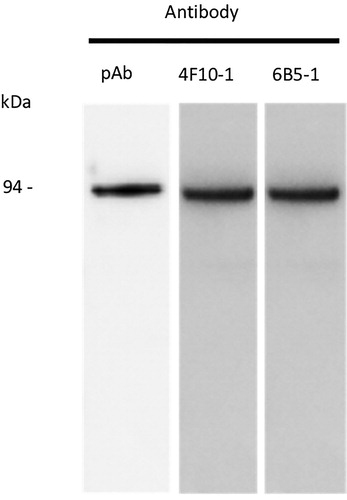
Figure 2. Slot blot of R.toxicus FH79 culture filtrate probed with anti R. toxicus mAbs 4F10 and 6B5.
Time course of expression
We assayed R. toxicus cells in liquid culture over the course of 11 days growth under optimal conditions, measuring the OD600 on each day of sampling. R. toxicus cells entered the stationary growth phase around day 8. Samples from the culture supernatant and centrifuged pellet were tested by ELISA assays. We observed absorbance readings that roughly paralleled the OD600 taken from each day of the experiment (). In the first three days, little to no reactivity to the antibody was detected. Reactivity in the pelleted cells increased significantly on day 4, when the bacterial concentration had increased and more cells were available to react. In the culture supernatant, the absorbance readings continued to increase gradually through the end of the experiment; however, in the pellet, there was a slight decline in absorbance through day 11, once the culture reached the stationary phase and less protein was detected in the pellet due to cell death. The target protein was observed at a higher concentration in the culture supernatant, due to continued secretion of the protein from live cells, presumably augmented by protein released from dead cells.
Figure 3. Time course of detection in ELISA assays with pAb in cultures of R. toxicus FH79 cells plotted against optical density (O.D.) of culture at 600 nm.
Specificity – inclusivity
We tested the specificity of the pAb and mAbs against extracts of cultured cells from 22 strains of R. toxicus from various regions of Australia on western blots with chemiluminescent detection. We chose western blotting to ensure that the antibodies reacted with the target 94 kDa protein and chose chemiluminescent detection to ensure sensitive detection. Recombinant protein was used as a positive control on blots. The pAb and both mAbs produced a 94 kDa band on chemiluminescent western blots with all R. toxicus strains tested (). One strain, FH 231, produced a very weak signal in one of the two replicate experiments.
Specificity – exclusivity
We tested the specificity of the pAb and mAbs against extracts of cultured cells from related strains of Rathayibacter spp. from a broad range of global origins, near-neighbour Clavibacter spp. as well as Pseudomonas spp. (Table 1S) on western blots with chemiluminescent detection. Recombinant protein was used as a positive control on blots. The hybridomas secreting the mAbs were selected after counter-screening for lack of reactivity against a pooled panel of the Rathayibacter spp. listed in Table 1S. None of the anti- R. toxicus pAb and mAbs produced a signal on chemiluminescent western blots with proteins from Rathayibacter spp. other than R. toxicus (), demonstrating their specificity for the protein expressed only by R. toxicus cells. The antibodies were thus determined to be highly specific for R. toxicus and suitable for diagnostic detection of the pathogen.
Table 2. Specificity–exclusivity testing of anti- R. toxicus antibodies on chemiluminescent western blots tested against cultures of Rathayibacter, Pseudomonas and Clavibacter spp.
Limit of detection (LOD)
We conducted colorimetric indirect ELISA assays with the pAb to determine the lower limit of detection of the antibodies in the assays. Dilution plating was conducted on the cultures used in the assays to calculate the number of colony-forming units (CFU), which was held constant in the replicate experiments. We calculated the limit of detection (LOD) in ELISA assays as the number of cells producing an absorbance reading equal to or greater than twice the negative control background reading (no primary antibody). In replicate ELISA assays, the LOD was 4 × 10⁴ CFU (). In parallel experiments on slot blots, measuring protein instead of CFU, the LOD was 5 ng (Figure 1S), which is approximately equivalent to 5 × 10³ cells. (CFU were not quantitated for this experiment). The limit of detection on slot blots was considered to be the amount of protein loaded that produced a band detected by chemiluminescent image capture for 30 s or less.
Table 3. Limit of Detection of R. toxicus FH79 cells in ELISA assays with pAb. Ratings are an average of 3 absorbance values per plate from two repeated experiments.
Discussion
Our proteomic approach successfully identified a highly antigenic protein secreted by R. toxicus with low similarity to orthologs in other Rathayibacter spp. and near-neighbours. Our approach stands in contrast to the strategy deployed in Australia, where antibodies generated for screening of exported hay and seed in Australia were generated against heat-killed intact R. toxicus cells (Masters et al., Citation2006). We were able to use a truncated recombinant form of the protein to generate antibodies that only reacted against all R. toxicus strains tested, and a protein fraction in culture medium secreted by R. toxicus FH 79. The antibodies detected both R. toxicus cells and secreted protein in immunoassays with a high level of sensitivity and specificity. The L.O.D. was approximately 4 × 10⁴ cells in ELISA format, comparable to other bacterial immunoreagents in indirect ELISA assays. Although not directly comparable, antibodies against heat-killed R. toxicus cells also exhibited a very high level of sensitivity, capable of detecting one seed gall in 1 kg of hay (Masters et al., Citation2006).
Although variants of the R. toxicus AYW_02955 gene with moderate homology exist in other Rathayibacter spp., neither the pAb or mAbs reacted with cultured cells of any Rathayibacter spp. other than R. toxicus. This may be due to lack of expression of the target protein in other Rathayibacter spp. or specificity of our antibodies for epitopes in regions of the protein unique to R. toxicus. We did not test the antibodies against near-neighbour Leifsonia spp., as sequences in published genomes of these species had a low level of similarity to the gene found in R. toxicus and had no overlapping regions rich in predicted epitopes. Thus our target protein may be useful as a “biomarker” for sensitive detection of R. toxicus in plant and environmental samples. The antibodies could be applied in ELISA or lateral flow “dipstick” formats for low-cost early detection protocols for field or lab applications.
The R. toxicus strains tested in our assays represent largely historical collections made in Australia over the past decades, and specific dates and locations are not available for all strains (). Genetic characterization of R. toxicus strains in two separate studies has each revealed at least 3 populations in distinct regions of Australia. Agarkova, Vidaver, Postnikova, Riley, and Schaad (Citation2006) applied PFGE and AFLP to identify 3 clades of R. toxicus collected from Western Australia (clade A) and South Australia (clades B,C). Arif et al. (Citation2016) utilized MLST and ISSR to identify three genetically distinct populations of R. toxicus in South Australia (populations RTI, RTII) and Western Australia (population RTIII). Table 2S identifies the clades and populations of those strains that were used in our studies. Our antibodies reacted with strains representing Clade A and population RT-III from Western Australia, as well as Clades B,C and population RT-II from South Australia (). Further, our antibodies detected strains collected in New South Wales, Australia, that were not genetically characterized by Agarkova et al. (Citation2006) or Arif et al. (Citation2016). It is likely that our antibodies will react with strains in population RT-I, as the identical AYW_02955 gene sequence is present in strains of population RT-I (Arif et al., Citation2015). Although they reacted positively against all tested strains, we cannot be certain that our reagents will react with all populations present in Australia, and plan to obtain and test additional strains in the near future.
Although we observed high levels of expression of the target protein in cultured cells under optimum growth conditions, R. toxicus cells in nature experience a very different environment. Bacterial galls in grass seed are dehydrated and the R. toxicus cells are presumably in cell cycle arrest for an extended period. We cannot predict the level of expression of our target protein under these conditions. Although we were able to detect the protein on slot blots in preliminary experiments with dehydrated R. toxicus FH79 cell pellets after more than one year (data not shown), these conditions do not necessarily mimic the environmental stress encountered by the bacterium in the field. The antibodies will therefore be subjected to further testing with field samples recently collected in Australia for the development of an immunoassay for R. toxicus in sample matrices that may be encountered from field-derived samples and commodities at U.S. ports of entry. Such assays would help to prevent the introduction of the pathogen as a contaminant in imported commodities and protect livestock production areas in North America.
Supplemental Material
Download MS Word (18.5 KB)Acknowledgements
Mention of trade names or commercial products in this publication is solely for the purpose of providing specific information and does not imply recommendation or endorsement by the U.S. Department of Agriculture. USDA is an equal opportunity provider and employer.
Disclosure statement
No potential conflict of interest was reported by the authors.
Data availability statement
The data that support the findings of this study are available from the corresponding author upon reasonable request.
Additional information
Funding
Related Research Data
References
- Agarkova, I. V., Vidaver, A. K., Postnikova, E. N., Riley, I. T., & Schaad, N. W. (2006). Genetic characterization and diversity of Rathayibacter toxicus. Phytopathology, 96, 1270–1277.
- Allen, J. (2012). Annual ryegrass toxicity–an animal disease caused by toxins produced by a bacterial plant pathogen. Microbiology Australia, 33, 18–21.
- Arif, M., Busot, G. Y., Mann, R., Rodoni, B., Liu, S., & Stack, J. P. (2015). Complete genome of the select agent Rathayibacter toxicus isolate SA03-04 from South Australia. Phytopathology, 105, S4.96.
- Arif, M., Busot, G. Y., Mann, R., Rodoni, B., Liu, S., & Stack, J. P. (2016). Emergence of a new population of Rathayibacter toxicus: An ecologically complex, geographically isolated bacterium. PloS one, 11, e0156182. doi.org/10.1371/journal.pone.0156182
- De Boer, S. H., & Copeman, R. J. (1980). Bacterial ring rot testing with the indirect fluorescent antibody staining procedure. American Potato J, 57, 457–465.
- Fennessey, C. M., McMahon, M. B., Sechler, A. J., Kaiser, J., Garrett, W. M., Tancos, M. A., … Schneider, W. L. (2018). Partial proteome of the corynetoxin-producing gram-positive bacterium. Rathayibacter Toxicus. Proteomics, 18(1-3), 1700350.
- Jespersen, M. C., Peters, B., Nielsen, M., & Marcatili, P. (2017). BepiPred-2.0: Improving sequence-based B-cell epitope prediction using conformational epitopes. Nucleic Acids Research, 45, 24–29.
- Masters, A., Colegate, S., Galvin, D., Gregory, A. R., Van Burgel, A., McKay, A. C., & Stevens, V. (2017). ELISA based correlation of Rathayibacter toxicus antigen and corynetoxins in pasture and hay. International Journal of Poisonous Plant Research, 4, 53–67.
- Masters, A. M., Gregory, A. R., Evans, R. J., Speijers, J. E., & Sutherland, S. S. (2006). An enzyme-linked immunosorbent assay for the detection of Rathayibacter toxicus, the bacterium involved in annual ryegrass toxicity, in hay. Australian Journal of Agricultural Research, 57, 731–742.
- Masters, A. M., Samarasinghe, B., Kalkhoven, M. J., den Hollander, G. L., & Palmer, D. G. (2011). Improvements to the immunoassay for detection of Rathayibacter toxicus in hay. Crop and Pasture Science, 62, 523–530.
- Miller, S. A., & Martin, R. R. (1988). Molecular diagnosis of plant disease. Annual Review of Phytopathology, 26, 409–432.
- Möller, S., Croning, M. D., & Apweiler, R. (2001). Evaluation of methods for the prediction of membrane spanning regions. Bioinformatics (Oxford, England), 17, 646–653.
- Murray, T. D., Schroeder, B. K., Schneider, W. L., Luster, D. G., Sechler, A. J., Rogers, E. E., & Subbotin, S. A. (2017). Rathayibacter toxicus and bacterial head blight diseases of grasses. Phytopathology, 107, 804–815.
- Nielsen, H. (2017). Predicting secretory proteins with SignalP. In D. Kihara (Ed.), Protein function Prediction (pp. 59–73). New York, NY: Humana Press.
- Norman, D. J., Zapata, M., Gabriel, D. W., Duan, Y. P., Yuen, J. M., Mangravita-Novo, A., & Donahoo, R. S. (2009). Genetic diversity and host range variation of Ralstonia solanacearum strains entering North America. Phytopathology, 99, 1070–1077. doi: 10.1094/PHYTO-99-9-1070
- Sechler, A. J., Tancos, M. A., Schneider, D. J., King, J. G., Fennessey, C. M., Schroeder, B. K., Murray, T. D., et al. (2017). Whole genome sequence of two Rathayibacter toxicus strains reveals a tunicamycin biosynthetic cluster similar to Streptomyces chartreusis. PloS One, 12, e0183005.
- Stokstad, E. (2004). Nurseries may have shipped sudden oak death pathogen nationwide. Science, 5666, 1959. doi: 10.1126/science.303.5666.1959a
- Swanson, J. K., Yao, J., Tans-Kersten, J., & Allen, C. (2005). Behavior of Ralstonia solanacearum race 3 biovar 2 during latent and active infection of geranium. Phytopathology, 95, 136–143.
- Tjosvold, S. A., Chambers, D. L., & Blomquist, C. L. (2008). Seasonal symptom expression, laboratory detection success, and sporulation potential of Phytophthora ramorum on rhododendron and camellia. In S. J. Frankel, J. T. Kliejunas, & K. M. Palmieri (Eds.), Proceedings of the sudden oak death third science symposium. Gen. Tech. Rep. PSW-GTR-214 (pp. 101–107). Albany, CA: U.S. Department of Agriculture, Forest Service, Pacific Southwest Research Station.
- U.S. Dept. of Agriculture. (2002). 7 CFR Part 331, The agricultural bioterrorism protection Act of 2002: Possession, use, and transfer of biological; agents and toxins; Interim and final rule. Federal Register, 67, 76908–76938.
- USDA ARS National Plant Disease Recovery System. (2015). Recovery plan for rathayibacter poisoning caused by Rathayibacter toxicus. https://www.ars.usda.gov/ARSUserFiles/opmp/RathayibacterPoisoning_March2015.pdf