
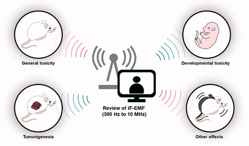
Abstract
Purpose
Many novel devices such as induction cookers or wireless power transfer produce electromagnetic fields (EMFs) in the intermediate frequency (IF) range (300 Hz to 10 MHz) and it is very meaningful for summarizing the bioeffects of IF-EMF research, particularly animal studies. This review takes into account experimental studies that used murine models to study the health effects of exposure to IF-EMF. The analyses included here use data available in the literature published from January 1988 to August 2021 including the animal studies about general adverse effects, tumorigenic effects, and effects on developmental stages. The studies that linked IF-EMF exposure during pregnancy or neonatal stage to behavioral and cognition changes were included. Additionally, this review also covers the effects of IF-EMF on gene expression patterns in the brain, behavior patterns associated with learning and memory, and immune function.
Conclusions
Although most studies have suggested that IF-EMF is harmless, some adverse effects have been reported after exposure at developmental stages and prolonged exposure. Compared to extremely low frequency (ELF) or radiofrequency (RF) EMF bands, studies on health effects with more diverse perspectives of IF-EMF have not been conducted. Therefore, performing more research should be necessary using the latest biomedical tools. From this point of view, a comprehensive review of IF-EMF studies, particularly animal studies, will provide a valuable basis for further risk analysis in humans.
Graphical Abstract
Introduction
The use of electromagnetic fields (EMFs) has become increasingly widespread since the twentieth century, which has resulted in a growing incidence of human exposure to EMFs. In turn, this has led to growing concerns regarding the potentially negative effects of EMFs on human health. In 1996, the World Health Organization (WHO) launched an international EMF project (The International EMF Project 2022) to address these concerns and protect public health. This project evaluated the scientific evidence for the potential human effects of EMFs in the frequency range up to 300 GHz. Further, this project promoted research on important issues that remain understudied and encouraged the development of international standards to limit EMF exposure.
Public concern is generally focused on exposure to extremely low frequency (ELF) EMFs which are oscillating fields defined as having frequencies below 300 Hz (e.g. electrical supply equipment including power lines) and exposure to 10 MHz to 300 GHz radiofrequency (RF) EMFs (e.g. microwave ovens, broadcasting equipment, and portable equipment with mobile phone and WiFi). The health effects of EMFs are very diverse and many studies have focused on the two aforementioned frequency bands. In contrast, very few studies have focused on the health effects of the intermediate frequency (IF) range (300 Hz to 10 MHz), which lies between the ELF and RF ranges. This is partly because there are fewer types of devices that generate EMF in this frequency range. However, there have been increasing concerns about the potentially harmful health effects of IF band technology used in homes and workplaces. These concerns include a potential link between IF-EMF exposure and employee disabilities (e.g. swelling, tingling sensation in the fingertips, and headaches).
One of the world's leading groups in research on exposure to IF-EMF is located in Korea. In 2000, the Ministry of Information and Communication in Korea established the Basic Plan for the Evaluation of EMF Effects in Humans. In accordance with this plan, Korean scientists jointly conducted animal experiments from 2002 to 2006 to analyze the effects of 20 kHz IF-EMFs. This study first sought to evaluate the relationship between chronic exposure to video display terminal (VDT) systems such as computer monitors and the occurrence of disease. Further, with the increasing adoption of wireless power transmission technology, more IF band devices are expected to be used in household and work environments, thus highlighting the need to evaluate the health risks associated with IF-EMF exposure. Nevertheless, when compared to ELF-EMF or RF-EMF, very few studies have evaluated the health effects of IF-EMF exposure in humans, and the majority of IF-EMF studies have been conducted by the Korea Institute of Radiological and Medical Science (KIRAMS), the Electric Power Research Institute in Japan, and Kuopio University in Finland using animal models. In addition to the scarcity of animal and cellular experiments studies on the effects of IF-EMF exposure, extremely few epidemiological studies have been conducted so far. However, in 2014, there was an initiative by an international joint project of the EU (GERoNiMO – Generalized EMF Research Using Novel Methods; GERoNiMO Citation2022) to conduct animal and cellular experiments on IF-EMF exposure, as well as research on IF-EMF exposure environments. Further, Japan has been conducting personal/environmental IF-EMF exposure assessments and epidemiological studies since 2018.
Animal experiments remain essential to understanding the fundamental mechanisms underlying health effects or pathophysiological changes by environmental health risk factors including EMF. In this review, we limited review articles to only rodent studies although many studies were conducted in other animal species, including chick, fish, rabbit, etc., because there are difficulties in interpreting IF-EMF health effects from the various species. Mice and rats have long served as the preferred species for biomedical research animal models due to their anatomical, physiological, and genetic similarity to humans. Especially over 95% of animal studies were conducted in mice (Vandamme Citation2014). Well-established animal care standards for rodents help researchers repeat consistent results with similar experimental conditions. Therefore, review limited in rodent studies may help interpret and confirm bio/health effects of IF-EMF from different exposing conditions. At this point, it is very meaningful to comprehensively summarize the current state of IF-EMF research, particularly animal studies, which would provide an important basis for future risk analyses in humans.
IF-EMF sources
The definition of the IF range varies from country to country and from institution to institution. The WHO (WHO 2005) defines a range between 300 Hz and 10 MHz, whereas the International Commission on Non-Ionizing Radiation Protection (ICNIRP) does not provide an explicit definition (ICNIRP Citation1998). The European Commission (SCENIHR) defines it between 300 Hz and 100 kHz (SCENIHR Citation2015). In Korea, the Korea Institute of Electromagnetic Engineering and Science (KIEES), an organization that standardizes terms related to EMFs, defines the range between 300 Hz and 10 MHz. Meanwhile, the Federal Office for Radiation Protection (BfS) in Germany only considers ELF and RF fields and sets the boundary at 100 kHz (Bodewein et al. Citation2019). In order to evaluate the bioeffects of examined studies from different countries, we reviewed the range of IF-EMF between 300 Hz and 10 MHz recommended by WHO.
The main sources of IF-EMF can be divided into the following areas.
Industry: Dielectric heater sealers, induction and plasma heaters, broadcast and telecommunication transmitters.
General public: Induction cookers, proximity readers, electronic article surveillance systems and other anti-theft devices, computer monitors, and TV sets.
Hospitals: MRI systems, electromagnetic nerve stimulators, electrosurgical instruments, and other medical devices.
Defense: Submarine communication transmitters and high frequency (HF) transmitters.
Human EMF exposure from IF devices is generally lower than the recommended limit established by the ICNIRP. However, some occupations such as oil and induction heater workers, as well as soldiers and technicians working in the vicinity of high-power broadcast equipment may be exposed to fairly high levels of IF-EMF (SCENIHR Citation2015).
Potential health effects of IF-EMF
Several thermal and non-thermal endpoints have been established to evaluate how IF-EMF (mainly electric fields) affect biological systems. Basic restriction for the general population and occupational exposure is determined based on the minimal exposure required to elicit any observable adverse effects (thermal or non-thermal). Exposure to high IF-EMF levels at relatively high frequencies can cause thermal damage (a slow process when tissues are kept hot for long periods); however, the most obvious risk in this frequency is that of cell membrane stimulation. This non-thermal effect is linked to changes in the membrane potential induced by external EMF exposure and may affect/stimulate peripheral nerves and muscle cells, to name a few examples. Another mechanism is electroporation, which is defined as a reversible or irreversible disruption of the cell membrane that occurs when IF-EMFs induce a large potential difference across the cell membrane. Although this phenomenon can cause tissue damage by electric shock, its therapeutic applications are also being studied, as short electric pulses could allow for better drug penetration in human tissues. External IF-EMF exposure can only have these effects on the human body at intensities much higher than normal environmental exposure.
Methods
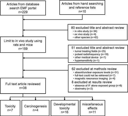
This review takes into account experimental studies that used murine models to study the health effects of exposure to IF-EMF. For the interpretation of health effects of IF-EMF, subjects of experiments were limited to mice and rats which are the most common experimental animals in biomedical research. The analyses included here use data available in the public domain, i.e. literature published in English from January 1988 to August 2021. The databases Electromagnetic Field-Portal (EMF-PORTAL Citation2022) and Public/Publisher MEDLINE (PubMed 2022) were searched using the terms ‘intermediate frequency,’ ‘intermediate frequency field,’ and the results were then filtered by the following terms: ‘experimental studies,’ ‘mouse,’ or ‘rat’. Total of 239 research articles were checked for the title and abstract as an initial search results. This review was limited to experimental studies investigating bio/health effects of IF-EMF on rodents (mice and rat); 80 studies were excluded for in vitro studies (n = 34), ex vivo studies (n = 4), and other species (n = 42). Studies investigating therapeutic outcomes were also excluded unless they reported specific bioeffects; tumor treating fields (n = 25), pulsed RF (n = 18), and EMF from other medical device (n = 7). A study investigating hypersensitivity was excluded. Next, we checked the method and result sections of the remaining 108 studies and excluded 55 studies with absent/unclear exposure levels (n = 51) or magnetic resonance imaging (n = 4). Furthermore, seven studies for which full-text articles could not be retrieved, six studies that did not have IF alone exposed group, and two studies investigating the dosimetry were excluded. Finally, 38 research articles were reviewed. Full-text copies of potentially relevant articles were obtained, and the results and reported outcomes were extracted ().
Figure 1. Flow diagram of research results review. Initially, 239 studies were considered and this review was limited to experimental studies investigating bio/health effect of IF-EMF on rodents (mice and rat).
Results
The review encompasses 38 experimental studies that used rats or mice to investigate the various bioeffects of IF-EMF, including effects on prolonged exposure, developmental stages, and tumorigenicity. The uniformity of IF-EMF magnetic flux density and numbers of cages or animals during the exposure are summarized in , if the description of IF-EMF exposing system was available. The exposure characteristics and biological systems investigated in experimental studies for the various bioeffects are shown in .
Table 1. In vivo IF-EMF exposure systems.
Table 2. In vivo animal studies investigating IF-EMF on toxicity.
Table 3. In vivo animal studies investigating IF-EMF on carcinogenicity.
Table 4. In vivo animal studies investigating IF-EMF on fetus.
Table 5. Other in vivo animal studies investigating bioeffects of IF-EMF.
Effects of long-term exposure
Spontaneous cancer incidence, hematologic, blood biochemical, and histological effects were observed after exposing rats to 20 kHz IF-EMFs for eight hours a day at a magnetic flux density of 6.25 μT (five days a week for 90 days). No cancer was observed in neither the non-exposed nor the IF-EMF-exposed group, and no changes in body weight and the weight of various organs (lung, liver, brain, heart, testis, and ovary) were observed. Additionally, no alterations in hematological parameters (white blood cell count, red blood cell count, hemoglobin concentration, hematocrit, mean red blood cell mass, mean hemoglobin concentration, and platelet count), blood biochemical parameters (total protein, hematologic nitrogen, creatine, glucose, total bilirubin, total cholesterol, and various enzymes), urine parameters (acidity, specific gravity, protein, ketone bodies, erythrocytes, leukocytes, glucose, bilirubin, and urobilinogen), and organ pathology were observed (Kim et al. Citation2006). Similarly, almost no effects were observed when the rats were evaluated after 12–18 months of long-term exposure under the same exposure conditions; however, the number of neutrophils increased in females in the exposed group after 12 months, and the number of lymphocytes decreased in males after exposure for 18 months (Lee et al. Citation2006). To confirm these findings, the exposure environment and time were kept the same and only the exposure magnetic flux density was increased from 6.25 μT to 30 μT. However, despite long-term exposure to 30 μT for 18 months, abnormalities in neutrophil count and lymphocyte count were not confirmed (Lee et al. Citation2010).
Another research group exposed mice to 10 kHz at high magnetic flux densities of 80, 280, and 1000 μT. The exposures were conducted for 22.6 hours a day for 14 or 90 consecutive days. However, no changes in animal behavior, body weight, morbidity, or biochemical and hematological parameters were observed (Robertson et al. Citation1996). Similarly, a study by a Japanese research team on rats did not identify changes in body weight, organ weight, or hematological parameters after exposure at 20 kHz for 22 hours a day at a magnetic flux density of 200 μT for 14 days or 13 weeks (Nishimura et al. Citation2016). Additionally, Svedenstål et al. exposed newborn, young, and adult mice to 20 kHz IF-EMFs for 40 days, after which an increase in body weight was observed (Svedenstål and Johanson Citation1998). Kumari et al. observed sperm cells from cauda epididymis after exposing mice to 7.5 kHz IF-EMF at 12 and 120 μT for 5 weeks. The 120 μT exposure group exhibited significant increase in sperm motility (Kumari, Capstick, et al. Citation2017). Herrala et al. assessed the genotoxic effects of IF-EMF in vitro and in vivo. Rat primary astrocytes were exposed to a magnetic flux density of 30 or 300 μT for 24 hours. Male C57BL/6 J mice were continuously exposed for five days at 12 or 120 μT. The results from comet assays and micronuclei counts showed no genotoxicity or co-genotoxicity of 7.5 kHz IF-EMFs at magnetic flux densities of up to 300 μT in vitro or in vivo (Herrala et al. Citation2018). Recently, Zhao et al. developed a wireless power transmission system and exposed mice to 47 kHz (270 μT), after which they calculated the peak value of the induced electric field strength and temperature rise of each organ in mice. The heart tissue structure did not change, but the liver, spleen, ovary, and testes were affected. Additionally, the liver showed the greatest morphological changes, which was consistent with the highest peak electric field strength of 1.43 V/m (Zhao et al. Citation2020) ().
Tumorigenic effects
The KIRAMS also conducted animal experiments to evaluate the effects of IF-EMF on tumor development. This research group estimated that humans would be most likely exposed to 20 kHz EMFs around the chest area, and therefore sought to link IF-EMFs to breast, lung, and skin cancer. First, 20 or more female rats per group were examined to evaluate the effect of IF-EMFs on the development of mammary tumors. Specifically, this study evaluated the effects of IF-EMF exposure alone and in combination with known environmental carcinogens such as dimethylbenz(a)anthracene (DMBA) on the incidence of mammary tumors. The organisms were exposed to IF-EMFs for 14 weeks, five days a week, for eight hours a day. IF-EMFs alone did not induce mammary tumors, and exposure to IF-EMFs in combination with DMBA did not significantly increase the incidence of mammary tumors caused by DMBA. Although the number of tumors per animal and tumor size were evaluated, the authors could not conclusively confirm the effect of IF-EMF co-exposure on tumorigenesis. Additionally, further analyses confirmed that 6.25 μT IF-EMF exposure did not adversely affect the development of mammary malignancies caused by DMBA. Therefore, the authors concluded that IF-EMFs did not induce cancer development, nor did they play a role in accelerating or exacerbating the progression of mammary tumors induced by known carcinogens. However, this study failed to evaluate the effect of IF-EMF exposure on genetic breast cancer predisposition, which is often seen in humans.
Next, the effect of IF-EMFs on lung cancer development was evaluated in both male and female mice. Lung tumors were induced by benzo(a)pyrene (BP), a carcinogen found in cigarette and automobile smoke. BP was injected into newborn mice within 24 hours of birth and pulmonary adenomas were observed 9 weeks later. Exposure to IF-EMF alone did not induce the development of pulmonary adenomas and co-exposure with BP did not affect the development of pulmonary adenoma produced by BP alone. Moreover, IF-EMF exposure did not affect the number of lung tumors per animal. Nevertheless, this study only observed the short-term effects of IF-EMF exposure on the development of pulmonary adenomas, and therefore the development of malignant tumors could not be monitored. Moreover, similar to the mammary tumor model, the effect of IF-EMF on the occurrence of lung cancer due to genetic alterations was not observed.
Lastly, DMBA was applied to the skin to evaluate the effect of tetradecanoyl phorbol ester (TPA) and IF-EMF co-exposure on the incidence of skin cancer. When the mice were co-exposed to DMBA and TPA, skin cancer occurred more quickly and the cancer became malignant. One week after DMBA exposure, TPA was applied to the skin of female adult mice and the mice were exposed to IF-EMFs for 19 weeks. TPA was applied to the skin once a week for 19 weeks. Exposure to IF-EMFs alone did not induce skin cancer, nor did DMBA exposure alone. In contrast, a 100% incidence of skin cancer occurred when DMBA and TPA were applied together; however, this effect was not exacerbated by IF-EMF exposure. Moreover, IF-EMF exposure did not change the number of skin tumors per animal. One of the limitations of this study was that it was difficult to identify whether IF-EMF treatment interacted with DMBA and TPA co-treatment, as the combination of these two compounds had already resulted in a 100% skin cancer incidence rate. Despite these limitations, the authors ruled out any association between IF-EMFs and the occurrence of skin cancer, as IF-EMFs alone did not induce any skin malignancies. Therefore, the authors concluded that an IF-EMF of 20 kHz at a magnetic flux density of 6.25 μT did not affect the occurrence of mammary tumors, lung adenomas, and skin tumors (Lee et al. Citation2007).
Svedenstål and Holmberg monitored lymphoma development in mice exposed to 5.24 Gy ionizing radiation (IR) and a 20 kHz IF-EMF (15 μT) but no effects were identified. Specifically, the authors reported that there was no statistically significant difference between the number of animals with lymphoma in the control and IF-EMF-exposed groups, as well as between the IR-exposed and IR + IF-EMF-exposed groups (Svedenstål and Holmberg Citation1993). Recently, Nishimura et al. investigated the carcinogenicity of 20 kHz IF-EMF in duplicate experiments in transgenic rasH2 mice and demonstrated that chronic exposure (200 μT, 22 hours/day for 26 weeks) did not have carcinogenic effects (Nishimura et al. Citation2019). Lerchl et al. assessed the long-term effects of IF-EMF in CD-ISG mice. Female CD-1 IGS mice were exposed to 20 kHz (360 μT, 24 hours/day) for 10 months, after which body mass analysis, behavioral tests, and histopathology revealed that long-term to this IF-EMF did not have any adverse effects on body mass, learning/memory behaviors, and tumor incidence. However, IF-EMF-exposed mice exhibited significantly higher alertness in behavioral tests compared to sham-exposed mice. The results of animal studies using rodent models confirm that IF-EMFs are not significantly carcinogenic (Lerchl et al. Citation2021) ().
Effects on developmental stages
In addition to the potentially tumorigenic effects of IF-EMFs, the impact of these frequencies on early development and gestation are among the most important public concerns. Early developmental stages are known to be especially vulnerable to environmental insult. The KIRAMS thus exposed pregnant mice to IF-EMF during the organogenesis stage to investigate its effects on fetal formation. The females were exposed to a 20 kHz IF-EMF at a magnetic flux density of 6.25 μT for eight hours a day from the 2.5th to the 15.5th day of gestation. Cesarean sections were performed on the 19th day of pregnancy to observe the fetuses. The number of implantations, the number of live and dead fetuses, the sex, weight, and length of the fetuses, the length of the head, and the width and weight of the placenta were then measured. Additionally, embryo resorption, embryonic death, and fetal death were also investigated. No statistically significant differences in the number of implantations, the number of live and dead fetuses, fetus sex, and placental weight were linked to IF-EMF exposure. Additionally, the number of fetuses with growth retardation showed a weak increasing tendency in the group exposed to IF-EMF, but there was no statistical significance. The body weight and head width were not affected by IF-EMF exposure. IF-EMF exposure was also not statistically linked to external or internal malformations. Moreover, no skeletal abnormalities were observed in response to IF-EMF exposure (Lee et al. Citation2009).
Several studies have been conducted to evaluate the effects of IF-EMFs on fetuses, including teratogenicity, reproduction, and development in mice and rats; however, these results have been inconsistent. Wiley et al. reported that exposure to a 20 kHz IF-EMF (up to 200 μT) had no influence on the number of implantations, fetal death, resorption, malformation, and body mass of CD-1 mice fetuses (Wiley et al. Citation1992). In contrast, Frölen et al. reported that CBA/S mouse fetuses exposed to IF-EMFs (20 kHz, 15 μT) had lower body masses during early gestation (days 1–5.5), as well as higher overall resorption rates (Frölen et al. Citation1993). Juutilainen et al. attempted to replicate former results by Frölen using CBA/Ca and CBA/S mice and reported that the resorption rate is not affected in CBA/Ca but increased in CBA/S mice (Juutilainen et al. Citation1997). Juutilainen et al. described difficulties in interpreting inconsistent results in two mouse strains and suggested the animal strains as a crucial variable in bioelectromagnetic research. Svedenstål and Johnson found no increase in resorption but significant decreases in body weight and length in living CBA/S mouse fetuses exposed to a 20 kHz IF-EMF (15 μT) during pregnancy (gestational days 0–5.5 or 0–7) (Svedenstål and Johanson Citation1995). Other studies (Huuskonen et al. Citation1993, Citation1998a, Citation1998b) also sought to evaluate the early adverse effects of IF-EMF, but there were no significant differences in the maternal weight, number of corpora lutea, implantations, resorptions, and number of dead and live fetuses in CBA/Ca and CBA/S mice when exposed to a 20 kHz IF-EMF (15 μT) from gestational days 1 to 18 ().
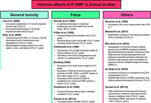
Figure 2. Adverse effects of IF-EMF in animal models in vivo. The magnetic flux density (µT) was rms value, but if it was a peak value, it was indicated as ‘peak’. TNFα: tumor necrosis factor-α; IL-6: interleukin 6; IL-1β: interleukin 1 beta; G: gestation; PD: postnatal day; CNP: 2′,3′-cyclic nucleotide 3′-phosphodiesterase; AChE: acetylcholine esterase; NGF: nerve growth factor; NMDA: N-methyl-d-aspartate; POMC: proopiomelanocortin.
Others
The effects of IF-EMFs on the brain and behavior of rodents have also been investigated. Win-Shwe et al. exposed mice to a 21 kHz IF-EMF at a magnetic flux density of 3800 μT for one hour every day for 2 weeks. The mice were allowed to recover for a week, after which the hypothalamus was removed and the expression of genes related to memory was determined. However, no abnormalities were observed (Win-Shwe et al. Citation2013). Furthermore, other studies have suggested that exposing mice to IF-EMFs during postnatal development may cause genetic/biochemical alterations. IF-EMFs transiently affected the N-methyl-d-aspartate (NMDA) receptor, which is related to crucial signaling pathways and inflammatory mediators in the hippocampus of young adult mice; however, the mice recovered after the termination of exposure without visible histopathological changes (Win-Shwe et al. Citation2015). Ohtani et al. exposed mice to very high magnetic flux density (84 kHz, 2.35 × 10⁴ μT) for 2 weeks (1 hour/day, 5 days/week); however, this exposure level did not affect transcriptional responses in the brain or liver (Ohtani et al. Citation2019). Kumari et al. exposed mice to a 7.5 kHz IF-EMF (12 or 120 μT for 5 weeks) and assessed behavioral and histopathological endpoints. The mice exposed to the IF-EMF for 48 hours in the 120 μT groups displayed impaired memory in passive avoidance tests and the Morris swimming assay, which coincided with a significant increase in the mRNA levels of the pro-inflammatory cytokine tumor necrosis factor alpha (TNFα). However, the IF-EMF did not affect body weight, spontaneous activity, motor coordination, level of anxiety or aggression, or histopathology of the hippocampus (Kumari, Koivisto, et al. Citation2017). Lerchl et al. assessed memory-associated tasks in the eight-arm maze in CD-1 mice exposed to a 20 kHz IF-EMF (360 μT) for 7 months, but no significant differences were observed (Lerchl et al. Citation2021).
Furthermore, three studies investigated the effects of IF-EMFs on hematological parameters and immune modulation in animals. Ushiyama et al. reported that whole-body exposure of Sprague-Dawley (SD) rats to a 21 kHz IF-EMF (3800 μT) for 14 consecutive days (1 hour/day) did not affect immune function in juvenile rats, including biochemical, hematological, and immunological parameters (Ushiyama et al. Citation2014). Kleijn et al. exposed Balb/c mice to ELF- and IF-EMF signals spanning from 20 to 5000 Hz for 1, 4, and 24 hours/day in short-term (1 week) and long-term (15 weeks) experiments to investigate the whole-body effects of these treatments on the level of stress regulation and immune response (de Kleijn et al. Citation2016). Ohtani et al. examined genotoxicity in the blood from mice exposed to high magnetic flux density (84 kHz, 2.35 × 10⁴ μT) for 2 weeks (1 hour/day, 5 days/week) and no adverse effects were observed in the hematological parameters, micronuclei assay and pig-a mutation assay (Ohtani et al. Citation2021).
Most in vivo studies compare the effects of IF-EMF-treated animals to those of sham-exposed animals; however, some studies have implemented specific research models to identify the health effects of IF EMF exposure. For instance, Juutilainen et al. exposed audiogenic seizure (AGS)-susceptible rats to 10 kHz and 28 kHz frequencies as well as a 100 Hz sinusoidal magnetic field. The time from the beginning of the sound stimulus to the onset of the seizure (seizure latency) and the duration of the convulsion was then measured. There was no difference between the sham and IF-EMF exposed groups (Juutilainen et al. Citation1988). Rusovan et al. studied the effects of IF-EMFs on the regeneration of the sciatic nerve in rats. Female SD rats with crush lesions of the right sciatic nerve were exposed to various IF-EMF frequencies (50–2000 Hz) at 100 μT, after which regeneration rate was measured with a pinch test and neurofilaments were characterized via immunohistochemistry. Interestingly, a 1000 Hz IF-EMF increased the regeneration of the sciatic nerve in rats (Rusovan et al. Citation1992). Maaroufi et al. investigated the possible relationship between iron levels, EMF exposure, and brain oxidative stress in young adult rats by exposing rats to high iron concentrations and/or a 150 kHz IF-EMF. Iron overload (IO) did not induce any oxidative stress in young adult rats and instead stimulated antioxidant defenses in the cerebellum and prefrontal cortex. However, IF-EMF exposure both alone or coupled with IO stimulated lipid peroxidation mainly in the cerebellum, thus demonstrating that IF-EMFs may be harmful to young adults by impairing the antioxidant defense system (Maaroufi et al. Citation2011) ().
Discussion
In this paper, initially 239 studies were considered and this review was limited to experimental studies investigating bio/health effects of IF-EMF on rodents. One hundred and eight articles were selected excluding in vitro and ex vivo experimental articles, articles not in mice and rat studies, articles on cancer treatment effect, pulsed RF and other medical device papers, and hypersensitivity articles. Finally, 38 research articles were targeted, excluding articles with uncertain exposure levels or magnetic resonance imaging, articles that were not full text, and articles with inaccurate dosimetry.
In vivo biological effects of IF-EMFs from 38 research articles can be divided into the following categories: analysis of (1) tumorigenesis; (2) animal studies after long-term exposure; (3) effects on developmental stages during gestation; (4) effects on sperm fertility; and (5) recent research that linked IF-EMF exposure during pregnancy or neonatal stage to behavioral and cognition changes. Studies on the effects of IF-EMFs have also evaluated gene expression in the brain after IF-EMF exposure, learning and memory behaviors, and immune function.
Most studies have reported that IF-EMFs do not induce any observable adverse effects. However, some studies have linked the adverse effects of IF-EMF exposure (15 and 66 μT) on early development in mice, including increased malformation rates, decreased fetus brain weight, increased placental resorption rate, increased fetus mortality, and reduced body mass and length of fetuses (Stuchly et al. Citation1988; Frölen et al. Citation1993; Dimberg Citation1995; Svedenstål and Johanson Citation1995; Huuskonen et al. Citation1998b). However, no adverse effects were reported in some studies using higher exposures (up to 200 μT) (Wiley et al. Citation1992; Dawson et al. Citation1998; Nishimura et al. Citation2011, Citation2012). In addition to developmental effects, some adverse effects were reported such as increased or decreased neutrophil count (6.5 μT), increased concentrations of some cytokines (10 μT), and decreased concentrations of testosterone and progesterone (270 μT), although no correlation was observed between the level of exposure of IF-EMF and the induction of adverse events (Lee et al. Citation2006; de Kleijn et al. Citation2016; Zhao et al. Citation2020). Changes in mRNA or protein expression in the brain at various inconsistent exposure levels of IF-EMF have also been reported (Rusovan et al. Citation1992; Maaroufi et al. Citation2011; Win-Shwe et al. Citation2015; Kumari, Koivisto, et al. Citation2017) (). Although various biological effects have been reported in some experimental studies, these results were not independently reproduced and did not depend on the degree of IF-EMF exposure. In addition, animal studies could not identify an exposure threshold of IF-EMF. Available animal studies have provided no confirmed evidence that IF-EMF is associated with bioeffects relevant to human health. Therefore, we concluded that IF-EMF exposure within ICNIRP limits (ICNIRP reference levels: 27 μT for the general public and 100 μT for occupational exposure; ICNIRP Citation2010) did not produce harmful effects on animals. We also suggest that future experimental studies should be continued to monitor health effects in animals related to IF-EMF to provide adequate and sufficient information for a meaningful safety assessment.
Nevertheless, our knowledge about the effects of IF-EMFs remains very limited compared to our understanding of the ELF or RF bands. In fact, the ICNIRP recommendations for IF-EMF were also based on research on the RF and ELF bands. In addition, based on the characteristics of IF-EMFs elucidated so far, these frequencies appear to mainly affect tumorigenesis, reproduction, and development. Moreover, unlike RF- and ELF-EMFs, the role of IF-EMF in degenerative brain disease, neurological diseases, and immune-related diseases is still unknown. Additionally, IF-EMF studies using cell systems are also very lacking, and studies adopting a mechanistic approach to evaluate the effects of IF-EMFs are extremely rare. Therefore, as the use of IF-EMF-based home appliances increases, evaluating the potential health impacts of these frequencies and their exact role in tissue and organ system becomes more urgent.
Conclusions
As the use of IF-EMF-based devices such as induction cookers and wireless power transfer increases, it is meaningful to comprehensively summarize the biological effects of IF-EMF studies conducted so far, especially animal studies. We reviewed a total of 38 experimental studies published from databases EMF Portal and PubMed between January 1988 and August 2021 that used rats or mice to investigate the effects of IF-EMF on prolonged exposure, developmental stages, tumorigenicity, effects on sperm fertility, and other effects that linked IF-EMF exposure during pregnancy or neonatal stage to behavioral and cognition changes. In addition, studies on gene expression in the brain, learning and memory behaviors, and immune functions by IF-EMFs have also been evaluated. Even though most studies have reported that IF-EMFs do not induce any adverse effects, some studies have linked harmful effects on early development in mice. The adverse effects on neutrophil count, cytokines, and the concentrations of testosterone and progesterone, and the alteration of mRNA or protein expression levels in the brain, were also reported. However, the health effects of IF-EMF exposure were not independently reproduced and were not dependent on the degree of IF-EMF exposure. Therefore, we concluded that IF-EMF exposure within ICNIRP limits (ICNIRP reference levels: 27 μT for the general public and 100 μT for occupational exposure) did not produce any harmful effects on animals. Of course, more research needs to be done using the latest biomedical tools; however, a comprehensive review of IF-EMF studies, especially animal studies, will provide a valuable basis for further risk analysis of IF-EMF in humans.
Author contributions
H.-J. Lee: conceptualization, investigation, and writing-original draft preparation. H. Jin: writing-original draft preparation, visualization, investigation, reviewing, and editing. Y.H. Ahn: conceptualization. N. Kim: conceptualization and supervision. J.K. Pack: .conceptualization H.-D. Choi: writing-reviewing and supervision. Y.-S. Lee: conceptualization, writing-original draft preparation, writing-reviewing and editing, and supervision. All authors read and approved the final manuscript.
| Abbreviations | ||
| EMFs | = | electromagnetic fields |
| IF-EMF | = | intermediated frequency electromagnetic field |
| ELF | = | extremely low frequency |
| RF | = | radiofrequency |
| WHO | = | World Health Organization |
| EAS | = | Electronic article surveillance |
| WPT | = | wireless power transfer |
| VDT | = | video display terminal |
| KIRAMS | = | Korea Institute of Radiological and Medical Science |
| EU | = | European Union |
| GERoNiMO | = | Generalized EMF Research using Novel Methods |
| ICNIRP | = | International Commission on Non-Ionizing Radiation Protection |
| SCENIHR | = | The Scientific Committee on Emerging and Newly Identified Health Risks |
| KIEES | = | Korea Institute of Electromagnetic Engineering and Science |
| BfS | = | Bundesamt für Strahlenschutz |
| HF | = | high frequency |
| PubMed | = | Public/Publisher MEDLINE (NLM journal articles database) |
| EMF-PORTAL | = | Electromagnetic Field-Portal |
| DMBA | = | dimethylbenz(a)anthracene |
| BP | = | benzo(a)pyrene |
| TPA | = | tetradecanoyl phorbol ester |
| IR | = | ionizing radiation |
| NMDA | = | N-methyl-d-aspartate |
| TNFα | = | tumor necrosis factor alpha |
| SD | = | Sprague-Dawley |
| AGS | = | audiogenic seizure |
| IO | = | iron overload |
Graphic_abstract.TIF
Download TIFF Image (261.4 KB)Disclosure statement
The authors declare no conflict of interest.
Additional information
Funding
Notes on contributors
Hae-June Lee
Hae-June Lee, a principle researcher in the Korea Institute of Radiological & Medical Sciences. Her research interests include the effects of IF, RF and ELF EMF on animals.
Hee Jin
Hee Jin is a Postdoctoral Researcher at the Ewha Womans University. Her research interests are the target elucidation of radiation-induced lung fibrosis and damage response of EMF.
Young Hwan Ahn
Young Hwan Ahn is a neurosurgeon with a PhD degree in medicine. He is a professor of neurosurgery at the Ajou University Medical Center. His research field is effects of EMF on brain, especially using animals.
Nam Kim
Nam Kim is a professor at the Department of Electronic Engineering, Chungbuk National University. He does research in holography, optical information processing, printed antennas, guideline suggestion from EMF exposure, bioinformatics, and 5G technology.
Jeong Ki Pack
Jeong Ki Pack is a professor emeritus at the Department of Electronic Engineering, Chungnam National University. His research interests are in bioelectromagnetics and electromagnetic wave engineering.
Hyung-Do Choi
Hyung-Do Choi is a principle researcher in the Electronics and Telecommunications Research Institute. He has carried out research in field of biological effects of RF radiation and developed RF radiation protection standards.
Yun-Sil Lee
Yun-Sil Lee is a professor in the college of pharmacy at Ewha Womans University. Her research focuses on radiation biology and bioeffects on EMF. She is working about basic radiation damage response in relationship with cancer development and pulmonary fibrosis.
Related Research Data
References
- Bodewein L, Schmiedchen K, Dechent D, Stunder D, Graefrath D, Winter L, Kraus T, Driessen S. 2019. Systematic review on the biological effects of electric, magnetic and electromagnetic fields in the intermediate frequency range (300 Hz to 1 MHz). Environ Res. 171:247–259.
- Chiang H, Wu R, Shao B, Fu Y, Yao G, Lu D. 1995. Pulsed magnetic field from video display terminals enhances teratogenic effects of cytosine arabinoside in mice. Bioelectromagnetics. 16(1):70–74.
- Dawson BV, Robertson IG, Wilson WR, Zwi LJ, Boys JT, Green AW. 1998. Evaluation of potential health effects of 10 kHz magnetic fields: a rodent reproductive study. Bioelectromagnetics. 19(3):162–171.
- de Kleijn S, Ferwerda G, Wiese M, Trentelman J, Cuppen J, Kozicz T, de Jager L, Hermans PW, Verburg-van Kemenade BM. 2016. A short-term extremely low frequency electromagnetic field exposure increases circulating leukocyte numbers and affects HPA-axis signaling in mice. Bioelectromagnetics. 37(7):433–443.
- Dimberg Y. 1995. Neurochemical effects of a 20 kHz magnetic field on the central nervous system in prenatally exposed mice. Bioelectromagnetics. 16(4):263–267.
- EMF-PORTAL. 2022. [accessed 2022 Mar 14]. www.emf-portal.org.
- Frölen H, Svedenstål BM, Paulsson LE. 1993. Effects of pulsed magnetic fields on the developing mouse embryo. Bioelectromagnetics. 14(3):197–204.
- [GERoNiMO] GERoNiMO – generalised EMF research using novel methods. 2022. [accessed 2022 Mar 14]. https://www.swisstph.ch/fr/projects/project-detail/project/geronimo-generalised-emf-research-using-novel-methods/.
- Herrala M, Kumari K, Koivisto H, Luukkonen J, Tanila H, Naarala J, Juutilainen J. 2018. Genotoxicity of intermediate frequency magnetic fields in vitro and in vivo. Environ Res. 167:759–769.
- Huuskonen H, Juutilainen J, Julkunen A, Mäki-Paakkanen J, Komulainen H. 1998a. Effects of gestational exposure to a video display terminal-like magnetic field (20-kHz) on CBA/S mice. Teratology. 58(5):190–196.
- Huuskonen H, Juutilainen J, Julkunen A, Mäki-Paakkanen J, Komulainen H. 1998b. Effects of low-frequency magnetic fields on fetal development in CBA/Ca mice. Bioelectromagnetics. 19(8):477–485.
- Huuskonen H, Juutilainen J, Komulainen H. 1993. Effects of low‐frequency magnetic fields on fetal development in rats. Bioelectromagnetics. 14(3):205–213.
- [ICNIRP] International Commission on Non-Ionizing Radiation Protection. 1998. Guidelines for limiting exposure to time-varying electric, magnetic, and electromagnetic fields (up to 300 GHz). Health Phys. 74(4):494–522.
- [ICNIRP] International Commission on Non-Ionizing Radiation Protection. 2010. Guidelines for limiting exposure to time-varying electric and magnetic fields (1 Hz to 100 kHz). Health Phys. 99:818–836.
- Juutilainen J, Björk E, Saali K. 1988. Epilepsy and electromagnetic fields: effects of simulated atmospherics and 100-Hz magnetic fields on audiogenic seizure in rats. Int J Biometeorol. 32(1):17–20.
- Juutilainen J, Huuskonen H, Komulainen H. 1997. Increased resorptions in CBA mice exposed to low-frequency magnetic fields: an attempt to replicate earlier observations. Bioelectromagnetics. 18(6):410–417.
- Kim SH, Lee HJ, Choi SY, Gimm YM, Pack JK, Choi HD, Lee YS. 2006. Toxicity bioassay in Sprague-Dawley rats exposed to 20 kHz triangular magnetic field for 90 days. Bioelectromagnetics. 27(2):105–111.
- Kim SH, Song JE, Kim SR, Oh H, Gimm YM, Yoo DS, Pack JK, Lee YS. 2004. Teratological studies of prenatal exposure of mice to a 20 kHz sawtooth magnetic field. Bioelectromagnetics. 25(2):114–117.
- Kumari K, Capstick M, Cassara AM, Herrala M, Koivisto H, Naarala J, Tanila H, Viluksela M, Juutilainen J. 2017. Effects of intermediate frequency magnetic fields on male fertility indicators in mice. Environ Res. 157:64–70.
- Kumari K, Koivisto H, Capstick M, Naarala J, Viluksela M, Tanila H, Juutilainen J. 2018. Behavioural phenotypes in mice after prenatal and early postnatal exposure to intermediate frequency magnetic fields. Environ Res. 162:27–34.
- Kumari K, Koivisto H, Viluksela M, Paldanius KMA, Marttinen M, Hiltunen M, Naarala J, Tanila H, Juutilainen J. 2017. Behavioral testing of mice exposed to intermediate frequency magnetic fields indicates mild memory impairment. PLOS One. 12(12):e0188880.
- Lee HJ, Choi SY, Jang JJ, Gimm YM, Pack JK, Choi HD, Kim N, Lee YS. 2007. Lack of promotion of mammary, lung and skin tumorigenesis by 20 kHz triangular magnetic fields. Bioelectromagnetics. 28(6):446–453.
- Lee HJ, Gimm YM, Choi HD, Kim N, Kim SH, Lee YS. 2010. Chronic exposure of Sprague-Dawley rats to 20 kHz triangular magnetic fields. Int J Radiat Biol. 86(5):384–389.
- Lee HJ, Kim SH, Choi SY, Gimm YM, Pack JK, Choi HD, Lee YS. 2006. Long-term exposure of Sprague Dawley rats to 20 kHz triangular magnetic fields. Int J Radiat Biol. 82(4):285–291.
- Lee HJ, Pack JK, Gimm YM, Choi HD, Kim N, Kim SH, Lee YS. 2009. Teratological evaluation of mouse fetuses exposed to a 20 kHz EMF. Bioelectromagnetics. 30(4):330–333.
- Lerchl A, Drees Née Grote K, Gronau I, Fischer D, Bauch J, Hoppe A. 2021. Effects of long-term exposure of intermediate frequency magnetic fields (20 kHz, 360 µT) on the development, pathological findings, and behavior of female mice. Bioelectromagnetics. 42(4):309–316.
- Maaroufi K, Save E, Poucet B, Sakly M, Abdelmelek H, Had-Aissouni L. 2011. Oxidative stress and prevention of the adaptive response to chronic iron overload in the brain of young adult rats exposed to a 150 kilohertz electromagnetic field. Neuroscience. 186:39–47.
- Nishimura I, Doi Y, Imai N, Kawabe M, Mera Y, Shiina T. 2019. Carcinogenicity of intermediate frequency magnetic field in Tg.rasH2 mice. Bioelectromagnetics. 40(3):160–169.
- Nishimura I, Oshima A, Shibuya K, Mitani T, Negishi T. 2012. Absence of reproductive and developmental toxicity in rats following exposure to a 20-kHz or 60-kHz magnetic field. Regul Toxicol Pharmacol. 64(3):394–401.
- Nishimura I, Oshima A, Shibuya K, Mitani T, Negishi T. 2016. Acute and subchronic toxicity of 20 kHz and 60 kHz magnetic fields in rats. J Appl Toxicol. 36(2):199–210.
- Nishimura I, Oshima A, Shibuya K, Negishi T. 2011. Lack of teratological effects in rats exposed to 20 or 60 kHz magnetic fields. Birth Defects Res B Dev Reprod Toxicol. 92(5):469–477.
- Ohtani S, Ushiyama A, Maeda M, Wada K, Suzuki Y, Hattori K, Kunugita N, Ishii K. 2019. Global analysis of transcriptional expression in mice exposed to intermediate frequency magnetic fields utilized for wireless power transfer systems. Int J Environ Res Public Health. 16(10):1851.
- Ohtani S, Ushiyama A, Wada K, Suzuki Y, Ishii K, Hattori K. 2021. No evidence for genotoxicity in mice due to exposure to intermediate-frequency magnetic fields used for wireless power-transfer systems. Mutat Res Genet Toxicol Environ Mutagen. 863–864:503310.
- PubMed. 2022. [accessed 2022 Mar 14]. https://pubmed.ncbi.nlm.nih.gov/.
- Robertson IG, Wilson WR, Dawson BV, Zwi LJ, Green AW, Boys JT. 1996. Evaluation of potential health effects of 10 kHz magnetic fields: a short-term mouse toxicology study. Bioelectromagnetics. 17(2):111–122.
- Rusovan A, Kanje M, Mild KH. 1992. The stimulatory effect of magnetic fields on regeneration of the rat sciatic nerve is frequency dependent. Exp Neurol. 117(1):81–84.
- [SCENIHR] Scientific Committee on Emerging and Newly Identified Health Risks. 2015. Potential health effects of exposure to electromagnetic fields (EMF); [accessed 2022 Mar 14]. https://ec.europa.eu/health/scientific_committees/emerging/docs/scenihr_o_04.1.pdf.
- Stuchly MA, Ruddick J, Villeneuve D, Robinson K, Reed B, Lecuyer DW, Tan K, Wong J. 1988. Teratological assessment of exposure to time-varying magnetic field. Teratology. 38(5):461–466.
- Svedenstål B-M, Johanson K-J. 1998. Leukocytes and micronucleated erythrocytes in peripheral blood from mice exposed to 50-Hz or 20-kHz magnetic fields. Electro Magnetobiol. 17(2):127–143.
- Svedenstål BM, Holmberg B. 1993. Lymphoma development among mice exposed to X-rays and pulsed magnetic fields. Int J Radiat Biol. 64(1):119–125.
- Svedenstål BM, Johanson KJ. 1995. Fetal loss in mice exposed to magnetic fields during early pregnancy. Bioelectromagnetics. 16(5):284–289.
- The International EMF Project. 2022. [accessed 2022 Mar 14]. https://www.who.int/initiatives/the-international-emf-project.
- Ushiyama A, Ohtani S, Suzuki Y, Wada K, Kunugita N, Ohkubo C. 2014. Effects of 21-kHz intermediate frequency magnetic fields on blood properties and immune systems of juvenile rats. Int J Radiat Biol. 90(12):1211–1217.
- Vandamme TF. 2014. Use of rodents as models of human diseases. J Pharm Bioallied Sci. 6(1):2–9.
- WHO. 2005. World Health Organization Fact Sheet; [accessed 2022 Mar 14]. https://www.who.int/peh-emf/publications/facts/EHS_Factsheet_296_English.pdf.
- Wiley MJ, Corey P, Kavet R, Charry J, Harvey S, Agnew D, Walsh M. 1992. The effects of continuous exposure to 20-kHz sawtooth magnetic fields on the litters of CD-1 mice. Teratology. 46(4):391–398.
- Win-Shwe TT, Ohtani S, Ushiyama A, Fujimaki H, Kunugita N. 2013. Can intermediate-frequency magnetic fields affect memory function-related gene expressions in hippocampus of C57BL/6J mice? J Toxicol Sci. 38(2):169–176.
- Win-Shwe TT, Ohtani S, Ushiyama A, Kunugita N. 2015. Early exposure to intermediate-frequency magnetic fields alters brain biomarkers without histopathological changes in adult mice. Int J Environ Res Public Health. 12(4):4406–4421.
- Zhao J, Wu Z, Yang T, Zhao Y, Wang L. 2020. Electromagnetic biological effect on mice in wireless power transmission system. IEEE Access. 8:205558–205567.