Abstract
As part of an analysis of type material of Amphora species described by Hustedt, the lectotypes of Amphora fontinalis, A. rugosa and A. schroederi and the holotype of A. subturgida were analysed. Light microscopy showed that all these tropical freshwater taxa are similar in frustule and valve outline, position of the raphe, appearance of the dorsal striae and thickening of the central dorsal virgae. Scanning electron microscopy allowed reliable differentiation between the four, based on the morphology of dorsal striae, dorsal areolae, conopeum, proximal raphe fissures, presence or absence of an inner longitudinal rib close to the raphe sternum, and degree of thickening of the central dorsal virgae. Previous reports of A. fontinalis from Tahiti and Moorea were confirmed while reports of this species from Australia and of A. subturgida from Mauritius, Papua and Israel were refuted. Based on our results, species descriptions are emended and the distributions of these four species are revised. Comparisons of these species and others of the subgenus Halamphora are presented.
Introduction
Amphora Ehrenberg is a very large and heterogeneous genus represented in marine, continental and estuarine environments. Its frustule resembles “a third of an orange” (Hendey,Citation1964), with both valves on the ventral side of the cell. A modern critical analysis of the whole genus is still necessary and references must often be made to classic works, such as Cleve (Citation1895), Van Heurck (Citation1885) and Peragallo & Peragallo (Citation1901). Over a century ago, Cleve (Citation1895) believed that Amphora should be split into several genera, considering that its members showed affinities to different naviculoid diatoms. However, to avoid taxonomic confusion and to allow his ideas to be tested, Cleve (Citation1895) maintained Amphora, but delimited nine subgenera, based on frustule and valve outline, girdle band striation, raphe position and stria type. This taxonomic arrangement has been followed by modern authors (e.g. Patrick & Reimer, Citation1975; Bérard-Therriault & Cardinal, Citation1986), although it is recognized that Cleve's subgenera need revision. The subgenus Halamphora Cleve was characterised as having elliptical to lanceolate frustules, a girdle composed of numerous, punctate bands, valves usually with rostrate or capitate ends and punctate striae, a straight or slightly curved raphe near the ventral margin, and a narrow raphe sternum.
This paper is part of a series devoted to the analysis of the type material of species of the subgenus Halamphora, which were described by Hustedt and which are easily misidentified when examined with light microscopy (LM). In their comprehensive paper on Amphora coffeaeformis (Agardh) Kützing, Archibald & Schoeman (Citation1984) set out the necessity of studying type material in order to fix the correct application of names. This opinion has been endorsed by Sala et al. (Citation1998), in a revision of material from Argentina reported as A. coffeaeformis, and by Clavero et al. (Citation2000) in their analysis of type material of Amphora tenerrima Aleem & Hustedt and Amphora tenuissima Hustedt. Type material of Amphora holsatica Hustedt (Sar et al., Citation2003) and Amphora tumida Hustedt (Sar et al.,Citation2004) were studied subsequently and it was concluded that particular ultrastructural features of the valves and frustules allow the specific limits of these taxa to be defined.
Amphora subturgida Hustedt, Amphora fontinalis Hustedt and Amphora rugosa Hustedt were described (Hustedt,Citation1938) from material collected from different localities on Java and Sumatra, while Amphora schroederi Hustedt had been described (Hustedt,Citation1921) from material collected in East Africa. Simonsen (Citation1987) examined the slides selected by him as the lectotype of A. fontinalis, A. rugosa and A. schroederi and Hustedt's holotype of A. subturgida with LM, but the photographs are inadequate to reveal details of the striae, areolae, conopeum, raphe and girdle bands, features that allow specific differentiation within the subgenus Halamphora.
Van Landingham (Citation1967) recognized all four species as valid, but gave no other references, which suggests that the taxa had not been cited in more general floras up to 1967. According to Gaul et al. (Citation1993) and Henderson & Reimer (Citation2003), A. fontinalis has been reported by Coste & Ricard (Citation1990) and A. subturgida by Coste & Ricard (Citation1982, Citation1984), Vyverman (Citation1991) and Ehrlich (Citation1995), whereas the absence of A. rugosa and A. schroederi from these catalogues (Gaul et al., Citation1993, Henderson & Reimer,Citation2003) suggests that (prior to 1999) there were no published studies about them.
In this study we carry out a revision of type material of A. fontinalis, A. rugosa, A. schroederi and A. subturgida with LM and scanning electron microscopy (SEM) to provide a comprehensive description of these species and to compare them with each other, as they are the most morphologically similar species within the subgenus Halamphora.
Materials and methods
Slides and samples in the Hustedt Collection at the Alfred Wegener Institut für Polar und Meeresforschung in Bremerhaven (BRM, Germany) were used for this study (). In order to obtain morphometric data on as many specimens as possible we included those illustrated in Simonsen (Citation1987) and other specimens found by us on type slides and in type material. Photomicrographs were obtained with an Axioplan D 7082 (LM) in bright field and with Nomarski differential interference contrast.
Material in samples used by Hustedt to prepare the type slides was also mounted for SEM according to Ferrario et al. (Citation1995). Photomicrographs were obtained with a SEM ISI-DS 130 and FEI Quanta FEG 200.
The measurements of areolae and striae were done according to Anonymous (Citation1975) and the terminology used is that suggested by Ross et al. (Citation1979), Cox & Ross (Citation1981) and Lee & Round (Citation1987). Details of frustule construction in the genus Amphora and of valve morphology viewed from different angles can be found in Schoeman & Archibald (Citation1979).
Results
Amphora fontinalis
We found several specimens (sample As271, slide U1/88) that match in valve outline and general appearance those described as A. fontinalis by Hustedt (Citation1938). There were only minor differences between our measurements and those given in the protologue, and we think that all the cited material belongs to the same species. shows the morphometric data from the protologue, Simonsen (Citation1987, pl. 69, figs 10–15) and the specimens we analysed. SEM analysis allowed us to establish that the specimens share the same fine structure.
Amphora fontinalis Hustedt emend. Sala, Sar, Hinz & Sunesen (Figs )
Figs 1–3. Amphora fontinalis type material: LM. Figs 1, 2. Frustule in ventral view, same specimen at different focus. Fig. 3. Frustule in dorsal view. Scale bars: 5 µm.
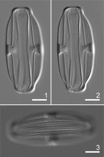
Figs 4–14. Amphora fontinalis type material: SEM. Note that valves acquire different aspects when seen from different planes of view. Fig. 4. Frustule in ventral view. Note the ventral striae interrupted at mid-valve and girdle bands. Fig. 5. Frustule in dorsal view. Note the valve mantles and girdle bands. Fig. 6. Valve in external view showing the narrow conopeum, the pattern of striation and the stauroid area. Fig. 7. Valve in internal view showing the dorsal striae and the thickened central stauroid area. Fig. 8. Detail of central part of valve. Note the dorsal striae composed of a single row of rectangular, sometimes round areolae, central striae with ‘ghost’ areolae. Fig. 9. Detail of valve apex showing terminal fissure and the conopeum broken near the pole. Fig. 10. Valve centre in internal view. Note structure of the stauroid area. Fig. 11. Valve centre of a tilted specimen. See the areolae occluded by hymenes and the ventral striae interrupted at mid-valve. Fig. 12. Valve pole of specimen in Fig. 11 showing the sternum and the helictoglossa. Fig. 13. Detail of frustule end in dorsal view. Note open girdle bands showing a double row of areolae. Arrowhead indicates the conopeum. Fig. 14. Girdle bands in dorsal internal view showing the areolae occluded by hymenes. Scale bars: Figs 4–7: 5 µm; Figs 8–13: 2 µm; Fig. 14: 1 µm.
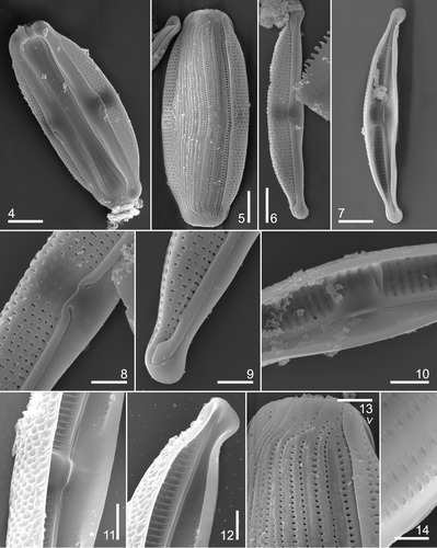
Table 1. Details of slides and samples examined of Amphora spp.
Table 2. Morphometric comparison of the analysed species showing combined data, data from the protologues, Simonsen (Citation1987) and our observations. Abbreviations: F: frustule; V: valve; C: centre; P: poles; LM: light microscopy; SEM: scanning electron microscopy. –, no data; *, measured from Hustedt's figures.
Hustedt (Citation1938). Systematische und ökologische Untersuchungen über die Diatomeen-Flora von Java, Bali und Sumatra. Systematischer Teil I, Schluβ. Arch. Hydrobiol., Suppl., 15: 414–415, pl. 24, figs 4, 5.
LECTOTYPE: BRM, Slide U1/88, designated by Simonsen (Citation1987: 234).
TYPE LOCALITY: Ranu Pakis, Java.
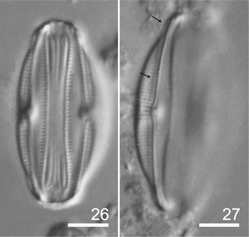
Light microscopy ()
Frustule elliptic, slightly constricted in the middle with gently protracted, truncate ends, 20–33 µm long, 8–15 µm wide. Valves nearly elliptical in ventral girdle view, 4–6 µm wide, with convex dorsal and ventral margins, poles ventrally deflected with protracted apices. Raphe distant from the ventral margin. Raphe branches slightly curved, with proximal ends bent towards the dorsal side. Central nodule conspicuous, merging with the dorsal central virgae and forming a stauroid structure. Dorsal striae slightly radial throughout, 19–23 in 10 µm at the centre, more closely spaced towards the ends, 20–24 in 10 µm, striae clearly punctate. Ventral striae short, interrupted at valve centre, 26–36.5 in 10 µm. Dorsal central virgae thickened. Intercalary bands numerous (about 20 in 10 µm) and distinctly porous.
Scanning electron microscopy ()
Valve vaulted, dorsal side convex with a high mantle, ventral valve side wide and plain over most of its surface, curving abruptly into a shallow mantle. Dorsal striae uniseriate, with 20–29 areolae in 10 µm, areolae rectangular to round, occluded internally by hymenes. ‘Ghost’ areolae visible externally at the proximal ends of the central striae. Virgae thickened internally only. Internal, dorsal stauroid structure conspicuous. Single ventral row of small linear areolae, occluded internally by hymenes, at some distance from the valve margin.
Raphe slightly curved, distant from ventral margin, opening internally in a distinct sternum, narrower on the ventral than the dorsal side. Internal proximal raphe ends deflected ventrally, terminating under a tongue-like expansion. Terminal ends deflected ventrally in poorly developed helictoglossae. External proximal and terminal fissures deflected dorsally; central pores dilated. Conopeum narrow, slightly indented or straight at the centre and slightly wider at the poles.
Girdle composed of numerous broad, open bands, with one row of ovoid or round poroids on the ventral side of the frustule and two rows on the dorsal side, 24–33 areolae in 10 µm.
Amphora rugosa
We found several specimens (sample As733, slide U1/60) that match in valve outline and general appearance those described as Amphora rugosa by Hustedt (Citation1938) but not in their morphometric parameters. shows that specimens analysed in this study with LM and SEM agree with those illustrated by Simonsen (Citation1987, pl. 342, figs 11–15), but are narrower and have more dorsal striae in 10 µm than those described in the protologue. Simonsen (Citation1987) pointed out that “none of the marked specimens on slide U1/60 is in a favourable position for photography. Therefore unmarked specimens were selected”. He attributed the differences in the striation pattern between the specimens found by him and those of protologue illustrations to a mistake in the scale given by Hustedt (1500/1 instead of 2000/1). Based on the above, we consider that Hustedt's, Simonsen's and our own specimens are conspecific and, in giving the dorsal areola density, we disallow the protologue measurements. SEM analysis allowed us to establish that all the specimens share the same fine structure.
Amphora rugosa Hustedt emend. Sala, Sar, Hinz & Sunesen (Figs )
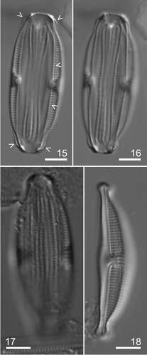
Figs 15–18. Amphora rugosa type material: LM. Figs 15–16. Frustule in ventral view. Same specimen, note the stauroid area in both focuses and the ventral striae in Fig. 15. Arrowheads indicate the conopeum. Fig. 17. Frustule in dorsal view. Note the punctate girdle bands. Fig. 18. General appearance of the valve. Scale bars: 5 µm.
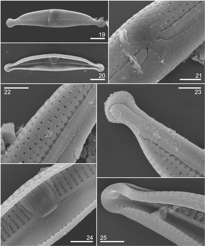
Figs 19–25. Amphora rugosa type material: SEM. Fig. 19. Valve in external view. Fig. 20. Valve in internal view. Fig. 21. Detail of central part of valve corresponding to specimen in Fig. 19. Note proximal raphe fissures bent first to the dorsal side and then to the centre, the conopeum interrupted at the centre and the ventral striation continuous at mid-valve. Fig. 22. Detail of the striae composed by a proximal short portion with two rows of small roundish areolae arranged in quincunx and a distal part with a single row of round or rectangular points. Fig. 23. Detail of valve apex showing the conopeum slightly broadened at the pole and the terminal fissure. Fig. 24. Valve centre showing the striae structure, the thickened central virgae and proximal raphe ends terminating under a tongue-like expansion. Note that dorsal central striae are shorter and narrower than the others. Fig. 25. Detail of valve pole showing the terminal raphe end ventrally deflected in poorly developed helictoglossa. Scale bars: Figs 19, 20: 5 µm; Figs 21–25: 2 µm.
Hustedt (Citation1938). Systematische und ökologische Untersuchungen über die Diatomeen-Flora von Java, Bali und Sumatra. Systematischer Teil I, Schluβ. Arch. Hydrobiol., Suppl., 15: 415–416, pl. 24, .
LECTOTYPE: BRM, Slide U1/60, designated by Simonsen (Citation1987: 234).
TYPE LOCALITY: Panjingahan, Sumatra.
Light microscopy ()
Frustule elliptic, constricted in the middle with slightly protracted, truncate ends, 28–45 µm long, 11.5–18 µm wide. Valves semi-elliptical, 5–8 µm wide, with convex dorsal margin and tumid ventral margin. Poles ventrally deflected with capitate apices. Raphe distant from the ventral margin. Raphe branches almost straight with proximal ends strongly bent towards the dorsal side. Central nodule conspicuous, merging with the dorsal central virgae in a stauroid structure. Dorsal striae slightly radial throughout, distinctly porous, 20 in 10 µm; central ones short, nearly the half of the other, 16–19 in 10 µm at the centre, 17–20 at the poles. Ventral striae short, continuous at mid-valve, 20–25 in 10 µm at the centre, 23–25 at the poles. Dorsal central virgae wider than the others. Conopeum not visible except towards the poles. Intercalary bands numerous, distinctly porous, 16–23 areolae in 10 µm.
Scanning electron microscopy ()
Valve vaulted, dorsal side convex, with a high mantle; ventral side wide, curved, plain over the major part of the surface, turned into a shallow mantle. Dorsal striae composed of a proximal short portion with two rows of small round areolae, and a distal part with a single row of round or rectangular openings in external view, with two rows of small round areolae internally. Central striae shorter and narrower than the others. Virgae thickened internally only, forming a stauroid structure in the middle of the valve. Continuous single row of small linear areolae at the centre of the ventral side of the valve. Raphe branches slightly curved, opening internally in a distinctive sternum. Internal proximal raphe ends bent ventrally, terminating under a tongue-like expansion. Terminal ends deflected ventrally in poorly developed helictoglossae. Proximal fissures bent at a right angle to the dorsal side with dilated central pores; terminal fissures curved to the dorsal side. Conopeum developed on the dorsal side of the valve, straight, interrupted at the centre and slightly broadened at the poles. Girdle composed of broad open bands, with double rows of poroids on the dorsal side.
Amphora schroederi
We found several specimens (sample A34 and slide 219/83) that match, in valve outline, general appearance and morphometric parameters, those described as A. schroederi by Hustedt (Citation1921). Nevertheless, the range of measurements is broader than that given in the protologue. shows morphometric data from the protologue, the specimens figured by Simonsen (Citation1987, pl. 69, figs 10–15, and specimens analysed in this study with LM and SEM. Hustedt (Citation1921) pointed out that the ventral valve side lacked structure, but all the specimens show ventral striae along the margin. This difference could be attributed to the position of the specimens analysed by Hustedt. SEM analysis allowed us to establish that all the specimens are coincident in fine structure.
Amphora schroederi Hustedt emend. Sala, Sar, Hinz & Sunesen ()
Figs 26–27. Amphora schroederi type material: LM. Fig. 26. Frustule in ventral view. Fig. 27. Valve general appearance. Arrows indicate the conopeum. Scale bars: 5 µm.
Hustedt (Citation1921). Bacillariales. Hedwigia, 63: 116, figs 16–18.
LECTOTYPE: BRM, Slide 219/83, designated by Simonsen (Citation1987: 55).
TYPE LOCALITY: Taveta River, East Africa.
Light microscopy ()
Frustule elliptic, constricted in the middle part with slightly protracted, truncate ends, 18–26.5 µm long, 9–12 µm wide. Valves nearly elliptical in ventral girdle view, 4–5.2 µm wide, the convex dorsal margin impressed at the centre and convex ventral margin, poles ventrally deflected with protracted apices. Raphe distant from the ventral margin. Raphe branches almost straight forming an obtuse angle, with proximal ends gently bent towards the dorsal side. Central nodule conspicuous. Dorsal striae indistinctly porous, slightly radial throughout, 16–24 in 10 µm at the centre, more closely spaced towards the ends, 22–27 in 10 µm. Ventral striae short, densely arranged, not interrupted at mid-valve. Dorsal central virgae wider than the others. Conopeum scarcely visible as a dark line across the dorsal striae. Intercalary bands numerous and distinctly porous.
Scanning electron microscopy ()
Figs 28–37. Amphora schroederi type material: SEM. Note that valves present different aspects when seen from different planes of view. Fig. 28. Valve in external view. Fig. 29. Valve in internal view. Fig. 30. Disarticulated frustule in ventral view showing ventral side of the valve, wide, structureless in a major part of the surface, with short striae continuous through the centre. Girdle with numerous open areolate bands. Figs 31 and 32. Detail of central part of valve from two different specimens. Note the dorsal striae composed of a proximal short portion biseriate and distal portion uniseriate. Fig. 33. Detail of valve apex showing the conopeum broadened at the pole. Fig. 34. Valve centre of specimen in Fig. 29 at higher magnification. Dorsal central striae are shorter than the others, with thickened central virgae, areolae occluded by hymenes and proximal raphe ends terminating under a tongue–like expansion. Fig. 35. Valve pole of specimen in Fig. 29 at higher magnification. Terminal raphe end is ventrally deflected in poorly developed helictoglossa. Fig. 36. Detail of the striae morphology in external view. Major part of the double row of areolae is placed under the conopeum, broken in the photographed valve. Fig. 37. Detail of portion of the striae with double row of areolae in internal view. Scale bars: Figs 28–30: 5 µm; Figs 31–36: 2 µm; Fig. 37: 0.5 µm.
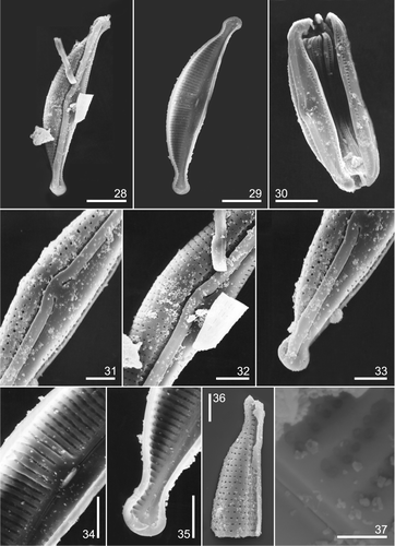
Valve vaulted, dorsal side convex with a high almost vertical mantle, ventral side wide and structureless over most of its surface, curving abruptly into a shallow mantle. Dorsal striae with a proximal very short portion of two rows of small roundish areolae arranged opposite one another or in quincunx, and a distal portion of a single row of round to rectangular areolae, 30–35 in 10 µm. Central striae shorter, without the biseriate portion. Virgae internally thickened, particularly in the middle of the valve. Single ventral row of small linear areolae, circular at the centre, occluded internally by hymenes, 20–26 in 10 µm at the centre, 24–29 at the ends. Raphe distant from the ventral margin, opening internally in a distinctive sternum, wider on the dorsal than on the ventral side. Internal proximal raphe ends deflected ventrally, terminating under a tongue-like expansion. Terminal ends bent ventrally in poorly developed helictoglossae. Proximal and terminal fissures dorsally bent, central pores dilated. Conopeum well developed on dorsal side of the valve, broadened at the poles. Girdle with numerous broad open bands, with one row of ovoid poroids on the ventral side, 28–34 in 10 µm.
Amphora subturgida
We found numerous specimens (sample As486 and slide U1/90), that match those described as A. subturgida by Hustedt (Citation1938) in valve outline, general appearance and morphometric parameters. The only conflict was in the density of the dorsal central striae, which was higher in the specimens located by us and those figured by Simonsen (Citation1987) from the holotype. In we present the morphometric data from the protologue, of the specimens figured in Simonsen (Citation1987, pl. 341, figs 5–8) and of the specimens analysed in this study with light and scanning electron microscopy. As in the previous cases, when we analysed this material with SEM we could establish that all the specimens found are coincident in fine structure.
Amphora subturgida Hustedt emend. Sala, Sar, Hinz & Sunesen (Figs )
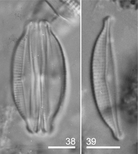
Figs 38–39. Amphora subturgida type material: LM. Fig. 38. Frustule in ventral view. Fig. 39. General appearance of the valve. Scale bars: 5 µm.
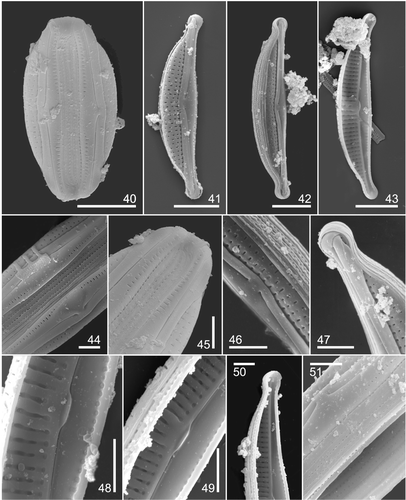
Figs 40–51. Amphora subturgida type material: SEM. Fig. 40. Frustule in ventral view. Fig. 41. Valve in external view. Fig. 42. Valve with girdle bands. Fig. 43. Valve in internal view. Fig. 44. Detail of central part of frustule in ventral view showing ventral striae, and girdle bands with a row of areolae. Fig. 45. Frustule pole of specimen in Fig. 40 at higher magnification. Note the open girdle bands and ventral striae. Fig. 46. Detail of central part of valve. Note the dorsal striae composed of a row of areolae throughout. Proximal raphe fissures dorsally bent and dilated in central pores. Fig. 47. Detail of valve apex showing the conopeum broadened at the pole and with a low cut neck at the end. Fig. 48. Detail of dorsal and ventral striae at the valve centre. Longitudinal rib close to the raphe-sternum interrupted at the valve centre and areolae occluded by hymenes. Fig. 49. Detail of valve centre showing thickened central virgae and proximal raphe ends terminating under a tongue-like expansion. Fig. 50. Valve pole showing terminal raphe end in a poorly developed helictoglossa. Fig. 51. Detail of central part of frustule without one of the valves, showing girdle bands in internal dorsal view with two rows of areolae and external ventral view with one row of areolae. Scale bars: Figs 40–43: 5 µm; Figs 44–51: 2 µm.
Hustedt (Citation1938). Systematische und ökologische Untersuchungen über die Diatomeen-Flora von Java, Bali und Sumatra. Systematischer Teil I, Schluβ. Arch. Hydrobiol., Suppl., 15: 116–117, pl. 24, figs 9–11.
HOLOTYPE: BRM, Slide U1/90, named on the label.
TYPE LOCALITY: Ajer Panas, Tjibodas, Java.
Light microscopy ()
Frustule elliptic, gently constricted in the middle part with slightly protracted, truncate ends, 14–25.2 µm long, 7–11 µm wide. Valves semi-elliptical with convex ventral margin, poles ventrally deflected with protracted apices. Raphe distant from the ventral margin. Raphe branches forming an obtuse angle. Central nodule conspicuous. Dorsal striae distinctly porous, parallel at valve centre, 18–24 in 10 µm, and radial at the ends, 20–27 in 10 µm. Ventral striae hardly visible. Dorsal central virgae slightly wider than the others. Conopeum scarcely visible as a dark line across the dorsal striae.
Scanning electron microscopy ()
Valve vaulted, dorsal side convex with a high mantle, ventral side wide and structureless over most of its surface with a shallow mantle. Dorsal striae uniseriate; areolae, 21–30 in 10 µm, transapically elongated. Single row of areolae close to the raphe-sternum delimited by an internal longitudinal rib. Virgae thickened internally only, more developed in the middle of the valve. Ventral striation composed of a single row of small elongate areolae, circular, sometimes inconspicuous, at the centre. Areolae internally occluded by hymenes. Raphe with internal proximal ends bent ventrally, terminating under a tongue-like expansion. Terminal ends deflected ventrally in poorly developed helictoglossae. Proximal and terminal fissures bent dorsally, central pores dilated. Conopeum well developed on the dorsal side of the valve, broadened at the poles, reaching the dorsal valve margin with a low cut neck at the ends. Girdle with numerous broad open bands. Bands with a row of small, round poroids on the ventral side, and two rows on dorsal side.
Discussion
Morphology and discrimination of the analysed species
Amphora fontinalis, A. rugosa, A. schroederi and A. subturgida are all tropical freshwater taxa that, in LM, are similar in frustule and valve outline, the position of the raphe (defining a wide ventral side), the appearance of the dorsal striae (clearly or indistinctly porous), and the variously thickened central dorsal virgae. On the basis of morphometric data obtained with LM, A. rugosa might be easily confused with A. fontinalis, but the former differs from A. subturgida and A. schroederi in length and width of the frustule and valve, and in the density of the girdle band areolae (). Amphora rugosa and A. fontinalis can be distinguished by their ventral striae, continuous in the former and interrupted in mid-valve in the latter. Amphora schroederi differs subtly from A. subturgida, having a more noticeably thickened central area on its dorsal side, a broader ventral valve side and the indistinctly punctate striae.
Discrimination between the four species is more reliable when they are analysed with SEM, since they differ in some ultrastructural features. One of these features is areola density, which is lower in A. rugosa than the other species. Nevertheless, the most striking differential characters are the morphology of the dorsal striae, dorsal areolae, conopeum, and proximal raphe fissures. The presence or absence of an inner longitudinal rib close to the raphe sternum and the degree of thickening of the central dorsal virgae are also useful characters. Amphora fontinalis and A. subturgida have uniseriate dorsal striae comprising rectangular or round areolae, but the former can be distinguished by its central striae with ‘ghost’ areolae, while the latter has a longitudinal rib close to the raphe sternum, interrupting the dorsal striae. Unlike the preceding species, A. rugosa and A. schroederi have a complex type of striae, similar in both taxa at the proximal short portion but dissimilar at the distal portion. Additionally, there are differences in the thickening of the virgae at the dorsal central area. This thickening is more developed in A. rugosa and A. fontinalis, where it forms a stauroid structure, less obvious in A. schroederi and indistinct in A. subturgida.
The conopeum of the four taxa differs in morphology and degree of development. In A. fontinalis, A. schroederi and A. subturgida it is continuous throughout, while in A. rugosa it is interrupted in the middle of the valve. Based on SEM analysis of several Amphora species of the subgenus Halamphora (Archibald & Schoeman,Citation1984; Schoeman & Archibald,Citation1984; Nagumo & Kobayasi, Citation1990; Sánchez Castillo,Citation1993; Krammer,Citation1997; Clavero et al.,Citation2000; Sala & Maidana,Citation2003; Sar et al.,Citation2003, Citation2004; this study), this feature is exclusive to A. rugosa.
Finally, we found some dissimilarity in the path of the central raphe fissures, which are more strongly bent to the dorsal side in A. fontinalis and A. rugosa than in the other two species.
Distribution
Amphora fontinalis was reported from Tahiti and Moorea, Polynesia (Coste & Ricard, Citation1990) and Australia (John,Citation1983). Although their published micrographs are inadequate to see some details of the valve structure, it is possible to determine that the LM illustrations in Coste & Ricard (Citation1990, pl. 1, figs 19, 20) correspond to this taxon. However, Coste & Ricard's specimen (Coste & Ricard, Citation1990, pl. 5, fig. 19), and those in John (Citation1983, pl. 62, figs 3–5) differ in valve outline and ventral stria arrangement, and we consider they correspond to other taxa that we could not identify with certainty from the available illustrations.
Amphora subturgida has been recorded by Coste & Ricard (Citation1982, 1984), Vyverman (Citation1991) and Ehrlich (Citation1995). Based on their eletron micrograph illustrations however, we believe that the specimens were misidentified. The material from Mauritius (Coste & Ricard, Citation1982, pl. 4, fig. 3, pl. 8, fig. 11; Coste & Ricard, Citation1984, fig. 67), Papua (Vyverman,Citation1991, pl. 118, figs b–d) and Israel (Ehrlich,Citation1995, pl. 38, figs 7, 10, non fig. 11) agree with the type material of A. tumida (Sar et al.,Citation2004). However, the specimen illustrated by Ehrlich (Citation1995, pl. 38, fig. 11) differs in stria morphology from A. tumida and does not agree with A. subturgida or any other known taxon.
Amphora fontinalis was abundant in samples from Java and Sumatra, living in running freshwater bodies, showing a most favourable development in alkaline waters (Hustedt,Citation1938) and was also present in Tahiti and Moorea, Polynesia (Coste & Ricard, Citation1990). Amphora rugosa was reported from Java and Sumatra, as living in running, alkaline freshwater bodies and Amphora subturgida from Java (thermal waters), Sumatra and Bali (Hustedt,Citation1938). Finally, Amphora schroederi was found living on mud in the Taveta River, East Africa (Hustedt,Citation1921).
Comparison with related species
Amphora fontinalis, A. rugosa, A. schroederi and A. subturgida form a group within the subgenus Halamphora based on the position of the raphe, defining a wide valve ventral side, and the presence of a dorsal stauroid structure. Amphora montana Krasske, A. submontana Hustedt and A. normanii Rabenhorst also share these features. The LM micrographs of the type material of A. montana (Lange-Bertalot et al.,Citation1996, pl. 37, figs 8–8′) and those of A. submontana (Simonsen,Citation1987, pl. 527, figs 9–10) are sufficient to differentiate both species from those treated in this paper. Their dorsal and ventral striae are more densely arranged and the ventral striae almost reach the raphe sternum. However, light micrographs of A. normanii (Schoeman & Archibald, Citation1978, figs 1–6), considered possible type material, are insufficient to delimit this species from the taxa studied in this paper. Considering the morphometric data given by Patrick & Reimer (Citation1975), Schoeman & Archibald (Citation1978) and Carter & Round (Citation1993), A. normanii could easily be confused with A. schroederi and A. subturgida. The ultrastructural analyses of A. normanii carried out by Schoeman & Archibald (Citation1978) and Carter & Round (Citation1993) are insufficient for adequate comparison between this species, A. schroederi and A. subturgida. This limitation is due to the fact that their descriptions were based on non-type material, and that the illustrated specimens (from England and USA) differ in dorsal stria structure and conopeum morphology. Thus, there are doubts about the true identity of A. normanii that can only be resolved through ultrastructural analysis of Rabenhorst's material.
Even with LM, the above taxa are easily distinguishable from the rest of the subgenus, as species with a narrow valve ventral side but lacking a dorsal stauroid structure. The latter taxa, whose type material has been studied with SEM, are: A. coffeaeformis (Archibald & Schoeman,Citation1984), A. castellata Giffen (Schoeman & Archibald,Citation1984), A. pseudoholsatica Nagumo & Kobayasi and A. holsaticoides Nagumo & Kobayasi (Nagumo & Kobayasi, Citation1990), A. subholsatica Krammer (Krammer,Citation1997), A. tenerrima and A. tenuissima (Clavero et al.,Citation2000), A. atacamae Frenguelli (Sala & Maidana,Citation2003), A. holsatica Hustedt (Sar et al.,Citation2003), A. tumida (Sar et al.,Citation2004). All of these have elliptical to lanceolate frustules, valves with rostrate or capitate ends, punctate striae, almost straight raphes, narrow sterna and girdles composed of numerous punctate bands. They therefore belong within the subgenus Halamphora as defined by Cleve (Citation1895). While the present analysis has improved our knowledge of this subgenus, before its delimitation can be confirmed, it is necessary to examine the ultrastructure of more species, not only within Halamphora, but also in the other subgenera of Amphora.
Acknowledgements
We wish to acknowledge Dr Richard Crawford, Curator of the Hustedt Collection, who kindly facilitated our investigation of the type material of the treated species. We are also grateful to two anonymous reviewers whose suggestions helped improve our paper, and to Dr Eileen Cox who helped us towards the final arrangement of our manuscript.
Related Research Data
References
- Anonymous . 1975 . Proposals for a standardisation of diatom terminology and diagnoses . Nova Hedwigia, Beih. , 53 : 323 – 354 .
- Archibald , REM and Schoeman , FR . 1984 . Amphora coffeaeformis (Agardh) Kützing: a revision of the species under light and electron microscopy . S. Afr. J. Bot. , 3 : 83 – 102 .
- Bérard-Therriault , L and Cardinal , A . 1986 . Les diatomées (Bacillariophyceae) benthiques de substrats durs des eaux marines et saumâtres du Québec. 6. Naviculales: Cymbellaceae et Gomphonemaceae . Naturaliste Can. , 113 : 405 – 429 .
- Carter , JR and Round , FE . 1993 . Studies on freshwater Amphora species V. A. montana and A. normanii . Diatom Res. , 8 : 1 – 11 .
- Clavero , E , Grimalt , JO and Hernández-Mariné , M . 2000 . The fine structure of two small Amphora species. A. tenerrima Aleem & Hustedt and A. tenuissima Hustedt . Diatom Res. , 15 : 195 – 208 .
- Cleve , PT . 1895 . Synopsis of the Naviculoid Diatoms. Part II . Kongl. Svenska Vetensk.-Akad. Handl. , 27 : 1 – 219 .
- Coste , M and Ricard , M . 1982 . Contribution a l’étude des diatomées d’eau douce des Seychelles et de l’Ile Maurice . Cryptogamie: Algologie , 3 : 279 – 313 .
- Coste , M and Ricard , M . 1984 . “ A systematic approach to the freshwater diatoms of Seychelles and Mauritius Islands ” . In Proceedings of the 7th International Diatom Symposium , Edited by: Mann , DG . 307 – 326 . Ko¨nigstein : Koeltz .
- Coste , M and Ricard , M . 1990 . “ Diatomées continentals des îles de Tahiti et de Moorea (Polynésie Française) ” . In Ouvrage dédié à la Mémoire du Professeur Henry Germain (1903–1989) , Edited by: Ricard , M and Coste , M . 33 – 62 . Königstein : Koeltz .
- Cox , EJ and Ross , R . 1981 . “ The striae of pennate diatoms ” . In Proceedings of the 6th International Diatom Symposium , Edited by: Ross , R . 267 – 276 . Königstein : Koeltz .
- Ehrlich , A . 1995 . Flora Palaestina. Atlas of the Inland-water Diatom Flora of Israel , Jerusalem : Publication of the Israel Academy of Sciences and Humanities . Geological Survey of Israel.
- Ferrario , ME , Sar , EA and Sala , SE . 1995 . “ Metodología básica para el estudio del fitoplancton con especial referencia a las diatomeas ” . In Manual de Métodos Ficológicos , Edited by: Alveal , K , Ferrario , ME , Oliveira , E and Sar , E . 1 – 23 . Concepción : Universidad de Concepción .
- Gaul , U , Geissler , U , Henderson , M , Mahoney , R and Reimer , C . 1993 . Bibliography on the fine-structure of diatom frustules (Bacillariophyceae) . P. Acad. Nat. Sci. Phila. , 144 : 69 – 238 .
- Henderson , MV and Reimer , CW . 2003 . Bibliography on the fine structure of diatom frustules (Bacillariophycae). II. (+Deletions, Addenda and Corrigenda for Bibliography I.) . Diatom Monographs , 3 : 1 – 372 .
- Hendey , NI . 1964 . An Introductory Account of the Smaller Algae of British Coastal Waters. Part V. Bacillariophyceae (Diatoms) , London : Her Majesty's Stationery Office .
- Hustedt , F . 1921 . “ Bacillariales ” . In Zellpflanzen Ostafrikas, gesammelt auf der Akademischen Studienfahrt 1910 Edited by: Schöder , B . Vol. 63 , 117 – 173 . Hedwigia
- Hustedt , F . 1938 . Systematische und ökologische Untersuchungen über die Diatomeen-Flora von Java, Bali und Sumatra nach dem Material der Deutschen Limnologischen Sunda-Expedition . Teil I. Systematischer Teil, Schluβ. Arch. Hydrobiol., Suppl. , 15 : 393 – 506 .
- John , J . 1983 . The diatom flora of the Swan River estuary, Western Australia . Bibliotheca Phycologica , 64 : 1 – 359 .
- Krammer , K . 1997 . Die cymbelloiden Diatomeen Eine Monographie der weltweit bekannten Taxa . Bibliotheca Diatomologica , 2 : 1 – 469 .
- Lange-Bertalot , H , Kúlbs , K , Lauser , T , Nörpel-Schempp , M and Willmann , M . 1996 . Diatom taxa introduced by Georg Krasske. Documentation and revision . Iconographia Diatomologica , 3 : 1 – 258 .
- Lee , K and Round , FE . 1987 . Studies on freshwater Amphora species. I. Amphora ovalis . Diatom Res. , 2 : 193 – 203 .
- Nagumo , T and Kobayasi , H . 1990 . “ Three Amphora species of the section Halamphora (Bacillariophyceae) found in Japanese brackish waters ” . In Proceedings of the 10th International Diatom Symposium , Edited by: Simola , H . 149 – 160 . Königstein : Koeltz .
- Patrick , RM and Reimer , CW . 1975 . The diatoms of the United States exclusive of Alaska and Hawaii. Vol. 2 . Monogr. Acad. Nat. Sci. Phila. , 13 : 1 – 213 .
- Peragallo , H and Peragallo , M . 1901 . Diatomées Marines de France et des Districts Maritimes Voisins Edited by: Tempère , J . Text. Micrographe-Editeur, à Grez-sur-Loing (S.-et-M.)
- Ross , R , Cox , EJ , Karayeva , NI , Mann , DG , Paddock , TBB , Simonsen , R and Sims , PA . 1979 . An amended terminology for the siliceous components of the diatom cell . Nova Hedwigia, Beih. , 64 : 513 – 533 .
- Sala , SE and Maidana , NI . 2003 . Morphology and taxonomy of Amphora atacamae Frenguelli (Bacillariophyceae) . Diatom Res. , 18 : 69 – 78 .
- Sala , SE , Sar , EA and Ferrario , ME . 1998 . Review of materials reported as containing Amphora coffeaeformis (Agardh) Kützing in Argentina . Diatom Res. , 13 : 323 – 336 .
- Sánchez Castillo , PM . 1993 . Amphora margalefii Tomàs var. lacustris P. Sánchez var. nova, a new brackish water diatom . Hidrobiologia , 269/270 : 81 – 86 .
- Sar , EA , Sala , SE , Hinz , F and Sunesen , I . 2003 . Revision of Amphora holsatica (Bacillariophyceae) . Eur. J. Phycol. , 38 : 73 – 81 .
- Sar , EA , Sala , SE , Hinz , F and Sunesen , I . 2004 . Revision of Amphora tumida (Bacillariophyceae) . Diatom Res. , 19 : 71 – 80 .
- Schoeman , FR and Archibald , REM . 1978 . The Diatom Flora of Southern Africa , Pretoria : Graphic Arts Division of the CSIR . N° 4. CSIR Special Report WAT 50.
- Schoeman , FR and Archibald , REM . 1979 . The Diatom Flora of Southern Africa , Pretoria : Graphic Arts Division of the CSIR . N° 5. CSIR Special Report WAT 50.
- Schoeman , FR and Archibald , REM . 1984 . Amphora castellata Giffen as observed with light and electron microscopes . Bacillaria , 7 : 111 – 134 .
- Simonsen , R . 1987 . Atlas and Catalogue of the Diatom Types of Friedrich Hustedt , Berlin : J. Cramer .
- Van Heurck , H . 1885 . Synopsis des Diatomées de Belgique Anvers : Texte. Martin Brouwers & Co., .
- Van Landingham , SL . 1967 . Catalogue of the Fossil and Recent Genera and Species of Diatom and their Synonyms. Part I. , Lehre : J. Cramer . Acanthoceras through Bacillaria.
- Vyverman , W . 1991 . Diatoms from Papua New Guinea . Bibliotheca Diatomologica , 22 : 1 – 224 .