Abstract
The purpose of this paper was to define the normal three-dimensional computed tomographic reconstruction of the equine cervical spine. Two millimetres thick transverse images of two foals were obtained. Images provided excellent anatomic detail of cervical spine and relevant anatomic structures were identified. Tridimensional reconstruction can be a valuable diagnostic aid for clinical evaluation of several spinal disturbances in foals. In addition, this technique can be used as a tool for teaching anatomy in veterinary schools.
Introduction
Computed tomography (CT) is an image diagnostic technique that utilises x-rays as its imaging source. The technique is based on a thin transversal section of the body, which is examined from multiple angles with a fine x-ray light beam. The transmitted radiation is calculated by a detector connected to a computer for analysis using a mathematic algorithm, which reconstructs the information as a tomographic image (Grossman Citation1990). When compared with conventional radiography, the digital image format of CT results in improved tissue contrast. Manipulation of the grey scale allows optimal visualisation of all tissues within the slice. Usually, patients are positioned horizontally in the CT gantry resulting in acquisition of the image in a transverse plane. Most computed tomographic machines can reorient this data to elaborate sagittal, dorsal, paraxial or oblique reformations (Losonsky et al. Citation1997).
The recent advances and refinements in CT technology involve the application of computer software for the generation of three-dimensional (3D) construction of an area of anatomic interest. This technique requires multiple thin section images and advantages of this procedure are that anatomical detail is improved and bony structures can be imaged with different degrees of rotation (Kraus et al. Citation1997). CT reconstruction has been applied previously for assessment of canine cervical and lumbar spine (Drees et al. Citation2009) and the anatomy of the immature California sea lion head (Dennison and Schwarz Citation2008).
The contribution of CT to equine anatomical and clinical knowledge is very limited due to the high cost and the lack of suitable design of this equipment for adult horses. The CT scanners used in veterinary medicine are designed to use in human patients; therefore, this type of machines is only suitable for foals due to size constraints of the CT gantry and scanning table. Due to all these limitations most of the studies on the horse are focused on technical procedures (Barbee et al. Citation1987), head diseases (Ragle et al. Citation1988; Allen et al. Citation1988; Vink-Nooteboom et al. Citation1998), distal extremities (Peterson and Bowman Citation1988; Ruohoniemi et al. Citation1997) or with 3D anatomy of the cervical articular process joints (Claridge et al. Citation2010).
This short communication reports a description of the normal equine cervical spine using 3D computed tomographic reconstruction. The use of 3D computed tomographic reconstruction contributes to the knowledge of the normal cervical vertebrae relations and allows the better understanding and clinical evaluation of several diseases that can cause incoordination and locomotor disturbances in horses.
Material and methods
The CT images were obtained from two clinically normal, neonatal 5-days-old Quarter horse foals that were pre-anesthetised with xylazine at 0.5 mg/kg, IV (Rompun®, Bayer HealthCare AG, Germany). The anesthesia was induced with a bolus of propofol at 2.0–2.5 mg/kg, IV (Diprivan®, AstraZeneca, UK) administered through a jugular catheter. The foals were intubated with a cuffed endotracheal tube to maintain an airway. Anesthetised foals were placed in sternal recumbence on the scanning table. Foals were monitored by counting respiratory and pulse rates. A series of 2-mm thick transverse images from the occipital condyles to the last cervical vertebra was obtained using a fourth generation CT equipment (General Electric Medical System, Milwaukee, WI). The parameters used for CT imaging were 120 kVp, 560 mAs. To better evaluate the cervical spine junction, a 3D reconstruction was performed with special emphasis on atlantoaxial and atlantooccipital areas.
Results
Computed tomography reconstruction enabled visualisation of all parts of the vertebrae, including articulations between vertebrae ( and A, B). The most representative sections were selected in order to show detailed anatomy of cervical spine structures. The cervical vertebrae 6th and 7th were not included because they showed many pitfalls in their imaging reconstruction. In a lateral view of the 3D reconstructed CT image of the cervical spine, the atlas or first cervical vertebra with its atypical structure was seen. It articulated with the skull cranially, showing modified articular processes also called cranial articular foveae, which articulate with the occipital condyles of the skull to form the atlanto-occipital joint. The transverse foramina located just laterals to the lateral masses were also seen. The axis, or second cervical vertebra showed an elongated and modified spinous process as its most prominent characteristic. In this view, the cranial articular surface and the caudal articular processes facing ventrolaterally were noted. At the root of its transverse process was identified the small transverse foramen. In the rest of vertebras, the dorsal and ventral tubercles of the transverse processes were identified and could be observed how the ventral tubercles were more prominent in the most caudal cervical vertebras. The cranial and caudal articular processes and their articular surfaces were shown. The ventral crest on the vertebral bodies was also visualised (). In the dorsal and ventral views of the cervical spine (), the occipital condyles could be seen joined to cranial articular foveae of atlas. The dens of axis and its attachment to the articular fovea on the internal surface of the ventral arch of the atlas were shown. The cranial articular processes of axis were fit into each caudal fovea of atlas. In the dorsal view, the spinous process of the axis was continued caudally to the articular processes by two ridges. The root of each wing of atlas was pierced by the notch alaris, which leads into the fossa of atlas. The transverse foramen was caudal to the lateral portion of atlas and each transverse process of the rest of cervical vertebras. In both views, the dorsal and ventral tubercles of transverse process of each vertebra were shown. In the ventral view, other structures such as the paracondylar process of occipital bone and the foramen magnum could be observed ().
Figure 1. Three-dimensional (3D) reconstructed CT image of the cervical spine viewed from the lateral aspect. 1. Dorsal arch of atlas. 2. Transverse process (wing) of atlas. 3. Transverse foramen of atlas. 4. Spinous process of axis. 5. Caudal articular process of axis. 6. Transverse process of axis. 7. Transverse foramen of axis. 8. Cranial articular process of third vertebra. 9.·Ventral tubercle of transverse process of third vertebra. 10. Dorsal tubercle of transverse process of third vertebra. 11. Caudal articular process of third vertebra. 12. Ventral crest on the body of cervical vertebrae. 13. Joint between articular processes.
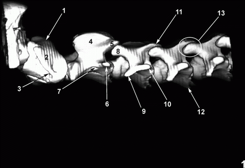
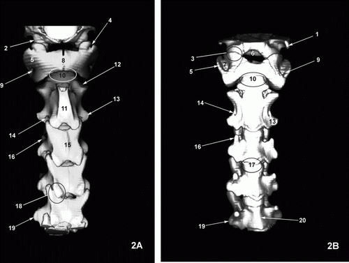
Figure 2. Three-dimensional (3D) reconstructed CT image of the cervical spine viewed from the dorsal (A) and ventral (B) aspect. 1. Paracondylar process of occipital bone (ventral view only). 2. Occipital condyles (dorsal view only). 3. Atlantooccipital joint (ventral view only). 4. Notch alaris (dorsal view only). 5. Wing of atlas. 6. Foramen magnum (ventral view only). 7. Fossa Atlantis (ventral view only). 8. Dorsal tubercle of atlas (dorsal view only). 9. Transverse foramen of atlas. 10. Dens of axis and atlantoaxial joint. 11. Spinous process of axis (dorsal view only). 12. Cranial articular process of axis. 13. Transverse process of axis. 14. Transverse foramen of axis. 15. Spinous process of third cervical vertebra (dorsal view only). 16. Ventral tubercle of transverse process of third vertebra. 17. Joint between vertebral bodies. 18. Joint between articular processes. 19. Dorsal tubercle of transverse process of fifth vertebra. 20. Ventral crest of fifth vertebra (ventral view only).
Discussion
Recently, the availability of used equipment and tolerable service cost has allowed a low, but steady, increase in the use of CT imaging in veterinary medicine (Barbee et al. Citation1987; Allen et al. Citation1988; Peterson and Bowman Citation1988; Ragle et al. Citation1988; Ruohoniemi et al. Citation1997; Vink-Nooteboom et al. Citation1998; Drees et al. Citation2009). CT can produce high-detail images with good soft tissue contrast. However, image quality is adversely affected by patient motion and, therefore, virtually all CT studies require to be performed on anesthetised patients. This technique is an excellent tool for the diagnosis of many diseases in small and large animal medicine.
Three-dimensional CT reconstruction is a useful procedure which is infrequently used in veterinary medicine. The collection of data requires multiple parallel thin sections, which are all, obtained at the same gantry tilt. Ideally, a very fast scanner such as the scanning beam ‘cine’ CT system can produce a rapid sequence of quality thin sections in order to reduce motion artefact (Grossman Citation1990). Advantages of this procedure are that the surface of bony structures can be imaged with different degrees of rotation without superimposition of soft tissues. The extent of bony lesions can be visualised with excellent detail by eliminating soft tissue CT numbers via computer so that the bone surfaces can be depicted as a 3D image. Three-dimensional CT reconstruction is accomplished by computerised post-processing and does not require anesthesia time in addition to that required for routine CT scanning (Kraus et al. Citation1997).
The use of CT with 3D reconstruction has been a valuable diagnostic aid for clinical evaluation of cervical vertebral malformations, degenerative changes of articular facets or cervical static stenosis in domestic animals (Drees et al. Citation2009), although only a few studies have dealt with anatomic descriptions of specific areas (Dennison and Schwarz Citation2008). CT has been established as one of the most useful procedures for the assessment of human spine and for the diagnosis of human lumbosacral diseases (Dorwart Citation1984; Dorwart and LaMasters Citation1985) – However, a few instances on the application of either CT or 3D reconstruction to any region of the spine of terrestrial mammals are available (Feeney et al. Citation1991; Jones et al. Citation1995; Claridge et al. Citation2010) and most of these have been performed in dead animals such as those describing the normal canine spinal anatomy by cadaver comparison to tomographic images (Feeney et al. Citation1991; Jones et al. Citation1995) or more recently studies by 3D anatomy of the equine cervical articular process joints in relation to the spinal cord. By contrast, we have attempted to describe our observations on recognisable structures in alive and neonatal animals, but further studies are necessary to define the ultimate limits of 3D CT reconstruction both for basic morphologic imaging and for interpretation of patient images with clinical signs. Much of this will, however, have to come from continued clinical experience and the ongoing comparison between imaged anatomy or pathology and case outcome or post-mortem studies.
On the teaching front, 3D imaging technology may facilitate teaching of anatomy to veterinary students by allowing the view of structures in a realistic, 3D manner. Thus, this technique is an anatomical tool that enables visualisation of bone, vascular network and selected soft tissue anatomy in a 3D image. This procedure eliminates the difficulties of visualising the structures of cervical spine in pictures or line drawings used in most student anatomy texts. Because experience with plain radiography suggests that there are variations between normal horses, more cases are required in order to determine variations in clinically normal foals.
Acknowledgements
We are very grateful to Marisa Mohamad Mingot and Jamal Jaber for their constructive comments.
References
- Allen , J , Barbee , D and Crisman , M . 1988 . Diagnosis of equine pituitary tumors by computed tomography. Part 1 . Compendium of Continuing Education for the Practicing Veterinarian , 10 : 1103 – 1105 .
- Barbee , D , Allen , J and Gavin , P . 1987 . Computed tomography in horses. Technique . Veterinary Radiology , 28 : 144 – 151 .
- Claridge , HA , Piercy , RJ , Parry , A and Weller , R . 2010 . The 3D anatomy of the cervical articular process joints in the horse and their topographical relationship to the spinal cord . Equine Veterinary Journal , 42 : 726 – 731 .
- Dennison , SE and Schwarz , T . 2008 . Computed tomographic imaging of the normal immature California sea lion head (Zalophus californianus) . Veterinary Radiology & Ultrasound , 49 : 557 – 563 .
- Dorwart , RH . 1984 . Computed tomography of the lumbar spine: techniques, normal anatomy, pitfalls, and clinical applications . Critical Reviews in Diagnostic Imaging , 22 : 1 – 42 .
- Dorwart , RH and LaMasters , DL . 1985 . Applications of computed tomographic scanning of the cervical spine . The Orthopedic Clinics of North America , 16 : 381 – 393 .
- Drees , R , Dennison , SE , Keuler , NS and Schwarz , T . 2009 . Computed tomographic imaging protocol for the canine cervical and lumbar spine . Veterinary Radiology & Ultrasound , 50 : 74 – 79 .
- Feeney , DA , Fletcher , TF and Hardy , RM . 1991 . Atlas of correlative imaging anatomy of the normal dog: ultrasound and computer tomography , Philadelphia , WB : Saunders .
- Grossman CB . 1990 . Magnetic resonance imaging and computed tomography of the head and spine. , 1st ed . Baltimore : Williams & Wilkins . p. 390 – 394 .
- Jones , JC , Cartee , RE and Bartels , JE . 1995 . Computed tomography anatomy of the canine lumbosacral spine . Veterinary Radiology & Ultrasound , 36 : 91 – 99 .
- Kraus , M , Mahaffey , M , Girard , E , Chambers , J , Brown , C and Coates , J . 1997 . Diagnosis of C5–C6 spinal luxation using three-dimensional computed tomographic reconstruction . Veterinary Radiology & Ultrasound , 38 : 39 – 41 .
- Losonsky , J , Abbott , L and Kuriashkin , I . 1997 . Computed tomography of the normal feline nasal cavity and paranasal sinuses . Veterinary Radiology & Ultrasound , 38 : 251 – 258 .
- Peterson , P and Bowman , K . 1988 . Computed tomographic anatomy of the distal extremity of the horse . Veterinary Radiology , 29 : 147 – 156 .
- Ragle , C , Koblik , P , Pascoe , J and Honnas , C . 1988 . Computed tomographic evaluation of the head trauma in a foal . Veterinary Radiology , 29 : 206 – 208 .
- Ruohoniemi , M , Kärkáinen , M and Tervahartiala , P . 1997 . Evaluation of the variably ossified collateral cartilages of the distal phalanx and adjacent anatomic structures in the finnhorse with computed tomography and magnetic resonance imaging . Veterinary Radiology & Ultrasound , 38 : 344 – 351 .
- Vink-Nooteboom , M , Junker , K , Van Den Ingh , T and Dik , K . 1998 . Computed tomography of cholesterinic granulomas in the choroid plexus of horses . Veterinary Radiology & Ultrasound , 39 : 512 – 516 .