Abstract
A three-year-old male Labrador retriever was presented with five-month history of tetraparesis with severe atrophy in the limbs. Prior to this five-month period, the dog had been healthy. At gross necropsy several muscles were atrophic reflecting decreased muscle mass. Histologically, there were atrophic type I and type II fibres, angulated type IIC fibres and an elevated oxidative activity that showed an altered mitochondrial pattern. The rapid progression of the clinical signs and the age at onset could suggest an atypical myopahty in this dog breed.
1. Introduction
A three-year-old male Labrador retriever was referred to the Veterinary Teaching Hospital of the University of Murcia (Spain) with five-month history of tetraparesis, more accentuated in the hind limbs. Prior to this five-month period, the dog had not presented any alteration in the gait. The owner could not report additional information about the rest of the litter. The dog had been vaccinated against distemper, rabies, hepatitis and leptospirosis. Canine leishmaniosis had been diagnosed by the referring veterinarian, and the dog was treated with meglumine antimoniate, allopurinol and prednisone. The dog improved slightly but it continued to be recumbent. The dog was referred to the Veterinary Teaching Hospital for evaluation. To our knowledge, this is the first report of a myopathy diagnosed in an adult Labrador retriever in this European region. It underlines the importance of this case because previously to this illness of five-month period, the dog was healthy with any myopathic clinical signs.
2. Case description
At presentation, physical and neurological examinations were carried out on the dog. The clinical signs included muscle atrophy in limbs, weakness, absent patellar reflex in the hind limbs and reduced flexor reflex. There was no ataxia, and the dog was conscious and alert. No megaesophagus was observed. Based on these findings, a neurological disease with signs of lower motor neuron dysfunction was suspected. A complete analysis was performed and the patient showed normal values except a slightly raised activity of the creatine kinase (CK), which indicates muscular damage, and low normal values of the aspartate aminotranspherase (AST), calcium and phosphorus. A normal radiograph was performed to discard hip dysplasia. A PCR for blood sample against Ehrlichia sp. and serology tests against Toxoplasma (IFI), Neospora caninum (IFI) and Leishmania infantum (ELISA) were negatives except one title toxoplasma (1:28 Ig G) and one title leishmania infantum (1:200). The dog was treated with allopurinol and clindamycin, but finally, the course of the disease was worst and the owners decided to euthanise the dog without further evaluation.
Immediately to his sacrifice, cerebrospinal fluid (CSF) was obtained with normal values of protein and nucleated cell count, and serology test for toxoplasma in CSF was negative. Striated muscles samples for histological examination were collected, frozen and cut. Serial cross-sections were stained with haematoxylin and eosin (HE), modified Gomori trichrome (GT), periodic acid–Schiff (PAS), nicotine adenine dinucleotide tetrazolium reductase (NADH-TR) (Dubowitz Citation1985) and myofibrillar adenosine triphosphatase after acid pre-incubation at pH 4.6 (Latorre et al. Citation1993).
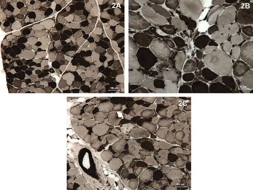
At necropsy, the dog showed atrophic signs in its four limbs, more accentuated in both hind limbs. Histologically, all the studied muscles (m. biceps femoris, m. vastus lateralis, m. semitendinousus, m. triceps and m. deltoideus) were atrophic, although the vastus lateralis and deltoideus muscles showed a higher atrophy degree. The muscles of the thoracic limb (triceps and deltoideus) had a scarce inflammatory infiltrated located into the endomysium (A, B) and some necrotic myofibres were observed (D, F). Throughout the sections of the muscles stained with HE, most of the myofibres had a wide range of structural changes within in their architecture: individual degenerative myofibres with intensely stained eosinophilic sarcoplasm, loss of cross-striation and large centrally located nuclei (A, B). GT stain and PAS reaction allowed evaluating the replacement of muscle fibres with fibrous tissue (C) and the normal accumulation of glycogen granules (D), respectively. ATPase reaction showed a high proportion of atrophic fibres belonged to both type I and type II fibres (). Inside type II fibres, type IIC fibres were increased in number with alterations in the fibre type pattern, such as a greater degree of angular atrophy () that also was presented in the type I (). On the other hand, there were globoid cells in both fibre types having a round shape ( and ). NADH-TR stain showed an elevated oxidative activity with dark blue colour, either diffuse or focal, and an altered mitochondrial pattern, completely disorganised and disorientated with abnormality in number and distribution of mitochondria (E, F).
Figure 1. Transverse sections of deltoideus (A–E) and vastus lateralis (F) muscles from a three-year-old dog with a Labrador retriever myopathy stained with histochemical and enzymatic reactions: (A, B) the haematoxylin and eosin stain showing the variability in myofibre size, the presence of an inflammatory infiltrated and the characteristic internal nuclei. (C) Modified Gomori trichrome stain demonstrating the increment of connective tissue. (D) Periodic acid–Schiff reaction showing a necrotic myofibre with pale appearance. (E, F) Increase of the oxidative activity and altered mitochondrial pattern is showed by nicotine adenine dinucleotide tetrazolium reductase reaction.
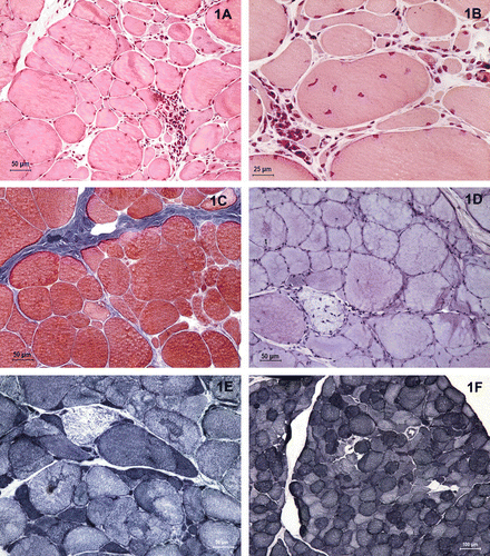
Figure 2. Transverse sections of triceps (A) and deltoideus (B–C) muscles from a three-year-old dog with a Labrador retriever myopathy. The ATPase reaction at pH 4·6 showing type I (dark), type IIA (pale) and type IIC (intermediate staining) myofibres with several grade of atrophic cells with a high proportion of angulated fibres in both type I and type IIC.
3. Discussion
Muscle lesions, characterised by myofibre atrophy, degeneration and regeneration and absence of nerve lesions, were consistent with the muscular dystrophy that afflicts Labrador retrievers (Gortel et al. Citation1996). The Labrador retriever myopathy, called recently centronuclear myopathy (Tiret et al. Citation2003), is an inherited disease that differs from the muscular dystrophies occurring in other breed dogs (Gortel et al. Citation1996). The first description of the disease was reported by Kramer et al. (1976), it is an autosomal recessive disorder. Later, it has been reported in several countries like Great Britain or USA, but to the authors’ knowledge this report describes the first case in an adult Labrador retriever in Spain. There is a great variation in the presentation and severity of clinical signs, but the loss of patellar reflex in his limbs, the stilted gait and the bunny hopping, as well as generalised skeletal muscle atrophy, especially of the proximal limb muscles (Bergman et al. Citation2002), are consistent findings (Gortel et al. Citation1996).
Normally, the affected dogs begin to show the clinical signs at three months of age, and they are gradually increased up to around one year of age (Gortel et al. Citation1996). After this time, progression of the disease is complete or reduces. In this unusual case, the dog was three years old and showed the signs in a short period of time that could be another form of presentation of the disease. The routine haematological and biochemical analyses are of little help in diagnosing Labrador retriever myopathy: only serum CK activity is often moderately elevated (Gortel et al. Citation1996; Bley et al. Citation2002; Green et al. Citation2005); in this case the CK activity was slightly raised due to the muscular damage and the AST level was at the highest point within the normal values, consecutive treatments could have cause hepatic damage. Thus, the diagnosis of this Labrador retriever myopathy was made on the basis of his signalment, clinical presentation and histological appearance of striated muscles. Most of the works only showed inherited myopathy affecting puppies (Gortel et al. Citation1996; Green et al. Citation2005; Cosford et al. Citation2008), and only in some cases the affected dogs were older than one year, but they showed previous symptoms (Bley et al. Citation2002). On presentation of this case, a three-year-old adult male was healthy five months earlier. For this reason, it may be a new variation of this disease.
Histopathologically, Labrador retriever myopathy has been well described (Gortel et al. Citation1996). Morphologic changes include muscle fibre group atrophy, presence of angular fibres, hypertrophic fibres in both types I and II, internal nuclei in several fibres, necrosis and fibrosis (Gortel et al. Citation1996; Bergman et al. Citation2002; Green et al. Citation2005). This case showed a severe degree of atrophic fibres with angulated cells in both types I and IIC; besides it observed some necrotic myofibres and an increment of connective tissue that included endomysial and perimysial fibrosis and fat infiltration. Although pathological changes may vary in centronuclear myopathy, the examined muscles showed a disorganised mitochondrial pattern, with a high number of myofibres with abnormality in number and distribution of mitochondria that is consistent with previous reports (Moore et al. Citation1987; Green et al. Citation2005).
With respect to the treatment, there is no definitive one for this condition and no medicament that improved muscle metabolism, and function was administered, such as L-carnitine (Davies et al. Citation2008). Moreover, the allopurinol was indicated for inducing a drastic and progressive reduction of Leishmania infantum load in lymph node aspirates but did not eliminate the parasite (Manna et al. Citation2009).
The pathogenesis of the Labrador retriever myopathy is unknown; muscular dystrophies are a heterogeneous group of inherited degenerative non-inflammatory muscle disorders (Cosford et al. Citation2008). Puppies affected with this autosomal recessive disease receive one defective gene from each one of the normal parents. A correct diagnosis is decisive to eradicate the disorder from the genetic lines of breeders. In addition, it is very difficult to screen the carrier parents and the affected puppies; therefore we are disabled for limiting the disease (Gortel et al. Citation1996).
In conclusion, this adult dog had clinical, biochemical and histochemical abnormalities consistent with a degenerative non-inflammatory muscle disorder that not only affects puppies with centronuclear myopathies. This case should alert clinicians to the possibility that other affected dogs were in the southwest of Europe.
References
- Bergman , RL , Inzana , KD , Monroe , WE , Shell , LG , Liu , LA , Engvall , E and Shelton , GD . 2002 . Dystrophin-deficient muscular dystrophy in a Labrador retriever . Journal of the American Animal Hospital Association , 38 : 255 – 261 .
- Bley , T , Gaillard , Cl , Bilzer , Th , Braund , KG , Faissler , D , Steffen , F , Cizinauskas , S , Neumann , J , Vögtli , T Equey , R . 2002 . Genetic aspects of Labrador retriever myopathy . Research in Veterinary Science , 73 : 231 – 236 .
- Cosford , KL , Taylor , SM , Thompson , L and Shelton , GD . 2008 . A possible new inherited myopathy in a young Labrador retriever . The Canadian Veterinary Journal , 49 : 393 – 397 .
- Davies , SE , Davies , DR , Richards , RB and Bruce , WJ . 2008 . Inherited myopathy in a Great Dane . Australian Veterinary Journal , 86 : 43 – 45 .
- Dubowitz , V . 1985 . “ Muscle biopsy ” . In Muscle biopsy: a practical approach , 2nd ed , Edited by: Dubowitz , V . 19 – 40 . Philadelphia : Baillière Tindall .
- Gortel , K , Houston , D.M , Kuiken , T , Fries , C.L and Boisvert , B . 1996 . Inherited myopathy in a litter of Labrador retrievers . The Canadian Veterinary Journal , 37 : 108 – 110 .
- Green , SL , Tolwani , RJ , Varma , S and Shelton , GD . 2005 . Absence of mutations in the survival motor neuron cDNA from Labrador retrievers with an inherited myopathy . The Veterinary Record , 157 : 250 – 254 .
- Kramer , JW , Hegreberg , GA , Bryan , G , Meyers , K and Ott , RL . 1976 . A muscle disorder of Labrador retrievers characterized by deficiency of type II muscle fibers . Journal of the American Veterinary Medical Association , 169 : 817 – 820 .
- Latorre , R , Gil , F , Vázquez , JM , Moreno , F , Mascarello , F and Ramírez , G . 1993 . Skeletal muscle fibre types in the dog . Journal of Anatomy , 182 : 329 – 337 .
- Manna , L , Vitale , F , Reale , S , Picillo , E , Neglia , G , Vescio , F and Gravino , AE . 2009 . Study of efficacy of miltefosine and allopurinol in dogs with leishmaniosis . Veterinary Journal , 182 : 441 – 445 .
- Moore , M.P , Reed , S.M Hegreberg , G.A . 1987 . Electromyographic evaluation of adult Labrador retrievers with type-II muscle fiber deficiency . American Journal of Veterinary Research , 48 : 1332 – 1336 .
- Tiret , L , Blot , S , Kessler , JL , Gaillot , H , Breen , M and Panthier , JJ . 2003 . The cnm locus, a canine homologue of human autosomal forms of centronuclear myopathy, maps to chromosome 2 . Human Genetics , 113 : 297 – 306 .