
Abstract
Most of the available scientific literature concerning claw vacuum drops and fluctuations (identical to teat-end vacuum) during machine milking has been published in the 1960s and 1970s. Fewer studies have been carried out more recently on modern types of dairy cows, and milking machines, and have been evaluated with modern statistical methods and software. Based on the accessible information, there are indications that reduced milking performance, leading to long machine-on time mainly as a consequence of a combination of vacuum drops and different types of fluctuations, may influence udder health in a negative way although scientific proofs are scarce. Based on an ISO note, the claw vacuum should remain within a range of 32–42 kPa during peak milk flow to ensure fast, complete and gentle milking. This range of vacuum pressure allows adequate liner movement and a sufficient pressure on the teat during the massage (d-) phase is guaranteed. Thus, liner slips caused by too low claw vacuum are avoided. On the other hand, the teat-end vacuum should not be much higher than 42 kPa as it can cause damage of teat tissue mainly during periods of low milk flow when it is almost identical with the system vacuum. Depending on the used system of milk transport through the tubes, the teat-end vacuum drops to some extent, intensified during periods of high milk flow and reduced during periods of low or absent milk flow at the start and end of milking. Hence, a general increase of system vacuum throughout milking is not suitable, as high teat-end vacuum acting on the teat during milking phases with low milk flow rates have to be avoided. However, a reduction of machine-on time and high vacuum load on the teat during low milk flow can potentially be achieved by early cluster removal, before complete cessation of milk flow. Ideally, this goal can be reached at quarter milking (as used in automatic milking systems) where each teatcup is individually removed based on the milk flow of the individual teat, and milking at very low or no milk flow can easily be avoided.
The history of machine milking, machine milking vacuum and related research
Fluctuations and drops in the teat-end vacuum occur regularly during machine milking as a result of various interactions between vacuum supply, teatcup liner movement and milk flow. Potential effects of these vacuum dynamics on milking performance, teat condition and udder health are continuously discussed by machine milking experts and dairy farmers. The purpose of this paper is to provide an overview of the scientific knowledge which has been created over the last five decades since the comprehensive introduction of machine milking in most industrialized countries with a significant dairy industry.
The functional basis of today's milking machines with vacuum evacuating the udder and a pulsating liner which rhythmically interrupts a milk flow was invented more than hundred years ago. However, machine milking was not wide spread during the first half of the twentieth century, and hand milking remained the most used method worldwide. During the 1950s with the economic growth, new strategies for good milking performance, efficient breeding through artificial insemination, increasing hygienic requirements for food including milk and reduced work load for the workers in agriculture were introduced in the industrialized countries. Increasing production and good milkability with high milk flow rates became a major goal of dairy breeding and genetics (Bruckmaier Citation1995; Moser Citation2010). Massive changes in the dairy industry have led to more efficient milking systems designed for harvesting increasing amounts of milk. Dairy herd improvement systems together with artificial insemination increased the efficiency of breeding for high-yielding dairy cows with the use of genetics from all over the world (Bruckmaier Citation1995; Moser Citation2010). On most dairy farms including small family farms, hand milking was replaced by machine milking (Rabold Citation1968; Bruckmaier Citation1995). The dairy industry promoted the intensive development of accurately working and highly efficient milking machines. Simultaneously, scientists became interested in the investigation of machine milking and its effects on milk yield, milkability, udder health and the quality of milk delivered to the consumer (Baxter et al. Citation1950; Wilson Citation1963; Bruckmaier Citation1995; Svennertsen-Sjaunja et al. Citation2003; Moser Citation2010). The newly established hygienic surveillance of cows and milk caused an increase in detection of udder infections and numerous clinical mastitis cases during the introduction period of machine milking (Wilson Citation1963). This led to the assumption that high vacuum levels and vacuum fluctuations occurring during the milking process had negative effects on teat condition and udder health. From the 1960s to present, one of the main goals of milking equipment engineers and a key subject of research was milking at a more constant teat-end vacuum to achieve fast milk removal while avoiding negative impacts on udder health being caused by the mechanical load on the teats (Baxter et al. Citation1950; Dodd Citation1963; Wilson Citation1963; Cowhig & Nyhan Citation1967; Cowhig Citation1968; Dodd & Neave Citation1968; Nyhan Citation1968; Reinemann et al. Citation2003). The 1960s and 1970s represented the most active period of research related to the effects of pulsation and vacuum including the impacts of irregular and cyclic vacuum fluctuations on milking performance, teat condition and udder health in dairy cows.
It is often assumed that various kinds of vacuum changes at the teat can disturb the correct function of the milking unit. They might either per se or in combination (vacuum drop, cyclic and irregular vacuum fluctuations) have an impact on milking performance (McDonald & Witzel Citation1968; Ambord & Bruckmaier Citation2010) and udder health (Nyhan & Cowhig Citation1967; Cowhig Citation1968; Nyhan Citation1968; Thiel et al. Citation1973; Zecconi et al. Citation1992). Negative impacts could comprise teat congestion or oedema, teat canal integrity and the facilitated physical movement of bacteria into the mammary system (Nyhan Citation1968; Reitsma et al. Citation1981; Bulletin of the International Dairy Federation 215 Citation1987).
The review summarizes the existing scientific work in this field of research. The used definitions and terms depend on the source of literature; consequently, we have standardized the terms and their definitions as far as possible. Our definitions follow those introduced by Nyhan and Cowhig (1967–1968), the Bulletin of the International Dairy Federation 215 (Citation1987) and the document no. 3918 of the International Organization for Standardization (Citation2007a).
Different types of vacuum dynamics at the teat end
The course of teat-end vacuum during machine milking is characterized by different types of vacuum changes.
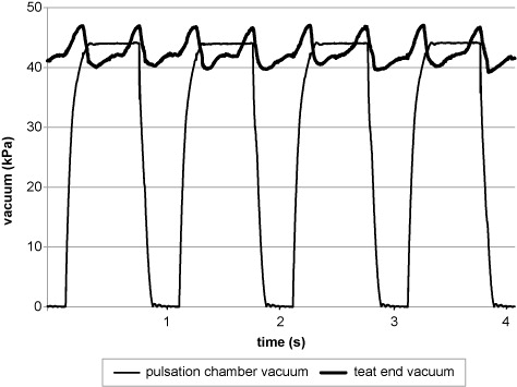
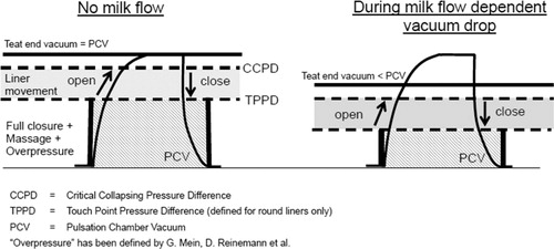
The functional principle of a two-chamber milking unit is based on the periodic extraction of milk from the mammary gland by vacuum. While there is permanent vacuum inside the teatcup liner and claw, the volume between the liner and the teatcup (pulsation chamber) is periodically subjected to either vacuum or atmospheric pressure by pulsation which leads to alternating opening or closing of the teatcup liner. The rhythmic movement of the liner between open and closed position occurs in most milking systems around 60 times per minute. If vacuum is present both inside and outside the liner then the liner is open and the applied vacuum removes milk from the teat. If atmospheric pressure is present in the pulsation chamber (outside the liner), it is collapsed and puts pressure on the teat tip, thus causing a cessation of milk flow despite the continued presence of vacuum at the teat. This period is called the liner-closed phase. It occurs during the absence of pulsation chamber vacuum, noting the pressure applied on the teat is only caused by the teat-end vacuum and is therefore subject to changes during dynamic changes of the teat-end vacuum (–).
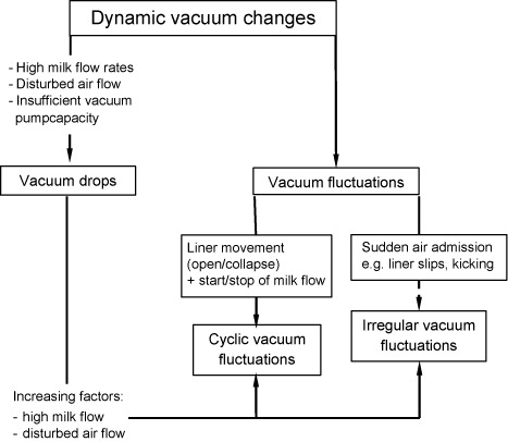
Vacuum drops
A drop of the teat-end vacuum during milking is mainly caused by the milk as soon as it starts to flow and has to be transported through the milk tube system (Ambord & Bruckmaier Citation2010). The vacuum drop is increased with high milk flow rates (Ambord & Bruckmaier Citation2010; Ströbel et al. Citation2013), small or blocked diameters of air inlets in the claw (Rasmussen et al. Citation2006; Ströbel et al. Citation2013), and is usually much more pronounced in a highline than lowline milk transport system (Ambord & Bruckmaier Citation2010). Vacuum drops are defined as:
a difference in the average vacuum (arithmetic average of all vacuum levels in the system), between a downstream and an upstream point in the milking system, or at any point in the milking system before and after an increase of airflow in the system other than through the vacuum regulator. (International Organization for Standardization Citation2007a)
However, while the peak flow rate is considerably reduced by low vacuum levels, the average milk flow rate is only slightly or not at all affected depending on the individual cow and its milk flow level (Ambord & Bruckmaier Citation2010). This effect is due to the continuous character of milk ejection within the udder with a lower vacuum level (Bruckmaier et al. Citation1994). If milk is removed from the udder very fast, the milk ejection can become the rate limiting factor of milking performance, thus causing an incomplete refill of the teat in the massage (= liner closed) phase of the pulsation (Ambord & Bruckmaier, Citation2009, Citation2010).
The degree of pressure inside the liner acting on the teat end during the massage phase of pulsation (atmospheric pressure in pulsation chamber) is also a crucial factor to guarantee undisturbed milking performance and proper teat condition. The pressure should be high enough to allow the massage effect on the teat end when the liner is closed. Blood and lymph vessels and the extracellular spaces in the teat tissue are dilated as the intra- and extravascular fluid accumulates in the teat tissue during milking (Hamann & Mein Citation1990; Zecconi et al. Citation1992; Koning & Ipema Citation2000; Neijenhuis et al. Citation2001). If the massage effect of the liner is insufficient, the fluid stays in the teat tissue, and changes of the pliability of the teat tissue cause a decrease in the resistance of the teat canal to bacterial invasion (O'Shea Citation1987; Neijenhuis Citation2004). Furthermore, there is a decrease of muscle contractions at the teat end, which facilitates penetration of bacteria and microorganisms into the teat canal (Bramley & Schultze Citation1991; Capuco et al. Citation1994). So, the main purpose of the adequate vacuum level is to guarantee the massage effect applied by the liner on the teat tissue in order to release the teat from congestion, blood and lymph which accumulated during the liner open (b-) phase of pulsation. However, too high pressure can lead to increased teat wall thickness (Hamann et al. Citation1993), tissue damage and the development of hyperkeratosis (Bade et al. Citation2007, Citation2009). On the other hand, based on practical experience, it seems possible that the claw vacuum transiently drops below 30 kPa during high milk flow without causing a compromised liner function as long as no liner slips occur (Besier & Bruckmaier 2014, unpubl.).
Vacuum fluctuations
Vacuum fluctuations occur suddenly during momentary increases and decreases of airflow in the milking system. Vacuum fluctuations are differentiated into two types – cyclic and irregular vacuum fluctuations – which will be explicitly specified in the following paragraphs. Fluctuations in vacuum are caused either by the recurrent movement of the liner and corresponding milk flow or by sudden excessive air inlets into the system (Nyhan Citation1968; Thiel et al. Citation1973).
Cyclic vacuum fluctuations
Cyclic vacuum fluctuations occur during the milking process in regular intervals inside the liner. They are induced by the liner movement (open and close) and mainly by the occurrence of milk flow from each teat when the liner opens (Thiel et al. Citation1973). The abrupt opening of the liner causes a sudden increase of the volume inside the liner. The claw vacuum increases and, at the same moment, a reverted pressure gradient develops in the claw and under the teat, causing air and milk to be propelled from the claw through the short milk tube to the teat end (Nordegren Citation1980). This happens particularly if the claw vacuum is very low due to a milk flow dependent vacuum drop. The collapse of the liner represents a sudden reduction of the volume inside the liner, which causes a transient small decrease of the vacuum level at a given amount of air in the system. The milk in transit flowing steadily into the claw within each pulsation cycle can amplify these fluctuations, especially if there are cows with high milk flow rates (Thiel et al. Citation1973). The reason for amplification is the higher inertness of the milk in the tube compared to the air which disturbs the airflow as soon as milk transits the short milk tube. Rasmussen et al. (Citation2006) tested the influence of blocked air inlets on vacuum fluctuations in automatic milking systems and expectedly found an increase in cyclic fluctuations with restrictions of the air inlets during milking. Highest vacuum fluctuations were observed during peak milk flow (Rasmussen et al. Citation2006).
Irregular vacuum fluctuations
Irregular vacuum fluctuations have been defined as an excessive decline and subsequent recovery of the vacuum level within a milking unit (Thiel et al. Citation1973). Irregular vacuum fluctuations are extraordinary events caused by liner slips, cluster changing or cluster fall off during milking. They are the consequence of an infrequent and unplanned air admission into the vacuum system (cf. ; Nyhan Citation1968; Thiel et al. Citation1973; Thiel Citation1978). The extent of irregular vacuum fluctuations depends on a combination of several factors, such as the type of milking system (highline or lowline; Beckley & Smith Citation1962; Østeras & Lund Citation1980; Reinemann et al. Citation1997) and the milk flow rate at the moment of occurrence (Schmidt et al. Citation1964; Thompson & Miller Citation1974), the extent and location of occurring sudden air admission (McDonald & Witzel Citation1968), obstruction to air flow in the system (Whittlestone Citation1962; McDonald & Witzel Citation1968; Thompson & Miller Citation1974), the internal diameter of the short and long milk tubes (McDonald & Witzel Citation1968), the type of pulsation (alternating or simultaneous pulsation; McDonald & Witzel Citation1968), functionality of the vacuum control valve (Wilson Citation1963), and the capacity of the vacuum pump (Beckley & Smith Citation1962). Abrupt changes of the vacuum level may range from transient to total vacuum loss, i.e., the amplitude between system vacuum and atmospheric air pressure (zero vacuum). As irregular vacuum fluctuations only occur as very short events during the milking process, they cannot be compensated by a general increase of the vacuum level in the whole milking system. The pressure acting on the teats throughout milking would be too high, especially during periods of low milk flow rates at the start and at the end of milking. The risk of negative impacts on the teat tissue concomitantly with an increased risk of intramammary infection would be considerably increased (McDonald & Witzel Citation1968; Thompson et al. Citation1978).
If irregular vacuum fluctuations occur in combination with cyclic vacuum fluctuations along with long milking duration, the risk for congestion of the teat tissue and new infections of the mammary gland has been shown to increase (Thiel et al. Citation1973; O'Callaghan et al. Citation1976; O'Shea et al. Citation1976).
The ISO 3918 (International Organization for Standardization Citation2007a) defines transient vacuum drops as:
a momentary difference in vacuum between a downstream and upstream point in a system, or at any one point in a system before and after an increase in airflow into the system other than through the vacuum regulator (vacuum valve).
Milk flow rates are decreased by irregular vacuum fluctuations (Schmidt et al. Citation1964). The increase of total milking time caused by high irregular vacuum fluctuations is most obvious in highline milking installations (Stanley et al. Citation1962; Langlois et al. Citation1981). The increased milking time and insufficient udder emptying can be conducive to irritated teat tissue and consequently lead to infected quarters with clinical mastitis while providing good conditions for bacterial invasion (Nyhan & Cowhig Citation1967). A large capacity vacuum pump, an increased diameter of vacuum lines and a high volume balance tank may help to avoid irregular vacuum fluctuations, if the reason is a milk flow dependent vacuum drop and hence reduced liner adhesion to the teats (Baker Citation1955). Following the recommendations of the International Organization for Standardization (Citation2007b):
the internal diameter and slope of the milkline shall be such that the vacuum drop between the receiver and any point in the milkline does not exceed 2 kPa with all units operating at the designed milk flow and airflow. (International Organization for Standardization Citation2007b)
Potential impact of vacuum fluctuations on udder health
Based on the existing literature, cyclic vacuum fluctuations are not supposed to have a negative impact on udder health as long as the vacuum level allows adequate liner movement and teat massage during the liner-closed phase (Nordegren Citation1980; Schlaiß Citation1994). This is certainly the case as long as the claw vacuum does not decrease below 30 kPa. As long as there is a positive pressure in the teat through the stored milk, the minimum claw vacuum needed to avoid liner slips can even be as low as 28 kPa (Besier & Bruckmaier, unpubl.). The minimum required claw vacuum depends certainly on the liner characteristics and the capacity of the claw. Thiel et al. (Citation1973) showed that the vacuum fluctuations caused by the cyclic movement of the teatcup liner (cyclic fluctuations) in combination with high or low milk flow rates (vacuum drop) did not lead to an increase of udder infection.
Several studies have shown that irregular vacuum fluctuations can have an impact on udder health and teat condition and can lead to increased infection rates (Wilson Citation1958; Beckley & Smith Citation1962; Stanley et al. Citation1962; Wilson Citation1963; Nyhan & Cowhig Citation1967; McDonald & Witzel Citation1968; Nyhan Citation1968). The question ‘why’ they can cause teat injuries or udder infection is not fully clarified. One possible reason could be the relation to abrupt and high vacuum changes. While cyclic vacuum fluctuations are limited to the effect of liner movement and milk flow within a closed system between teat, liner and milk tube, irregular fluctuations are usually caused by an uncontrolled leaking of atmospheric air into the system, and the sudden vacuum difference can be more than 40 kPa. It seems that injury of the mucosal lining of the teat sinus and the epithelium and sub-epithelial tissue of the teat canal is most evident after milk flow has ceased and milking is executed on empty teats (Peterson Citation1964). Obviously, an important reason for udder infection caused by irregular vacuum fluctuations is the physical movement of pathogens into the teat canal during jet flow (Schmidt et al. Citation1964; Nyhan Citation1967). Towards the end of milking, the teat cistern is not refilled with milk from the alveolar system of the mammary gland during the massage phase. If the teat is not refilled with milk quickly enough after the respective quarter is emptied, the compressed teat is re-opening when the liner opens. However, in this case, it opens via the influx of air or milk from outside the teat instead of a refill with milk from the mammary tissue, thus increasing the risk of cross-contamination and infection (Rasmussen Citation1993).
Another factor which can facilitate the movement of bacteria is the flooding of the teatcup liner and consequently the reflux of milk from the teat into the gland cistern due to the sudden dramatic vacuum loss during irregular vacuum fluctuations such as caused by liner slips. Depending on capacity and flow characteristics of the claw, bacteria can then also be spread from an infected quarter or from a contaminated claw to the teats of uninfected quarters (Nyhan Citation1968; Thompson & Miller Citation1974).
In several experimental trials, Nyhan and Cowhig (1965–1967) showed a significant correlation between irregular vacuum fluctuations and disturbed udder health in dairy cows. During the studies, cows were milked at different vacuum levels, and teats were exposed to Staphylococcus aureus. The results showed a relationship between irregular vacuum fluctuations, low reserve air capacity in the milking system and a high number of udder infections and cases of clinical mastitis, respectively. (Nyhan & Cowhig Citation1967; Nyhan Citation1968). Thiel et al. (Citation1973) repeated the trials previously conducted by Nyhan and Cowhig (Citation1967) and Nyhan (Citation1968) to show that both irregular and cyclic vacuum fluctuations can have an impact on udder health. In their study, 20 cows were milked in the same way as in the experiments of Nyhan and Cowhig (Citation1967). During the milking process, the teats were exposed to four irregular vacuum fluctuations per minute. These were combined with large cyclic vacuum fluctuations during pulsation cycles. Additionally, teats were exposed to Streptococcus agalactiae and Streptococcus dysgalactiae, and also to different pulsation rates and fast liner movement. Half-udder-clusters were used in this experiment. One cluster half was exposed to irregular vacuum fluctuations with the intention to increase new infection rate. The other half of the cluster was used as control unit at a stable vacuum level. These studies did not show any effect of either irregular or cyclic vacuum fluctuations per se to increase the number of udder infections. However, if the two types of fluctuations occurred concomitantly, the number of successful experimental infections increased. These findings were confirmed in further studies by O'Callaghan et al. (Citation1976) and O'Shea et al. (Citation1976), who also showed that irregular vacuum fluctuations per se did not cause new udder or quarter infections and that they did not necessarily have a negative impact on teat condition when occurring solely. In their experiments, dairy cows were machine-milked with various combinations of large and small irregular and cyclic vacuum fluctuations (half-udder-clusters with vacuum supply of either unstable or stable vacuum level; claw of different sizes to give large (small claw) or small (large claw) cyclic vacuum fluctuations) and exposure of teats to bacterial suspension before and immediately after each milking (O'Callaghan et al. Citation1976; O'Shea et al. Citation1976).
Influences of teat-end vacuum on teat-end thickness, hyperkeratosis and udder health
Teat tissue damage can be caused by machine milking at very high teat-end vacuum levels combined with long milking duration (Whittlestone Citation1962). Gleeson et al. (Citation2003) studied teat sinus injury and removal of teat canal keratin during milking in two groups of Holstein cows that had been milked for 112 days and a whole lactation, respectively. They concluded that over-milking of teats for 5 minutes resulted in an increase of teat sinus injuries and excessive removal of teat canal keratin (Gleeson et al. Citation2003). During milking, teat tissue gets stretched by the vacuum acting on the teat. The stretching causes micro fissures in the skin which is responding with an increased production of keratin. The excessive growth of keratin is termed hyperkeratosis, which is, from a histological point of view, a hyperplasia of the stratum corneum of the teat canal. It results in a visible thickening of the skin surrounding the external teat orifice which is termed teat-end callosity (Mein et al. Citation2003; Neijenhuis Citation2004). The teat-end callosity can reach different degrees, from smooth and soft skin at the teat canal end to very rough, callused rings, then classified as hyperkeratosis. High-yielding and multiparous cows show increased degrees of hyperkeratosis than low-yielding or younger cows. A high degree of callosity and hence hyperkeratosis is associated with an increased risk of clinical mastitis. The ISO recommends in a note (cf. International Organization for Standardization Citation2007b, p. 14) that milking should be conducted with a teat-end vacuum between 32 and 42 kPa during the peak flow period. This is presumably only mentioned in a note because milking systems are also capable to milk with a lower (or higher) vacuum level than in the given range. The limiting factor for minimum teat-end vacuum is the occurrence of liner slip and cluster fall off and not a compromised liner movement. The limit for a high vacuum is the compressive load acting on the teat with vacuum levels >42 kPa during periods of low milk flow and the absence of vacuum drops. The recommendation of the ISO note is therefore an advice for a range of vacuum that can guarantee quick and gentle milking at a low risk of liner slip or teat injury.
Besides the gentle milking with a milking vacuum at an ideal level, machine-on time should be as short as possible. Thus, the time of cluster removal is also a matter of discussion. In an experimental study, Rasmussen (Citation1993) showed that automatic cluster removal at a milk flow level of 400 g/min has no negative influence on milking performance and milk yield but shortens milking duration and reduces stress on the teat tissue. Thus, a longer period of milking at very low milk flow rates or even over-milking in certain quarters towards the end of milking can be avoided by a timely cluster removal. A quarter-specific milking with individual teatcup removal can totally avoid effects of milking on empty teats. However, this equipment is currently almost exclusively available for automatic milking systems.
As the teat canal has already been described as first line of defence against invading pathogens, there is a second barrier with bactericidal characteristics. This is built by blood-derived immunological factors such as leucocytes, immunoglobulins and other soluble factors in the milk. The main mechanisms for physical defence are binding of bacteria to keratin, desquamation of bacteria-coated keratin by the shearing forces associated with milk flow through the teat canal (Williams & Mein Citation1986) and desiccation of the canal lumen by evaporation from the external teat orifice (Capuco et al. Citation1992, Citation1994; Myllys et al. Citation1994; Neijenhuis Citation2004). Through an excessive removal of keratin from the teat canal, the teat loses its ability of inhibiting pathogens to enter. Thus, susceptibility of the mammary gland to new infection is increased (Capuco et al. Citation1992; Neijenhuis et al. Citation2001). Congestion, increased teat-end callosity, hyperkeratosis, teat orifice erosion or cyanotic teat ends seem to lead to an increased risk of udder infection. Damaged tissue regions provide good physical conditions (Langlois et al. Citation1981; Natzke et al. Citation1982; Bulletin of the International Dairy Federation 215 Citation1987; Hamann & Mein Citation1990; Zecconi et al. Citation1992; Neijenhuis Citation2004) and allow direct adherence of pathogens to colonize the teats and to invade the udder (Neave et al. Citation1969; Bramley et al. Citation1979; Myllys et al. Citation1994).
Among others, Isaksson and Lind (Citation1992) examined teat tissue reactions to pressure before, during and after milking. Machine milking caused circulatory teat changes defined as hyperemia. A thickening of teat walls during machine milking is obvious but does not last very long after the end of milking. No long-term impact on udder health by this teat congestion/oedema was detected. The authors assume that the reactions resemble to the physiological teat reactions which would be caused by calf nursing as well (Isaksson & Lind Citation1992) where pressure applied to the teat cistern by the calf's palate and tongue can vary from >55 kPa to negative (Rasmussen & Mayntz Citation1998). So, the characteristic changes of the teat tissue during machine milking are apparent, but seem to be reversible, already during later phases of the milking process (Isaksson & Lind Citation1992).
Shearn and Hillerton (Citation1996) investigated the relationship between hyperkeratosis and the incidence of new infection rate of the mammary gland and used a scoring system to evaluate hyperkeratosis in dairy cows of 25 herds. The scores ranged from 0, which describes normal orifices without dislocations, extrusions or abnormal growths, to 6 with severe hyperkeratosis, where the whole orifice appears enlarged with fronds >4 mm. Like Isaksson and Lind (Citation1992), they did not find a significant relationship between the mean somatic cell count and the degree of hyperkeratosis. In contrast to the results of Shearn and Hillerton (Citation1996), Zecconi et al. (Citation1992) found a strong correlation between teat tissue thickening caused by machine milking and udder infection rate. The possibility of development of clinical mastitis was twice as high, if teat thickness was increased by more than 5% during milking (Zecconi et al. Citation1992, Citation1996; Neijenhuis Citation2004).
Further parameters influencing the degree of hyperkeratosis are milk yield and machine-on time (Mein & Thompson Citation1993; Shearn & Hillerton Citation1996). There is evidence of increasing sensitivity of teats for developing hyperkeratosis and teat-end eversion towards the end of milking, i.e., the last 0.5 minute before cluster detachment (Rasmussen Citation1993). Most importantly, there are indications that the risk of new infections, which might also be due to vacuum fluctuations, is at highest when milk flow in the respective quarter has ceased (Cousins et al. Citation1973; Thompson et al. Citation1978). As already mentioned before, an early removal of the milking unit can decrease machine-on time, on the one hand, and increase average milk flow rate, improve teat condition and reduce the change of teat-end thickness during milking, on the other hand. No obvious negative effects of the early cluster-detachment on milk yield or milk composition and no effect on the incidence or prevalence of subclinical mastitis did appear in the conducted experiments.
High-yielding cows with long machine-on times often show a higher degree of hyperkeratosis; hence, the period of applied pressure on the teat is longer compared to the pressure-period applied on teats of low-yielding cows with shorter machine-on times. In addition, high-yielding cows show often higher milk flow rates. High milk flow results also increased shear forces affecting the teat canal. This leads to excessive stress for the teat tissue and an increased risk for teat tissue damage and hyperkeratosis. The highest severity of hyperkeratosis was observed in months 3 and 4 postpartum by Shearn and Hillerton (Citation1996), with a decline towards the end of lactation (Shearn & Hillerton Citation1996). Obviously, the risk of hyperkeratosis was highest during the period of highest milk production with long machine-on times.
Hyperkeratosis occurs – at least to a low degree – in most dairy herds. During milking, keratin production and the development of hyperkeratosis are stimulated by the mechanical load acting on the teat, including the partial removal of the keratin layer caused by the velocity of milk flow through the teat canal and the shear stress due to milk flow (Williams & Mein Citation1986). Up to a certain level, this process seems to be a normal physiological response. Therefore, it should not be considered as a teat and teat canal risk factor in general. If the load applied on the teat end during milking is very high (average claw vacuum >42 kPa at very short intervals between milkings), the teat orifice is increasingly surrounded by a ring of keratin and consequently hyperkeratosis develops to a pathological degree (Mein et al. Citation1987, Citation2003; Neijenhuis Citation2004). Higher scores of hyperkeratosis and increasing cell counts within a dairy herd are either an indicator of bad herd management or the malfunction of one or all elements of the milking system and irregularities during milking process (Capuco et al. Citation1994; Shearn & Hillerton Citation1996).
Conclusions
In conclusion, irregular vacuum fluctuations during milking combined with cyclic vacuum fluctuations and very high teat-end vacuum levels seem to increase the incidence of udder infection and to reduce milk flow rate. There is an impact on proper liner movement; the degree of massage on the teat end is decreased. There is no unique explanation of the factors causing a higher infection rate, but some attempts to explain the cases. Irregular or cyclic vacuum fluctuations per se seem not to have any negative impact; it is rather the combination of several parameters, like the simultaneous existence of vacuum fluctuations at high vacuum levels and long machine-on times or over-milking, which are responsible for the occurrence of udder infection, oedema or hyperkeratosis. These factors facilitate invasion of pathogens into the udder and affect immune mechanisms of the udder in a negative way. Therefore, concluding from the results of this literature research, it is recommended to maintain the teat-end vacuum between 32 and 42 kPa as mentioned in the ISO note 5707 (Citation2007b, p. 14), to remove clusters as early as possible (e.g., at a remaining milk flow rate of 400–600 g/min) and to use liners with adequate sizes that fit the dairy herd they are milking.
Overall, it has to be mentioned that a considerable portion of the literature used for this review is from the 1960s, 1970s or 1980s, and there are not many newer investigations. Thus, it is necessary to conduct new studies on the influence of vacuum drops and fluctuations, high vacuum levels and long milking durations on milking performance and udder health of dairy cows.
Circumstances under which experiments had been conducted during the 1960s have changed. Experimental equipment as well as the dairy cows themselves have changed until today. Nowadays, collected data from the experiments is analyzed by computers with an appropriate software, and already in the 1990s dairy cows gained much higher milk yields (up to 35 kg/day in average with an average milk yield of 2.7 kg/min) than in the 1960s (1.7 kg/min; Bruckmaier Citation1995). Hence, these factors are not comparable and it is likely that similar investigations conducted today would show different results.
For the aspects of hyperkeratosis, experimental work and research has very much advanced during the last couple of years. The database concerning this issue is very good and broad, and can be used by practitioners on farms to improve milking routine and to lower economic losses caused by mastitis in dairy herds.
Disclosure statement
No potential conflict of interest was reported by the authors.
References
- Ambord S, Bruckmaier RM. 2009. Milk flow-controlled changes of pulsation ratio and pulsation rate affect milking characteristics in dairy cows. J Dairy Res. 76:272–277. 10.1017/S0022029909003963
- Ambord S, Bruckmaier RM. 2010. Milk flow-dependent vacuum loss in high-line milking systems: effects on milking characteristics and teat tissue condition. J Dairy Sci. 93:3588–3594. 10.3168/jds.2010-3059
- Bade R, Reinemann DJ, Mein GA. 2007. Sources of variability in compressive load applied to bovine teats. Paper presented at the Proceedings of 46th National Mastitis Council Annual Meeting, San Antonio, TX; p. 212–213.
- Bade R, Reinemann DJ, Zucali M, Ruegg PL, Thompson PD. 2009. Interactions of vacuum, b-phase duration, and liner compression on milk flow rates in dairy cows. J Dairy Sci. 92:913–921. 10.3168/jds.2008-1180
- Baker DT. 1955. The proper use of milking machines to prevent mastitis. Proc Amer Vet Med Assoc. 1955:63.
- Baxter ES, Clarke PM, Dodd FH, Foot AS. 1950. Factors affecting the rate of machine milking. J Dairy Res. 17:117–127. 10.1017/S0022029900005719
- Beckley MS, Smith FF. 1962. Vacuum stability in the pipeline milker. J Dairy Sci. 45:700.
- Bramley AJ, King JS, Higgs TM, Neave FK. 1979. Colonization of the bovine teat duct following inoculation with Stapylococcus aureus and Escherichia coli. Br Vet J. 135:149–152.
- Bramley AJ, Schultze WD. 1991. Effect of milking without pulsation on teat duct colonization with Streptococcus agalactiae and penetrability to endotoxin. J Dairy Sci. 74: 2982–2988. 10.3168/jds.S0022-0302(91)78484-1
- Bruckmaier RM. 1995. Hat die Melkbarkeitsprüfung noch eine Zukunft? [Is milkability testing still used in the future?] Schweizer Fleckvieh. 5:104–111.
- Bruckmaier RM, Schams D, Blum JW. 1994. Continuously elevated concentrations of oxytocin during milking are necessary for complete milk removal in dairy cows. J Dairy Res. 61:323–334. 10.1017/S0022029900030740
- Bulletin of the International Dairy Federation 215. 1987. Machine milking and mastitis. Brussels: IDF.
- Capuco AV, Bright SA, Pankey JW, Wood DL, Miller RH, Bitman J. 1992. Increased susceptibility to intramammary infection following removal of teat canal keratin. J Dairy Sci. 75:2126–2130.
- Capuco AV, Mein GA, Nickerson SC, Jack LJW, Wood DL, Bright SA, Aschenbrenner RA, Miller RH, Bitman J. 1994. Influence of pulsationless milking on teat canal keratin and mastitis. J Dairy Sci. 77:64–74. 10.3168/jds.S0022-0302(94)76929-0
- Cousins, CL, Thiel CC, Westgarth DR, Higgs TM. 1973. Further short-term studies of the influence of milking machine on the rate of new mastitis infections. J Dairy Res. 40:289–292. 10.1017/S0022029900014618
- Cowhig MJ. 1968. Factors affecting milking performance. Paper presented at the Proceedings of the Symposium on Machine Milking 1968, Shinfield; p. 15–25.
- Cowhig MJ, Nyhan JF. 1967. Vacuum fluctuation and udder infection. Research Report. Animal Husbandry and Dairying.
- Dodd FH. 1963. Research in machine milking. Paper presented at Alfa Laval Symposium on Machine Milking, Hamra, December 12–14; p. 18–28.
- Dodd FH, Neave FK. 1968. An evaluation of current knowledge. Paper presented at the Proceedings of the Symposium on Machine Milking, Shinfield; p. 61–69.
- Gleeson DE, Kilroy E, O'Callaghan E, Fitzpatrick E, Rath M. 2003. Effect of machine milking on bovine teat sinus injury and teat canal keratin. Irish Vet J. 56:46–50.
- Hamann J, Mein GA. 1990. Measurement of machine-induced changes in thickness of the bovine teat. J Dairy Res. 57:495–505. 10.1017/S002202990002954X
- Hamann J, Mein GA, Wetzel S, 1993. Teat tissue reactions to milking: effects of vacuum level. J Dairy Sci. 76:1040–1046. 10.3168/jds.S0022-0302(93)77432-9
- International Organization for Standardization. 2007a. ISO 3918. Milking machine installations – vocabulary. Geneva: ISO.
- International Organization for Standardization. 2007b. ISO 5707. Milking machine installations – construction and performance. Geneva: ISO.
- Isaksson A, Lind O. 1992. Teat reactions in cows associated with machine milking. J Vet Med A. 39:282–288. 10.1111/j.1439-0442.1992.tb00184.x
- Koning CJAM, Ipema AH. 2000. Milking characteristics of two liners. Paper presented at the Proceedings of International Symposium on Robotic Milking, Lelystad; p. 58–59.
- Langlois BE, Cox Jr JS, Hemken RH, Nicolai Jr J. 1981. Milking vacuum influencing indicators of udder health. J Dairy Sci. 64:1837–1842. 10.3168/jds.S0022-0302(81)82773-7
- McDonald JS, Witzel DA. 1968. Vacuum fluctuation at the teat end during mechanical milking. J Dairy Sci. 51:543–548. 10.3168/jds.S0022-0302(68)87026-2
- Mein GA, Thompson PD. 1993. Milking the 30.000-pound herd. J Dairy Sci. 76:3294–3300.
- Mein GA, Williams DM, Reinemann DJ. 2003. Effects of milking on teat-end hyperkeratosis: 1. mechanical forces applied by the teatcup liner and responses of the teat. Paper presented at the Proceedings of 42nd National Mastitis Council Annual Meeting, Fort Worth, TX; p. 114–123.
- Mein GA, Williams DM, Thiel CC. 1987. Compressive load applied by the teatcup liner to the bovine teat. J Dairy Res. 54:327–337.
- Moser P. 2010. Vom “schönen” Stier zur “eleganten” Kuh [“Beautiful” bulls becoming “elegant” dairy cows] [WWW Document]. Landwirtsch. Informationsdienst LID – Landwirtsch. Für Medien Schulen Konsum; [cited 2013 August 11]. Available from: http://www.lid.ch/de/medien/dossier/artikel/infoarticle/23270/
- Myllys V, Honkanen-Buzalski T, Virtanen H, Pyörälä S, Müller H-P. 1994. Effect of abrasion of teat orifice epithelium on development of bovine staphylococcal mastitis. J Dairy Sci. 77:446–452. 10.3168/jds.S0022-0302(94)76972-1
- Natzke RP, Everett RW, Bray DR, 1982. Effect of overmilking on udder health. J Dairy Sci. 65:117–125. 10.3168/jds.S0022-0302(82)82160-7
- Neave FK, Dodd FH, Kingwill RG, Westgarth DR, 1969. Control of mastitis in the dairy herd by hygiene and management. J Dairy Sci. 52:696–707. 10.3168/jds.S0022-0302(69)86632-4
- Neijenhuis F, 2004. Teat condition in dairy cows. Utrecht: Faculty of Veterinary Medicine, Utrecht University.
- Neijenhuis F, de Koning K, Barkema HW, Hogeveen H. 2001. The effects of machine milking on teat condition, in: ICAR technical series. Paper presented at the Physiological and Technical Aspects of Machine Milking, ICAR, Rome; p. 33–40.
- Nordegren S A. 1980. Cyclic vacuum fluctuations in milking machines [dissertation]. Stuttgart: Faculty IV Agricultural Sciences II, University of Hohenheim.
- Nyhan JF. 1968. Effect of vacuum fluctuation on udder disease. Paper presented at the Proceedings of the Symposium on Machine Milking 1968, Shinfield; p. 71–82.
- Nyhan JF, Cowhig MJ. 1967. Inadequate milking machine vacuum reserve and mastitis. Vet Rec. 81:122–124. 10.1136/vr.81.5.122
- O'Callaghan E, O'Shea J, Meaney WJ, Crowley C. 1976. Effect of milking machine vacuum fluctuations and liner slip on bovine mastitis infectivity. Ir J Agric Res. 15:401–417.
- O'Shea J. 1987. Machine milking factors affecting mastitis – a literature review (Bulletin No. 215), Machine Milking and Mastitis. Brussels: International Dairy Federation.
- O'Shea J, O'Callaghan EJ, Meaney WJ, Crowley C. 1976. Effect of combinations of large and small irregular and cyclic vacuum fluctuations in the milking machine on the rate of new udder infection in dairy cows. Ir J Agric Res. 15:377–399.
- Østeras O, Lund A. 1980. The correlation between milk flow, vacuum fluctuations and decrease in vacuum in the long milk tube at the claw in different milking machines. An introductory examination. Nord Vet Med. 32:281–290.
- Peterson KJ. 1964. Mammary tissue injury resulting from improper machine milking. Am J Vet Res. 25:1002–1009.
- Rabold K. 1968. Milking performance and technique of assessment. Proceedings of the Symposium on Machine Milking, Shinfield; p. 141–154.
- Rasmussen MD. 1993. Influence of switch level of automatic cluster removers on milking performance and udder health. J Dairy Res. 60:287–297. 10.1017/S0022029900027631
- Rasmussen MD, Frimer ES. Horvàth Z. Madsen NP, Klastup O, Jensen NE. 1986. The milking system separate milk and air transport. Influence on yield, milk ability, milk quality, udder health, and health, and vacuum conditions. Ber. Statens Husdyrbrugs-forsø. 619:1–50.
- Rasmussen MD, Mayntz M. 1998. Pressure in the teat cistern and the mouth of the calf during suckling. J Dairy Res. 65:685–692.
- Rasmussen MD, Wiking L, Bjerring M, Larsen HC. 2006. Influence of air intake on the concentration of free fatty acids and vacuum fluctuations during automatic milking. J Dairy Sci. 89:4596–4605.
- Reinemann DJ, Mein GA, Davis-Johnson M. 2003. Milking machine research: past, present and future. Paper presented at the Proceedings of 42nd National Mastitis Council Annual Meeting, Fort Worth, TX; p. 110–113.
- Reinemann DJ, Rasmussen MD, Mein GA. 1997. Characteristics of vacuum changes in the short milk tube and liner mouthpiece during milking. Paper presented at the ASAE Meeting, St. Joseph, MI; p. 6.
- Reitsma SY, Cant EJ, Grindal RJ, Westgarth DR, Bramley AJ, 1981. Effect of duration of teat cup liner closure per pulsation cycle on bovine mastitis. J Dairy Sci. 64:2240–2245.
- Schmidt GH, Switzer KO, Guest RW, Guthrie RS. 1964. Effect of teat-end vacuum fluctuations on milking rate and mastitis. J Dairy Sci. 47:761–765.
- Schlaiß G. 1994. Einfluss von Modifizierter Zitzengummibewegung auf Milchabgabeparameter und Zyklische Vakuumschwankungen [Effect of modified movement of the liner on milkability parameters and cyclic vacuum fluctuations] [ Ph.D. dissertation]. Stuttgart-Hohenheim: Hohenheim University.
- Shearn MFH, Hillerton JE. 1996. Hyperkeratosis of the teat duct orifice in the dairy cow. J Dairy Res. 63:525–532.
- Stanley DE, Kesler EM, Bortree AL. 1962. Effect of a fluctuating milking vacuum on certain measures of udder health. J Dairy Sci. 45:1343–1347.
- Stewart WE, Schultz LH. 1958. The rate of machine milking of dairy cows. II. Effect of vacuum and pulsation rate. J Dairy Sci. 41:849–856.
- Ströbel U, Rose-Meierhöfer S, Öz H, Brunsch R. 2013. Development of a control system for the teat-end vacuum in individual quarter milking systems. Sensors. 13:7633–7651.
- Svennertsen-Sjaunja K, Bruckmaier RM, Hamann J, Woolford M. 2003. Stimulation of milk ejection – a basic need or just a time consuming routine? Paper presented at the IDF World Dairy Summit and Centenary conference on 100 years with liners and pulsators in machine milking, Bruges; p. 419–430.
- Thiel CC. 1978. Influence of the milking machine on intramammary infection. Paper presented at the Proceedings of 17th National Mastitis Council Annual Meeting, Louisville, KY; p. 242–246.
- Thiel CC, Cousins CL, Westgarth DR, Neave FK. 1973. The influence of some physical characteristics of milking machine on the rate of new mastitis infections. J Dairy Res. 40:117–120.
- Thompson PD, Miller RH. 1974. Retrograde flow of milk within machine-milked teats. J Dairy Sci. 57:1489–1496.
- Thompson PD, Schultze WD, Sauls JN, Arapis SC. 1978. Mastitis infection from abrupt loss of milking vacuum. J Dairy Sci. 61:344–351.
- Whittlestone W.G., 1962. The relationship between milking machine practice and bovine mastitis. Aust Vet J. 38:114–118.
- Williams DM, Mein GA. 1986. The bovine teat canal: information from measurement of velocity of milk flow from the teat. J Dairy Res. 53:179–185.
- Wilson CD. 1958. Factors that predispose to mastitis with special reference to milking technique. Vet Rec. 70:159–166.
- Wilson CD. 1963. Man, machines, and mastitis. Vet Rec. 75:1311–1317.
- Zecconi A, Bronzo V, Piccinini R, Moroni P, Ruffo G. 1996. Field study on the relationship between teat thickness changes and intramammary infections. J Dairy Res. 63:361–368.
- Zecconi A, Hamann J, Bronzo V, Ruffo G. 1992. Machine-induced teat tissue reactions and infection risk in a dairy herd free from contagious mastitis pathogens. J Dairy Res. 59:265–271.