
Abstract
(The objective of this study was to describe the clinical, ultrasonographical, and pathological findings of traumatic reticuloperitonitis (TRP) with its complications in a flock of sheep. Thirteen sheep in a flock died within one month's duration before two sheep were admitted to the Firat University Veterinary Medical Teaching Hospital. Three additional dead sheep were submitted for necropsy. A tentative diagnosis of TRP on two of the sheep was made based on clinical and ultrasonographic findings. A needle perforating the reticulum was removed from each sheep by surgery. The two sheep improved after the surgery. The necropsy revealed fibrous adhesions and a fistular connection between the dorso-ventral face of the reticulum to diaphragma and pericardium with 3 cm syringe needles in the other three sheep. There were congophylic amyloid deposits in the glomerular tufts, Bowman capsule and vascular walls of the liver, kidneys and spleen. Additionally, there was centrilobular necrosis indicating heart failure.Taken together; TRP is rarely seen in sheep; however, TRP and its complications may cause economic losses in sheep flocks. Ultrasonographic examination of the reticular area in sheep may be used as an ancillary diagnostic technique.
1. Introduction
Traumatic reticuloperitonitis (TRP) resulting from perforation of the reticular wall by a sharp foreign material causes a fatal digestive system disease. It may result in local or diffuse peritonitis and foreign bodies may also penetrate into the thoracic cavity and the adjacent abdominal anatomic structures including the liver and spleen. Although TRP is reportedly common in cattle, its occurrence has rarely been documented in small ruminants (Radostits et al. Citation2007; Jones & Smith Citation2008). Ultrasonographic examination of reticulum has been used extensively to detect morphological changes in cattle with TRP, whereas there is no available information about the use of ultrasound in sheep with TRP.
The objective of this study was to describe the clinical, ultrasonographical and pathological findings of TRP with its complications in a sheep flock.
2. Material and methods
Two Akkaraman sheep were examined at the Firat University Veterinary Medical Teaching Hospital because of deaths, anorexia, lethargy and weight loss found in a flock of sheep. Three additional dead sheep from the same flock were submitted to the Pathology Department for necropsy. After the necropsy, representative tissue samples from the pericardium, myocardium, liver, kidneys, brain, lungs, eyes and visceral organs were fixed in a formalin solution (10%), processed routinely, embedded in paraffin, sectioned at 5 μm and stained with hematoxylin and eosin (H&E). Selected sections from the visceral organs were also stained with Congo Red (CR), periodic acid–Schiff (PAS) and Gram stains.
A routine physical examination together with an ultrasonographic examination of the reticular area by using a 3.0 MHz convex probe (3C-RS, Loqic Book XP, GE) was performed in the two sheep. The tentative diagnosis of TRP was made based on clinical and ultrasonographic findings. Surgery was performed by a left flank rumenotomy on the two sheep in standing position, under local infiltration analgesia using 2.0% lidocaine hydrochloride (Vilcain, Vetas, Istanbul, Turkey). The diagnosis was confirmed and a needle perforating the reticulum was removed from each sheep during the rumenotomy. Postoperative therapy included a penicilline-streptomycine combination (Vetimisin, Vetas, Istanbul, Turkey) and it was administered for five days. The two sheep became appetent one day post-surgery and made steady improvements the following days.
3. Results
The flock consisted of 200 Akkaraman (White Karaman) sheep and 100 of the sheep had been bought by the current owner seven months prior. They had been living mostly outdoors for the past five months. History revealed that 13 sheep died within one month prior to admission to our hospital. The flock's owner had made postmortem observations of three of them himself. His observations were similar to TRP and he found to have syringe needles in those sheep during the postmortem observations. The affected sheep showed anorexia, lethargy and weight loss before death. Mortality only occurred in the sheep that were bought from the other flock.
3.1. The postmortem findings
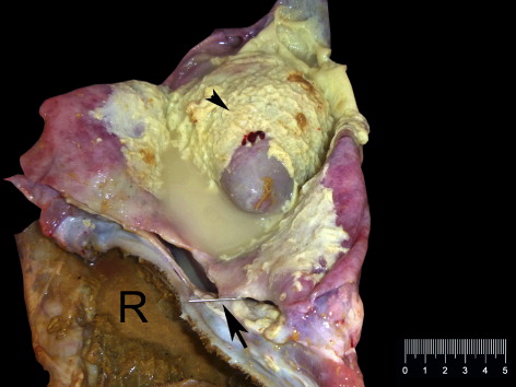
Necropsy revealed the fibrous adhesions and a fistular connection between the dorso-ventral face of reticulum to diaphragma and pericardium with 3 cm syringe needles in all three cases. The fistular connection was present between the reticulum and the apex of pericardium in the first case. The pericardium and epicardium were markedly thickened and the pericardial sac contained approximately 250 ml turbid, yellow tinged fibrio-purulent exudate in the first case (). In the second case, the sheep was approximately four months pregnant. There were adhesions between reticulum, diaphragm and the heart having a fistular connection with three syringe needles. The pericardial sac contained 200 ml of clear, sero-fibrinous exudate and the needles had penetrated to the pericardium. The lungs were congested and contained many tiny abscess in visceral surfaces approximately 2 cm in diameter and most lobes were adhered to the thoracic cage. The pericardium and epicardium were moderately thickened. In the third case, there was a fistular connection between the reticulum and pericardial sac having black hemorrhagic exudate.
The kidneys were swollen and pale in colour and the cut surfaces showed pin-point brown discoloration upon Lugol's iodin solution for the amyloid in all cases. There was bilateral keratitis characterized by cloudy appearance of the corneal surface in two of the three sheep.
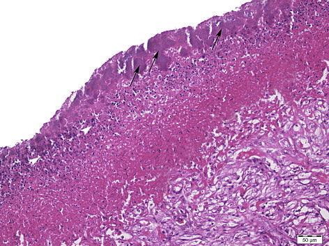
Microscopically, the epicardium and pericardium were moderately expanded by fibrous tissue proliferation measuring between 500 and 1000 µm in thickness containing capillaries and fibroblasts. Multifocally, aggregates of Gram- positive, coccoid bacterial colonies were attached to the free surfaces of the pericardial and epicardial tissues and were surrounded by degenerated and necrotic neutrophilic, scant mononuclear infiltrate and coagulative necrotic material ().
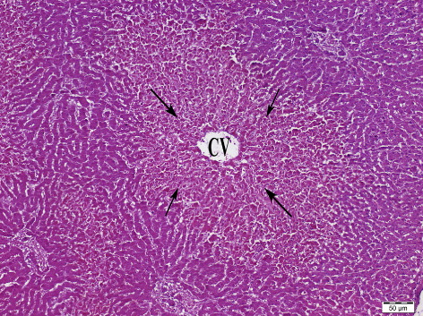
In all the cases, there was randomly distributed myocardial cell degeneration characterized by sarcoplasmic swelling, and vacuolation or myocytic necrosis characterized by sarcoplasmic eosinophilia, loss of cross-striations and pyknosis and karyorhexis. A number of Sarcocystis sp. brachizoides measuring 30–50 µm were present throughout the heart in all cases. Liver sections indicated multifocal severe and diffuse centrilobular hepatocellular necrosis with no inflammatory reactions in all the cases (). There was mild to moderate vascular hyperemia in the portal traits.
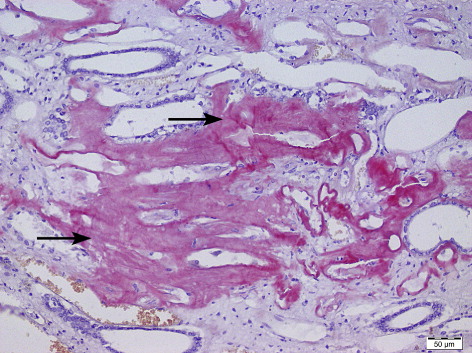
The renal cortex and medulla showed multifocal to linear interstitial nephritis characterized by lymphocyte, macrophage and plasma cell infiltrations with varying severity in all three cases. Many tubules contained intraluminal hyaline casts. The tubular epithelium showed mild to moderate hydropic degeneration. In the first case, there were congophylic amyloid deposits in the glomerular tufts, Bowman capsule and vascular walls of the liver, kidneys and spleen. In the second case, there was also medullar amyloid material showing positive staining with CR (). All the amyloid materials gave birefringence under cross-polarized light. In the eyes, there was a diffuse corneal ulcer and midstromal vascular infiltrations together with scant neutrophils.
3.2. Clinical and ultrasonographic findings
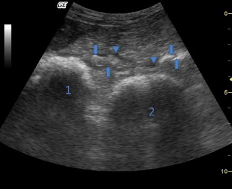
Two of the sheep that were brought to our hospital were depressed. One of them was approximately four months pregnant. The sheep’ rectal temperature, pulse and respiration rates were 39.5°C and 40.0°C, 82 beats/minute and 112 beats/minute, 28 breaths/minute and 32 breaths/minute, respectively. Ruminal contractions were reduced in both of them. Ultrasonography of the reticulum revealed echogenic fibrin deposits interspersed by small anechogenic fluids pockets between the caudoventral reticular wall and cranial wall of the rumen ().
4. Discussion
There are few case reports about TRP and its complications in sheep and goats (Akkoç Citation2007; Torki et al. Citation2011). In this report, we determined traumatic reticulopericarditis in three sheep by necropsy and TRP in two sheep by clinical and ultrasonographic examinations. The tentative diagnosis of TRP was further confirmed by the surgery. The other sheep deaths in the flock might also be due to TRP and its complications as the history and the postmortem observations of the owner were similar to TRP and its complications. This study described several TRP cases during in short time in the same flock. All affected sheep in the flock had been bought from another sheep flock that grazes nearby in garbage. The investigation of the pasture was not performed but the needles that were found during either the postmortem examination or surgery indicated a contamination of the pasture by syringe needles. Contamination of the pastures by syringe needles is dangerous not only for animal health but also for public health. Some human infectious diseases may be spread because of environmental contamination of medical waste. Producers, therefore, should be careful in regards to this type of contaminated pastures.
Incidences of TRP are low in sheep because ingestion of sharp non-food items such as nails and needles is extremely rare (Radostits et al. Citation2007; Jones & Smith Citation2008). Sheep flocks commonly graze on pasture in our area. Contaminated pasture with sharp materials and pica in the flock may have been responsible for the many TRP cases in this sheep flock.
Ultrasonography is used to diagnose TRP in cattle; however, ultrasonography cannot identify foreign bodies that cause TRP. Morphological changes in the reticular area can be identified by using an ultrasonographic examination. Fibrinous tissue deposits interspersed with fluid pockets are frequently determined in the reticular area and these are seen as echogenic areas cavitated by hypoechogenic or anechogenic areas during ultrasonographic examination. Solely fibrinous tissue deposits as seen in echogenic areas and reticular abscesses that have an echogenic capsule may also be observed in cattle with TRP. Although the caudoventral reticular wall is the most frequently affected, morphological changes may be seen in cranial or lateral to reticulum in cattle with TRP (Braun et al. Citation1993, Citation1994; Braun Citation2003). To our knowledge, there is no other study showing ultrasonograhic examination findings in sheep with TRP. In this report, ultrasonographic examination revealed echogenic fibrin deposits interspersed by small anechogenic fluids pockets between the caudoventral reticular wall and cranial wall of the rumen. These ultrasonographic findings are similar to those of cattle with TRP.
Amyloid is a pathologic substance deposited between cells in various tissues and organs of the body in a wide variety of clinical settings including chronic inflammatory and neoplastic processes (Johnson & Jamison Citation1984; DiBartola et al. Citation1989; Fernández et al. Citation2003). Amyloidosis was determined in cattle with mastitis, pneumonia, TRP, traumatic pericarditis, salpingitis, and metritis (Johnson & Jamison Citation1984; Yamada et al. Citation2006). Although amyloidosis was reported in sheep with gangrenous pneumonia, purulent polyarthritis, pseudotuberculosis and abdominal abscess (Ménsua et al. Citation2003), to the best of our knowledge amyloidosis has not been reported together with TRP (Ménsua et al. Citation2003). In this report the accumulation of amyloid material in the kidneys of the sheep that were found in the necropsies may have resulted from the inflammatory process due to TRP and traumatic pericarditis. This finding indicates that TRP and its complications may also cause amyloidosis as a complication in sheep like other inflammatory processes.
5. Conclusions
Although TRP is rarely seen in sheep, TRP and its complications may cause economic losses in sheep flocks. Ultrasonographic examination of the reticular area in sheep may be used to detect morphological changes in TRP cases as a part of a clinical examination. TRP and its complications may also cause amyloidosis as a complication in sheep as well.
Disclosure statement
The authors declared no potential conflicts of interest with respect to research, authorship and/or publication of this article.
References
- Akkoç A. 2007. Traumatic reticulopericarditis in a saanen goat. Turk J Vet Anim Sci. 31:283–285.
- Braun U. 2003. Ultrasonography in gastrointestinal disease in cattle. Vet J. 166:112–124.10.1016/S1090-0233(02)00301-5
- Braun U, Fluckiger M, Gotz M. 1994. Comparison of ultrasonographic and radiographic findings in cows with traumatic reticuloperitonitis. Vet Rec. 135:470–478.10.1136/vr.135.20.470
- Braun U, Gotz M, Marmier O. 1993. Ultrasonographic findings in cows with traumatic reticuloperitonitis. Vet Rec. 133:416–422.10.1136/vr.133.17.416
- DiBartola SP, Tarr MJ, Parker AT, Powers JD, Pultz JA. 1989. Clinicopathologic findings in dogs with renal amyloidosis: 59 cases (1976–1986). J Am Vet Med Assoc. 195:358–364.
- Fernández A, Ménsua C, Biescas E, Luján L. 2003. Clinicopathological features in ovine AA amyloidosis. Res Vet Sci. 75:203–208.
- Johnson R, Jamison K. 1984. Amyloidosis in six dairy cows. J Am Vet Med Assoc. 185:1538–143.
- Jones SL, Smith BP. 2008. Large animal internal medicine. In: Smith BP, editor. Traumatic reticuloperitonitis. Diseases of the alimentary tract. 4th ed. St. Louis: Mosby-Elsevier; p. 849–850.
- Ménsua C, Carrasco L, Bautista MJ, Biescas E, Fernández A, Murphy CL, Weiss DT, Solomon A, Luján L. 2003. Pathology of AA amyloidosis in domestic sheep and goats. Vet Pathol. 40:71–80.
- Radostits OM, Gay CC, Hichcliff KW, Constable PD. 2007. Veterinary medicine. In: Radostits OM, Gay CC, Hichcliff KW, Constable PD, editors. Traumatic reticuloperitonitis. Diseases of the rumen, reticulum and omaum. Diseases of the alimentary tract-II. 10th ed. Edinburgh: Saunders-Elsevier; p. 337–345.
- Torki E, Dezfoli MRM, Sasani F, Baghban F, Shahabi M, Motaghinejad M. 2011. Traumatic reticulo-pericarditis (TRP) in sheep: a report of 4 cases in a herd. Slov Vet Res. 48:45–50.
- Yamada M, Kotani Y, Nakamura K, Kobayashi Y, Horiuchi N, Doi T, Suzuki S, Sato N, Kanno T, Matsui T.. 2006. Immunohistochemical distribution of amyloid deposits in 25 cows diagnosed with systemic AA amyloidosis. J Vet Med Sci. 68:725–729.