
ABSTRACT
Sheared heel is a medial displacement caused by repetitive overloading on the palmar area, traditionally managed with a straight bar shoe. There are no reports of using z-bar shoes in horses with a sheared heel. The present study aims to investigate the effectiveness of z-bar shoeing and time required for sheared heel management. 3 show-jumping horses with medial palmar displacement on both front hooves (n = 6) were studied. The horses underwent lameness examination before study and radiography was performed on the affected feet. Z-bar shoes fitted with the large open space on the medial side, were applied for 48 weeks at 6 weeks intervals. Prior to shoeing, the affected hooves were placed on a clay mould and the impressions obtained were divided into 6 parallel lines. Each of the 6 lines was measured for medial/lateral ratio. There was no change in length ratios of the first, second and third lines at the toe to quarter region. A significant increase in length ratios of the fourth and fifth lines but marginal increase in the sixth line were noted 30 weeks post-shoeing. The z-bar shoes promote the expansion of the affected palmar area within 30 weeks of shoeing in horses with sheared heel.
Introduction
A sheared heel is a hoof-wall defect that affects the sport performance of horses. It is characterized by a proximal displacement of the unilateral heel bulb relative to the adjoining heel bulb (O'Grady Citation2003). Palmar hoof wall displacement is generally attributed to disproportionate distribution of weight-bearing on the entire hoof during the landing phase of leg movement. The degree of hoof wall deviation depends on the amount of repetitive overloading on the affected hoof area, particularly in the heel region (Redden Citation2003). Even though limb deformity and chronic lameness, such as navicular disease, are prone to cause displacement of the heel bulb, poor farrier practice is thought to be the underlying cause of sheared heels. Thus, the use of an appropriate trimming and shoeing method is crucial for horses with sheared heels (O'Grady and Castelijns Citation2011).
Appropriate farrier practice should improve hoof wall displacement and reduce the weight loading on the displaced hoof region. It has been reported that straight bar shoeing combined with specific trimming could improve hoof conformation. Specifically, trimming to lower the hoof wall from the toe, quarter to heel is proposed to produce the gap between the hoof wall and shoe on the affected side of the hoof (O'Grady Citation2012). The trimmed space is believed to allow the displaced hoof wall to grow distal to the shoe during weight-bearing. Also, the straight bar shoe increases the ground surface area of the hoof, in turn, allowing the affected hoof portion to be unloaded (O'Grady and Castelijns Citation2011; O'Grady Citation2012).
The z-bar shoeing method has also been used for the management of palmar foot pain, for example, unilateral palmar abscess (Dutton et al. Citation2009), quarter crack repair (O'Grady Citation2001) and navicular syndrome (Chanda et al. Citation2019). The large open portion of the shoe is supposed to eliminate the weight-bearing on the affected palmar area, which, in turn, reduces repeated trauma and eventually promotes the healing process.
Although the use of the straight bar shoe has been reported for sheared heel management, the outcome of this shoeing method is not fully satisfied. The z-bar shoe in which the empty part of the shoe could allow the affected region to be unloaded may be useful for the sheared heel management. However, there is a scarcity of reports on the efficacy of z-bar shoeing for this purpose. Moreover, the treatment time for this therapeutic shoeing practice in horses with a sheared heel has yet to be established. The aims of the present study were to investigate the effectiveness of using the z-bar shoeing method and determine the treatment time needed for horses with a sheared heel.
Materials and methods
Animals
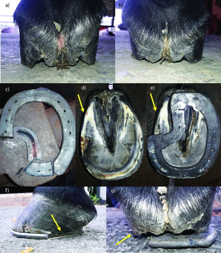
Three healthy show-jumping horses (1 gelding and 2 mares, age 9–14 years and weighing 300–450 kg) suffering from a hoof-wall defect on both forelimbs ((a,b)) (6 total affected hooves) were studied. The horses were housed in 4 × 5x4 m3 standard stalls and fed commercial pellets three times a day. Hay and water were freely accessed. The horses were undergone low to moderate intensity exercise 4–5 days a week. The daily exercise programme includes 20 min of trotting, 10 min of walking and 15 min of jumping skill practice. They had been routinely shod with custom metal shoes by a professional farrier at six-week intervals prior to the study.
Figure 1. Illustration of the sheared heel and z-bar shoeing on the affected hoof. The hoof with sheared heel shows a heel length disparity on the left forelimb (a) and right forelimb (b). The z-bar shoe is created to allow the affected area located in the large open portion of the shoe (c). The hoof is trimmed, particularly on the affected area (yellow arrow) (d). The z-bar shoe is applied with regard to the affected palmar area (e) and leaves the deviated hoof wall away from the ground (yellow arrow) (f and g).
Lameness examination
The horses underwent a lameness examination by an experienced veterinary surgeon before commencement of the study. In short, horses were trotted 30 m on the hard ground, followed by compression using a hoof tester onto the sole, quarter, and heel of the affected hooves. The fetlock flexion test, digital flexor tendon palpation and carpal flexion test were then performed on the affected limbs. Finally, dorsopalmar (DP) radiographs were taken to evaluate the conformation of bone structure. The gait analyses were conducted before and after each routine shoeing until the end of the study. The lameness score ranged from 0 to 5, following the American Association of Equine Practitioners (AAEP) (Anon Citation1991). A score of 0 meant no lameness and 5 meant non-weight bearing or unable to move.
Shoeing procedure
The z-bar shoe was designed according to a method reported previously (Chanda et al. Citation2019). Briefly, the custom metal shoes (Mustadfors Bruks, Dal Langed, Sweden) were cut at approximately one-third or one-half of the quarter region on the side of the displaced palmar portion. The intact branch and the cut branch of the metal shoe were then welded with another piece of metal to form the z-bar shoes ((c)). The affected hooves were trimmed, particularly in the deviated palmar area, to move the affected hoof wall away from the ground ((d)). The trimmed hooves were then placed on a small clay mould to generate the hoof ground surface figures ((b)) then they were fitted with z-bar shoes ((e–g)) by professional farriers for 9 consecutive shoeing periods (48 weeks) at 6-week intervals. The healthy hooves were shod with custom shoes according to the fundamental shoeing protocol (Poynton Citation2005).
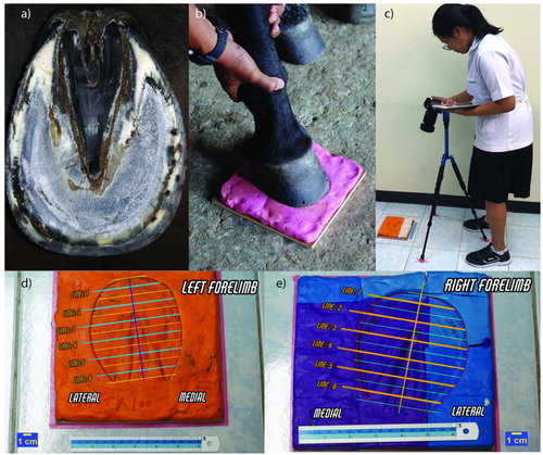
Figure 2. The determination of hoof ground surface dimension before shoeing. The trimmed hoof (a) is placed on the clay mould to determine hoof ground dimension, figure (b). The figure is then photographed at a constant distance by digital camera (c). The figure images are divided into six parallel lines and finally quantified for the medial/lateral ratio using the Image J software program (d and e).
Determination of surface dimension of hoof wall
The digital photography has been reported to evaluate the hoof conformation (Leśniak et al. Citation2017). The ground surface figures, alongside a 6-inch ruler for measurement, were photographed by a Sony A7 II digital camera (ILCE-7M2, Minato, Tokyo, Japan) at a constant distance of 80 cm for all captions ((c)). The images were subject to several computer programs for data preparation. First, the circumferences of each hoof ground surface figure were marked using Procreate software program (version 4.3.9) (Savage Interactive, Tasmania, Australia). Then, the hoof surface figures were divided horizontally into 6 parallel lines and perpendicular to the vertical midline using AutoCAD software program (version O.49.0.0) (Autodesk, Inc., California, U.S.A). The first line was situated cranial to the toe and the sixth line was located caudal to the heel bulb on the ground surface figures ((d,e)). Lastly, each parallel line was measured in centimetres for a certain length originating from the vertical midline to both medial and lateral sides using Imag J program (version 1.52a) (LOCI, University of Wisconsin, U.S.A). The distances of both ranges of each line were used to calculate the medial/lateral ratio for further analyses.
Statistical analysis
Statistics were analysed using GraphPad Prism 7.05 (GraphPad Software, Inc, San Diego, U.S.A). The changes in length ratios of 6 lines during 6 consecutive shoeings (0–30 weeks) were analysed using one-way ANOVA with repeated measurements followed by the Tukey multiple comparison test. The length ratios of the fourth, fifth and sixth lines were further analysed and compared between the sixth (30 weeks) and ninth (48 weeks) shoeing periods using the Mann–Whitney test. The data were expressed as mean ± SEM and p < 0.05 was considered statistically significant.
Results
Lameness examination
Three horses demonstrated the sheared heel on the medial portion of both front feet and they exhibited no foot pain after the lameness examination conducted prior to the study. The radiographic images of 6 affected hooves revealed a medial displacement of the third phalanx (Figure S1a-f) in which the movement of that third phalanx resulted in a disparity of the joint space (Figure S1b and e). Considering each horse individually, two horses showed no lameness before and after fitting with z-bar shoe throughout the study period (data not shown). Although one horse also demonstrated no foot pain before and after the shoeing for 30 weeks, unfortunately, it suffered an accident before the 36 weeks scheduled shoeing and then revealed severe lameness. This injured horse was excluded from the study after 30 weeks post-shoeing.
Determination of surface dimension of hoof wall
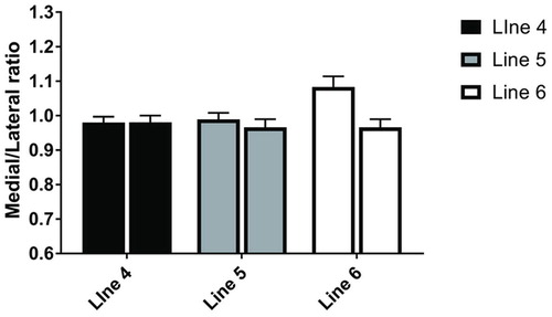
The medial/lateral ratios of the first ((a)), second ((b)) and third imaginary lines ((c)) did not change throughout the six consecutive shoeing period (0-30 weeks). The ratios of the three lines were close to 1 (0.98 ± 0.03–1.08 ± 0.05). A significant increase in length ratios of the fourth ((d)) and fifth ((e)) parallel lines was observed at 30 weeks post-shoeing compared to the control (0 week) (4th line; 0.94 ± 0.02 vs. 0.98 ± 0.02) (5th line; 0.87 ± 0.03 vs. 0.99 ± 0.02) (p < 0.05 for both). There was a marginal increase in the length ratio of the sixth line ((f)) at 30 weeks post-shoeing compared to the control (0 wk) (6th line; 0.89 ± 0.05 vs. 1.08 ± 0.03, p = 0.09). illustrates the apparent improvement of the hoof ground surface dimension, particularly in the displaced palmar regions, at 30 weeks post-shoeing. Two horses (n = 4) were continuously fitted with z-bar shoes for 3 additional consecutive shoeing periods (36, 42 and 48 weeks). The medial/lateral ratios of the fourth, fifth and sixth parallel lines were compared and no significant change was observed in length ratios of those lines between the sixth (30 weeks) and ninth (48 weeks) shoeing ().
Figure 3. The single line graphs illustrate the changes in length ratios of each line: 1st line (a), 2nd line 2 (b), 3rd line 3 (c), 4th line 4 (d), 5th line 5 (e) and 6th line (f) throughout the six shoeings (at 0, 6, 12, 18, 24 and 30 weeks) at six-week interval (n = 6). *P < 0.05, significant difference of length ratios from the pre-shoeing period.
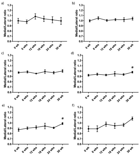
Figure 4. Each horse demonstrated proximal displacement of the medial palmar region (yellow arrow) on both front feet before fitting with z-bar shoe (upper row) (1st horse; a and b, 2nd horse; c and d, 3rd horse; e and f). After 30 wks of regular z-bar shoeing, the displaced hooves exhibited an expansion of the affected palmar region (white arrow) (lower row) from pre-shoeing (a vs. g; b vs. h; c vs. i; d vs. j; e vs. k and f vs. l).
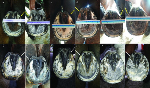
Figure 5. The bar graphs demonstrate no further changes in length ratio at palmar region compared between 30 weeks post-shoeing (left bar) and 48 weeks of shoeing (right bar) of each line (4th line, 5th line, and 6th line respectively).
Discussion
The sheared heel is a clinical condition defined by the proximal displacement of the palmar hoof wall. The deviation causes a reduction in the solar surface dimension of the hoof that gives rise to attenuated exercise performance. One finding of the study is a marked increase in length ratios of the transecting lines located on quarter to heel regions. This result suggests that the affected palmar hoof walls expanded, resulting in an improvement of the ground surface area on the affected hooves after fitting with the z-bar shoe. More importantly, a significant change of the palmar surface dimension was noticed at 30 weeks post-shoeing and no further change in the surface dimension of palmar region occur after shoeing for three more six-week periods with the z-bar shoe. Our findings indicate that z-bar shoeing has the therapeutic effect of improving the hoof ground surface area. It is also assumed that the horses should be routinely shod with z-bar shoes for at least 30 weeks for sheared heel therapy.
Considering the weight-bearing mechanism, the palmar area of the foot sustains a mechanical loading greater than the dorsal region. Heel area would play an essential role in reducing the compression during the loading phase of the stride. Because the heel portion of the foot is more elastic than a quarter and toe region, the reported expansion of the wall at the heel region may diminish the concussion when the foot is loading (Yoshihara et al. Citation2010; Back and Pille Citation2013; Brunsting et al. Citation2019). A reduction of the hoof surface dimension is due to the proximal deviation of the palmar hoof wall accompanying the repetitive overweight bearing on the affected palmar portion (O'Grady and Castelijns Citation2011). The radiographic images in the present study demonstrate a medial deviation of the third phalanx, which is related to the medial displacement of the hoof wall (Supplement file 1). The medial movement of the distal phalanx dampens the digit alignment resulting in narrowing the joint space on the lateral side. Nonalignment of the digits may lead to severe damage to their surrounding structure by, for instance, joint capsule distortion, supporting ligament desmitis and bone fracture due to mechanical overloading (O'Grady Citation2001; O'Grady and Castelijns Citation2011). Leaving the affected hoof portion unloaded along with a proper trimming will likely tackle this problem. For this reason, the z-bar shoeing in which the large open portion was created on the side of the deviated palmar hoof wall has been implemented to eliminate the repetitive overloading and allow a free movement of that affected hoof wall.
According to differences in hoof conformation among horses, the variety of changes in the ground-surface dimension was also observed between hooves. The length ratio of the dimension changes was adopted for evaluation of movement of the palmar hoof wall in response to z-bar shoeings. The relative values from the proportion of length ratio were used to correct the errors in using an absolute value from the measurement. Since the caudal region is more viscoelastic than the cranial part of hoof, as expected, an increasing length ratio of the fourth and fifth parallel lines, as well as a marginal increase in the sixth parallel line over the deviated region were noticeable. More importantly, the degree of the change in length ratio of the fifth line is greater than that of the fourth line, and the most considerable change was observed at the sixth imaginary line despite illustrating no statistical difference (4th line; 4.08%, 5th line; 12.12% and 6th line; 17.59% respectively). It is plausible that sixth line located within the heel bulb region reported as the most elastic portion of the foot, the striking variation of dimension change amongst hooves could be a contributing factor causing a marginal difference in this area. The z-bar shoeing caused not only an expansion of the defected hoof wall but also improvement in the heel bulb distortion (Supplement file 2). The horses performed low to moderate exercise training four to five days a week during the shoeing periods. The exercise training included 10 min of walking followed by 15–20 min of trotting and ending with five more min of walking. This finding provided evidence of the therapeutic effect of the z-bar shoeing method for the sheared heel. Of note, the proposed time course of the z-bar shoeing for sheared heel would offer an advantage for the farrier with regards to determining the shoeing period for each case. However, the exact time of the shoeing in the affected horse may vary among hooves depending on the severity of the hoof wall displacement, the farrier practice and the shoeing interval. Hence, the farrier must perform shoeing in close collaboration with the veterinarian to solve those problems and facilitate a satisfactory recovery from the hoof wall defect (Moyer Citation2003; Kummer et al. Citation2009; O'Grady Citation2012). The limitation of this study is the restrict number of sample size. Further study with a larger sample may be required to validate the potential using of the z-bar shoeing practice in horse with sheared heel. As the exercise pattern of show jumping correlates strongly to the fluctuation of high impact loading on the horse’s hooves, the question arose as to whether the horses fitted with z-bar shoes can participate in full exercise at show jumping events. The impact of z-bar shoeing on the show-jumping performance in horses with sheared heel needs to be further investigated.
Conclusions
The z-bar shoe has potential for use as therapeutic shoeing in the horse suffering from the sheared heel. Affected hoof should be regularly shod with z-bar shoes for at least 30 weeks to gain benefits of the shoeing. A close collaboration between veterinary practitioners and professional farriers is required to achieve the desired outcome.
TAAR_1814785_Supplementary_Material
Download Zip (3.2 MB)Acknowledgments
The authors would like to thank COL. Vithai Laithomya, the commander of the Royal Stable Unit, Royal Thai Army, and Mrs Vaewratt Kamonkon, the president of the Horse Lover’s Riding Club, Thailand, for allocation of their horses in this study. The great farrier practice performed by SM1 Apichai Pongaree, and SM1 Somchai Thanoorat is also appreciated.
Disclosure statement
No potential conflict of interest was reported by the author(s).
References
- Anon. 1991. Guide for veterinary service and judging of equestrian events. 4th ed. Lexington: AAEP.
- Back W, Pille F. 2013. The role of the hoof and shoeing. In: WB Saunders, editor. Equine locomotion. 2nd ed. London: Elsevier; p. 147–174.
- Brunsting J, Dumoulin M, Oosterlinck M, Haspeslagh M, Lefère L, Pille F. 2019. Can the hoof be shod without limiting the heel movement? A comparative study between barefoot, shoeing with conventional shoes and a split-toe shoe. VET J. 246:7–11. doi: 10.1016/j.tvjl.2019.01.012
- Chanda M, Senarat W, Thongkam E, Kanthavichit K, Puangthong C. 2019. Unilateral perineural anaesthesia on the lame leg facilitates the selection of z bar shoeing technique for the treatment of navicular syndrome. J Appl Anim Res. 47:154–158. doi: 10.1080/09712119.2019.1588736
- Dutton D, Lashnits K, Wegner K. 2009. Managing severe hoof pain in a horse using multimodal analgesia and a modified composite pain score. Equine Vet Educ. 21:37–43. doi: 10.2746/095777308X382669
- Kummer M, Gygax D, Lischer C, Auer J. 2009. Comparison of the trimming procedure of six different farriers by quantitative evaluation of hoof radiographs. VET J. 179:401–406. doi: 10.1016/j.tvjl.2007.10.029
- Leśniak K, Williams J, Kuznik K, Douglas P. 2017. Does a 4–6 week shoeing interval promote optimal foot balance in the working equine? Animals. 7:29. doi: 10.3390/ani7040029
- Moyer W. 2003. Hoof wall defects: chronic hoof wall separations and hoof wall cracks. Vet Clin Equine Pract. 19:463–477. doi: 10.1016/S0749-0739(03)00003-8
- O'Grady S. 2001. Quarter crack repair: an overview. Equine Vet Educ. 13:216–219. doi: 10.1111/j.2042-3292.2001.tb00093.x
- O'Grady SE. 2003. Shoeing management of sheared heels. In: Norman Edward Robinson, editor. Current therapy in equine medicine. 5. Elsevier; p. 528–532.
- O'Grady SE. 2012. Farriery for the hoof with a sheared heel. Vet Clin Equine Pract. 28:381–392. doi: 10.1016/j.cveq.2012.05.002
- O'Grady S, Castelijns H. 2011. Sheared heels and the correlation to spontaneous quarter cracks. Equine Vet Educ. 23:262–269. doi: 10.1111/j.2042-3292.2010.00219.x
- Poynton AP. 2005. Shoe and shoeing method. Google Patents US6732807B2.
- Redden RF. 2003. Hoof capsule distortion: understanding the mechanisms as a basis for rational management. Vet Clin Equine Pract. 19:443–462. doi: 10.1016/S0749-0739(03)00027-0
- Yoshihara E, Takahashi T, Otsuka N, Isayama T, Tomiyama T, Hiraga A, et al. 2010. Heel movement in horses: comparison between glued and nailed horse shoes at different speeds. Equine Vet J. 42:431–435. doi: 10.1111/j.2042-3306.2010.00243.x