Abstract
Our aim was to study the possible relationship between psychological stress and granulocyte activation primarily in healthy students during an examination period (n = 11) and also in chronically anxious patients (n = 15). We employed cell surface markers: lactoferrin, l-selectin, αMβ2-integrin and CD15s and flow cytometry to detect changes in the activation state of granulocytes, with the start of the stressed state in students at the beginning of an examination period, which was associated with elevated blood plasma cortisol level, and following relaxation hypnosis in both students, during their examination term, and patients. The ratios of all four types of marker-carrier granulocytes increased at the start of the examination period in students; an especially dramatic (ca. 5-fold) enhancement was observed in the proportion of lactoferrin-bearing cells relatively to the pre-examination term value. After hypnosis, the percentage of lactoferrin-exposing granulocytes decreased considerably both in students and in patients, by about half; a similar decrease was observed in the ratio of CD15s-carrier cells in patients. No significant alteration was observed during the study in state or trait anxiety levels, and in total or differential leukocyte counts. Thus, granulocyte activation could be associated with stress, while relaxation may facilitate reducing activation of these cells. In both groups of subjects, granulocyte surface lactoferrin appeared to be a sensitive “stress indicator”. This needs further evaluation.
Introduction
It was Hans (János) Selye, a Hungarian-born scientist (1907–1982) who first discovered a relationship between the neuro-endocrine system and the immune system in stress; he found that the thymus and the lymph nodes were shrunken in rats exposed to stressors, while the adrenal glands became enlarged (Selye Citation1936). In the past decades, much has been learnt about the interactions between the neuro-endocrine system: the hypothalamic-pituitary-adrenal axis, the sympathetic-adrenomedullary system and the immune system (Chrousos Citation1995). It has become evident, that the whole psycho-neuro-immuno-endocrine system functions as one integrated control unit of the organism, the functioning of which is influenced by stressful events or inflammatory/immunogenic stimuli alike (Chesnokova and Melmed Citation2002). An intense, prolonged stress response usually leads to an immunosuppression characterized typically by down-regulation of the specific T-cell response (Glaser and Kiecolt-Glaser Citation2005). In contrast, the number and the ratio of neutrophil granulocytes could increase in intensive and/or chronic stress; “stress neutrophilia” was observed after both strenuous exercise and following spaceflight (Gabriel and Kindermann Citation1998; Stowe et al. Citation1999).
Neutrophils have a basic role in different inflammatory reactions; however, few investigations have dealt with the possible activation of granulocytes in stress, and especially in psychological stress. Increased activation of neutrophils was shown in a brief mental stress experiment as well as after watching a horror film, by the nitro-blue tetrazolium test (Ellard et al. Citation2001; Mian et al. Citation2003). In another study, the mild stress of a public speaking task led not only to increased blood counts of all leukocyte types, but it resulted also in an elevated number of ICAM-1/CD54-carrier granulocytes (Goebel and Mills Citation2000). In an examination stress model, positive association was found between neutrophil phagocyte functions and both trait anxiety and perception of threat (Wadee et al. Citation2001). In the last few decades, some psychological interventions (e.g. relaxation, hypnosis with immune suggestion, conditioning) were studied for their efficacy in reducing stress, and at the same time, in modulating the immune system (Miller and Cohen Citation2001; Gruzelier et al. Citation2001; Kiecolt-Glaser et al. Citation2001; Gruzelier Citation2002). However, most of these and other stress investigations focused either on routine leukocyte data and/or on the functions related to lymphocytes; psycho-neuro-immunological assessment of stressed state generally does not include functional granulocyte tests (Vedhara et al. Citation1999).
The aim of our study was to investigate whether granulocytes could be activated by psychological stress in healthy students, and if this activation might be moderated by relaxation-imagery hypnosis. Additionally, we compared the post-hypnotic granulocyte changes in students to those of chronically anxious patients. Cell surface appearances of four granulocyte activation markers: lactoferrin, l-selectin, αMβ2-integrin and CD15s were tested in a stressed state (before hypnosis, at the start of the examination period) and in a relaxed state (after the third weekly hypnosis); in students, data were collected in the resting state before the examination term, as well. From the cell surface activation markers of granulocytes, lactoferrin was chosen first, as the most reliable marker, since almost no lactoferrin molecules are present at the plasma membrane in resting human neutrophils (Afeltra et al. Citation1997). Therefore, the appearance of lactoferrin on these leukocytes is linked to their activation, when this characteristic component of the specific, secondary granules of neutrophils gains access to the cell surface and to the extracellular milieu, and further on, to the blood plasma following degranulation (Schettler et al. Citation1991). Since cell surface lactoferrin has been used rarely as a granulocyte activation indicator, two widely assayed markers: l-selectin and αMβ2-integrin were also included in our investigations; both of these membrane receptors are considered as markers of early neutrophil activation (Videm and Strand Citation2004). Finally, we were interested also in detecting CD15s, an unusual and seldom tested cell surface activation marker, known as an oligosaccharide ligand to selectins; its appearance on the cell surface is probably due to mobilization from the azurophilic, primary granules upon neutrophil stimulation (Suzuki et al. Citation2000). The determination of two flow cytometry parameters: the percentage of marker-bearing granulocytes (activated granulocytes) and the mean fluorescence intensity (average marker density) enabled us not only to detect changes in granulocyte activation during stressed and relaxed states in students and in patients, but it facilitated also the comparison of the four activation markers as “stress indicators”.
Methods
Subjects and study design
We recruited 11 healthy students: (physiotherapy college students with a mean ± SD age of 23 ± 1.79 years (1 male and 10 females) and 15 anxious patients (mean age ± SD: 44 ± 11.96 years; 1 male and 14 females). The patients had mixed anxiety and depression disorder (according to the criteria of ICD-10: F41.20). Also, they were diagnosed to have chronic, tension type headache (G44.20) in agreement with the points of the Headache Classification Subcommittee of the International Headache Society (Citation2004). The main requirements for students were a minimum age of 18 years and good health. Patients were eligible if they had mild anxiety, were between 18 and 75 years of age and had no severe medical problems that would inhibit their full participation in the study (e.g. heart failure, respiratory problems, serious diabetes mellitus, schizophrenia, infectious diseases).
Subjects completed a medical history and life style questionnaire and underwent physical and neurological examination before and after the study. In both groups, three subjects were regular smokers, and two seldom smoked. While all students had regular physical activity, most patients took little exercise. Patients continued their usual medication during the program; the most often used medicines were: antidepressants (nine subjects), anxiolytics (nine subjects), antihypertensive tablets (five subjects); all other types of drugs were taken by three or less participants. The study protocol was approved in advance by the Regional Medical Ethics Committee; participants signed the informed consent form before the start of the study.
Both students and patients participated in three relaxation-imagery hypnosis group sessions (one/week, duration: 20 min), with each series preceded by a hypnotizability test (). Students were familiar with the basics of the relaxation techniques, and had moderately difficult examinations during the investigation period. According to our previous clinical experience, relaxation sessions are mostly beneficial for stressed patients with chronic tension headache. In the induction part of the hypnotic sessions, subjects heard standard suggestions using the Harvard Group Scales text, like feeling heaviness, warmth and full relaxation. In the relaxation-imagery hypnosis part, subjects were encouraged to see healthy and optimally functioning granulocytes in blood, which regained their relaxed state after a successful battle against some microorganisms. The imagery suggestions followed the general rules of the formulation of suggestions during hypnotherapy, but the specific imagination used in this study is an original idea of the authors based on a published report (Hammond Citation1990; Hall et al. Citation1996).
Figure 1 Flow diagram of stress-hypnosis study on granulocyte activation markers in healthy students and anxious patients. Students: blood samples were collected and Spielberger's psychotests were completed (A) in the resting period: before the examination (exam) term; (B) in a stressed state: at the start of the exam term (before the first hypnosis); (D) in a relatively relaxed state: after the third hypnosis, during the exam term. Patients: psychotest completed before the study selected patients for a chronic mild anxiety group; blood samples were collected only (B) in the anxiety state (before the first hypnosis) and (D) in a relatively relaxed state, after the third hypnosis. On the first occasion a hypnotizability test was performed in both groups.
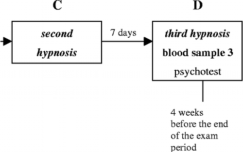
Blood samples were taken from the study participants in the morning hours (mostly between 7.30 and 9.30 a.m.) A/ in the resting state, before the examination period (students); B/ before the first and D/ immediately after the third hypnosis sessions (students and patients) (). Psychological test forms were filled in similarly, except for the chronically anxious patients, who completed the first psychotest at their first appearance in order to check their anxiety level.
Psychological tests
Hypnotizability of the participants was measured with the Harvard group test consisting of 12 hypnotic suggestions followed by completion of the standard questionnaire (Shor and Orne Citation1962). In our clinical practice we determined the following ranges: score 0–3: not susceptible; score 4–8: medium susceptible; and score 9–12: highly susceptible to hypnosis. Spielberger's test was used to examine the state and trait anxiety of the participants (Spielberger Citation1970). In the Hungarian version of the state-trait anxiety inventory, the normal value for state anxiety is 41.21 and for trait anxiety is 43.72; the scores of hospitalised neurotic and psychosomatic patients are 50.48 and 54.24, concerning state and trait anxiety, respectively (Sipos and Sipos Citation1983). In everyday clinical practice, the ranges for both state and trait scales are: < 50: normal; 50–60: mild; 60–70: medium; 70–80: high level of anxiety.
Blood samples, leukocyte counts and cortisol assay
Venous blood was drawn from antecubital veins in the morning (mostly between 7.30 and 9.30 a.m.) into three EDTA-containing Vacutainer tubes for cell counts and general hematology (4 ml), for blood plasma separation (7 ml) and for granulocyte activation analysis (2 ml), according to standard clinical protocols. The hematology tubes were sent to the central clinical laboratory, where they were processed during the next few hours. Blood counts were determined according to standard protocols using an automatic cell counter. Normal reference range for total leukocyte count is between 3.9 and 11.1 × 10⁹/l and for the neutrophil fraction: 44.0–68.0%, as given by the main clinical laboratory. The tubes for the cortisol assay were collected into ice; these blood samples were centrifuged within an hour, and then the plasma samples were stored at − 70°C until analysis. Cortisol was determined in duplicates by a single radioimmunoassay using a standard kit (DSL-2100 ACTIVE Cortisol Coated-Tube RIA Kit; Diagnostic Systems Laboratories, Webster, TX, USA). The intra-assay coefficient of variation was < 8%. The normal reference range of cortisol values is between 160 and 620 nmol/l as stated by the clinical endocrine laboratory. For granulocyte assays, the blood sample tubes were stored at room temperature and they were processed within 12 h of collection.
Immune labelling and flow cytometry
Cells surface granulocyte tests were performed by an indirect immunofluorescent method using a flow cytometer. Whole blood samples were used, because cell isolation techniques are known to activate leukocytes. Thus, whole blood aliquots (100 μl) were treated with (20 μl) saturating concentrations of either a polyclonal anti-lactoferrin antibody (200 mg/l, ICN), or a monoclonal anti-l-selectin (47 mg/l) or anti-αMβ2-integrin antibody (10 mg/l) (DAKO) or a monoclonal anti-CD15s antibody (50 mg/l) (BD Pharmingen). Negative controls contained normal rabbit serum or isotype-specific normal mouse Ig (DAKO). Following incubation (20 min, room temperature) and washing (two times with Hank's solution; sedimentation: in Eppendorf tubes using Heraeus Biofuge pico, 13,000 rpm), appropriate secondary antibodies: anti-rabbit (10 mg/l) or anti-mouse (20 mg/l) antibodies conjugated with FITC (DAKO) were added to the blood cells (20 μl aliquots of reagents to 180 μl aliquots of blood cells). After incubation and washing (performed as before), hemolysis was carried out with a lysis buffer (Biodesign, Lysing kit). One millilitre of lysis reagent was added to a 100 μl aliquot of blood cells; after vigorous vortexing and about 15 min incubation, sedimentation and a washing step followed (6000 rpm, Hank's solution). Finally, blood cells were resuspended in 200 μl Hank's buffer.
During cytometry using computer-assisted FACStar Plus Becton-Dickinson equipment, granulocytes were gated on the basis of their characteristic forward- and side-scatter features (average cell size: between those of lymphocytes and monocytes, by forward scatter; and a granular surface by side scatter). Measurements were preceded by standard equipment calibrations, and each detection series started with setting the background intensity level using the appropriate negative controls (as above). Generally, 10,000 events were measured per tube in this gated cell population. From the recorded results, the percentage of the labelled (activated) granulocytes (% of marker-bearing cells) and the mean fluorescence intensity (average marker density) are presented. The final cytometry results were from a minimum of 14 patients and a minimum of seven students in each case; all assays were performed in duplicate.
Data analyses
All dependent variables are given as means ± standard error (SEM). Primarily, the granulocyte data were used for pairwise comparisons separately in the student group (regarded as an auto-control statistical unit), and separately in the patient group, to examine the effect of stress or the influence of the relatively relaxed state in a normal and in a pathological group of subjects. Significance of differences between the values was determined using mostly the paired t-test or a non-parametric rank-sum test (Wilcoxon- or Mann–Whitney test in case of non-normal distributions). Paired t-test was performed also when analysing the cortisol data.
When statistical significances were examined between the values of the two different groups: the student and the patient groups, either one-way analysis of variance (one-way ANOVA) with all-pairwise multiple comparison procedures (Tukey Test) was applied, or Kruskal-Wallis one-way analysis of variance on ranks was used with Dunn's method of all-pairwise multiple comparison procedures (the latter in case of non-normal data distribution). Differences were considered statistically significant if p was less than or equal to 0.05.
Results
Stressed students and anxious patients
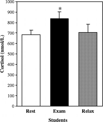
Our primary goal was to investigate granulocyte activation in healthy individuals with increasing psychological stress, and to detect possible reversal of this activation in a relatively relaxed state, following relaxation-imagery hypnosis. For this purpose, college students were tested three times: 1/ before an examination period, 2/ at the onset of the examination term, before hypnosis and 3/ after hypnosis, in the middle of the examination period. Plasma cortisol concentration increased significantly as the examination period started, and tended to decrease after the third hypnosis, when it was not different from the level before the examination period ().
Figure 2 Effect of examination stress and relaxation (after hypnosis) on blood plasma cortisol concentration in healthy students. As the examination (Exam) period started, cortisol concentration in blood plasma increased significantly in students (*p < 0.05 vs. Rest, n = 10). Relaxation had no statistically significant effect. Normal reference range for blood plasma cortisol: 160–620 nmol/l. Data are group means ± SEM.
As expected, the students and patients differed in their mean values on the state and trait anxiety inventories. Students had scores in the normal range ( < 50) throughout the study. In contrast, as expected, those of the chronically anxious patients were in the range of mild level of anxiety (between 55 and 60; clinical range for mild anxiety: 50 and 60) (). The state anxiety level of the pre-hypnotic patients was significantly higher than all three of the values for the students. In addition, the anxiety level of the patients after hypnosis was significantly greater than that of the students at the onset of the examination term. Trait anxiety levels were significantly greater in the patients both before and after hypnosis than in the students at the beginning of the examinations and after hypnosis. No significant changes in anxiety level were seen either in the students or in the patient group during the investigation.
Table I. State and trait anxiety scores in students and patients (group means±SEM).
The mean hypnotizability level of the students (7.91 ± 0.58) was not significantly different from that of the patients (6.47 ± 0.42). According to clinical practice, these scores in the range of 4–8 indicate medium susceptibility.
Cell surface appearance of activation markers
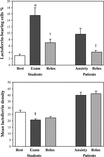
A low percentage of the granulocytes was labelled with anti-lactoferrin antibodies in the “resting students” one week before the examinations (). The percentage of lactoferrin-carrier cells rose dramatically (about 5-fold; p < 0.05) upon the start of the examination term, and it was approximately halved after the third hypnosis (p = 0.001). In anxious patients, the elevated initial percentage of lactoferrin-bearing granulocytes similarly decreased, by about half, after the last hypnosis (p < 0.05). Concerning the mean signal intensity (reflecting average surface marker density), no changes were observed in patients. In contrast, a small but significant decrease was observed in the relatively low mean density value of students when the examination period started (p < 0.01); this reduced value was not altered after the hypnosis. The patients' density values were significantly greater than those of the students (p < 0.05).
Figure 3 Effect of stress and relaxation on the appearance of lactoferrin on the cell surface of granulocytes in healthy students and in anxious patients. In students, the percentage of lactoferrin-bearing cells significantly increased from before the examination period (Rest; n = 8), to the start of the examinations (Exam; n = 9; *p < 0.05), and it declined after the final relaxation hypnosis (Relax; n = 9; †p = 0.001). Lactoferrin density decreased with the start of examinations (§p < 0.01). In patients, the percentage of lactoferrin-bearing cells decreased after relaxation hypnosis (Anxiety; n = 14 vs. Relax; n = 15; ‡p < 0.05). The patients' density values are significantly higher than those of the students p < 0.05). Data are group means ± SEM.
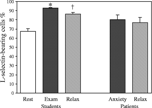
Concerning l-selectin, a different picture was seen, since in the students most granulocytes carried this adhesion receptor in the initial “resting” phase (). With the onset of examinations, this high proportion further increased (p < 0.001), and showed a slight reduction after hypnosis (p < 0.01) (). In contrast, the relatively high percentage of l-selectin-bearing cells did not change after hypnosis in the patients. No alterations were observed in the values of average receptor density either in students, or in patients.
Figure 4 Effect of stress and relaxation on l-selectin presence on the cell surface of granulocytes in healthy students and in anxious patients. In students, the percentage of l-selectin-carrier cells increased from Rest (n = 7) upon the start of the examinations (Exam; n = 11; *p < 0.001), and decreased slightly after the third hypnosis (Relax; n = 10; †p < 0.01). Data are group means ± SEM.
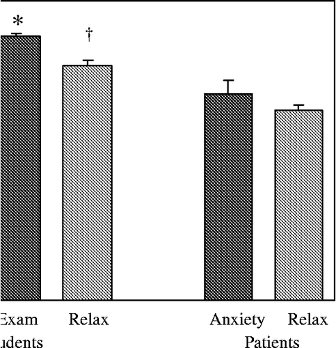
In the case of αMβ2 integrin, again a pronounced increase was seen in students at the start of the examinations, when the percentage of receptor-carrier cells reached almost 100% (1.5-fold greater than at rest; p < 0.001); this was followed by a slight decrease after the third hypnosis (p < 0.05) (). The receptor density showed no statistically significant changes. In patients, no significant changes were detected.
Figure 5 Effect of stress and relaxation on aMb2 integrin presence on the cell surface of granulocytes in healthy students and in anxious patients. In students, the percentage of integrin-bearing cells increased from Rest (n = 9) upon the start of the examinations (Exam; n = 7; *p < 0.001), and decreased moderately after the last hypnosis (Relax; n = 10; †p < 0.05). The tendency for integrin density to increase at the start of the examination period was not statistically significant in students. Data are group means ± SEM.
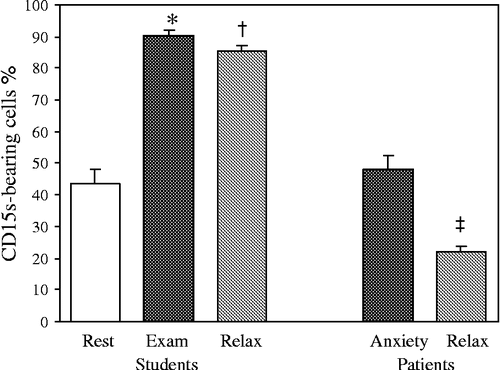
The observed change for CD15s (sialylated Lewis-X antigen, a selectin ligand) in the percentage of marker-exposing granulocytes was similar to that for the other markers in students: a 2-fold increase was seen at the beginning of the examination term (p < 0.001; ), followed by a limited reduction after hypnosis (p < 0.05). In patients, this parameter showed a significant reduction (by half) after the last hypnosis (p < 0.001; ), while the initial, pre-hypnosis value was not different from the resting value in the students. The density value decreased after the hypnosis in the patients in a similar fashion (p < 0.001), with no significant alterations in the students. The percentage of marker-bearing cells was significantly greater in students at the start of the examinations and after the final hypnosis than in patients at the start and after hypnosis (p < 0.05; ).
Figure 6 Effect of stress and relaxation on CD15s presence on the cell surface of granulocytes in healthy students and in anxious patients. In students, the percentage of CD15s-bearing cells significantly increased from Rest (n = 8) upon the start of the examinations (Exam; n = 9; *p < 0.001), and decreased slightly after the third hypnosis (Relax; n = 10; †p < 0.05). In patients, both the percentage of marker-carrier cells and the mean marker density were reduced after hypnosis (Anxiety; n = 14 vs. Relax; n = 14; ‡p < 0.001). The percentage of marker-carrier cells was greater in students during examinations and after hypnosis than in patients in both conditions (p < 0.05). Data are group means ± SEM.
Leukocyte counts
In both study groups, the values for total leukocyte count and for neutrophil ratio were within the normal reference ranges (total leukocyte count: 3.9–11.1 × 10⁹/l, neutrophil fraction: 44.0–68.0%), so no further inflammatory assays were done (). The total leukocyte counts of the students and patients did not change during the study, and there were no significant differences between any data groups for students and patients. However, the patients before hypnosis had a higher percentage of neutrophils than the students before and at the start of the examinations (p < 0.05; ); in addition, the neutrophil percentage was greater in the post-hypnotic patients than in the students at the onset of the examinations (p < 0.05).
Table II. Total leukocyte count and neutrophil granulocyte percentage in students and patients (group means±SEM).
Discussion
Our results suggest activation of granulocytes in the psychologically stressed state. In students, all four cell surface activation markers: lactoferrin, l-selectin, αMβ2 integrin and CD15s appeared on a significantly greater percentage of leukocytes at the beginning of the examination period than before. Furthermore, the decreased average density of lactoferrin at the onset of the examinations probably also reflects granulocyte activation in stressed students, and is probably due to its enhanced release to the blood plasma (Schettler et al. Citation1991). Initially, our mildly anxious patients were characterized by a relatively high percentage level of lactoferrin-carrier cells (almost 10%) indicating a stimulated state of the granulocytes. On the other hand, the significant decreases in the percentage of the marker-bearing cells after relaxation hypnosis suggest reduced activation of granulocytes. In students, the proportion of activated granulocytes decreased in the case of all markers; in patients, only the appearance of two markers, lactoferrin (percentage of carrier cells) and CD15s (both cytometric parameters), were reduced after hypnosis. Therefore, the mechanisms of granulocyte activation and the return to the resting state may be partially different between these patients and the students.
The differences in appearance of certain granulocyte markers at the cell surface in patients and in students might be related to several factors, for example differences in stress/anxiety type, in age, in gender, in physical activity, medication. It is interesting to note that excessive exercise, namely weight lifting (multiple repetitions of maximum squats) and marathon running increase the appearance (density) of l-selectin and β2 integrin on neutrophils, respectively (Kappel et al. Citation1991; Miles et al. Citation1997). In addition, increased plasma and urine concentrations of lactoferrin and myeloperoxidase observed in marathon runners after a race also suggest neutrophil activation after exhaustive exercise (Suzuki et al. Citation2003). Since moderate exercise, e.g. a 3 h run, does not affect β2 integrin appearance on these leukocytes (Kappel et al. Citation1991), physical activity probably did not influence the results of our students tested in the morning. Smoking can also lead to neutrophil granulocyte activation, as shown in a study on 60 year-old men with a median number of 35 pack-years, where enhanced blood plasma levels of myeloperoxidase, a neutrophil-monocyte/macrophage marker, and of specific human neutrophil lipocalin were measured (Ekberg-Jansson et al. Citation2001). Increased appearance (mean density) of l-selectin was also reported in smokers (Patiar et al. Citation2002). In each of our groups, the small number of smokers was the same; so, while a slightly elevated appearance of cell surface markers might be detected in smokers, this habit presumably did not have an impact on our in-group and inter-group comparisons.
The existence of psychological stress at the start of the examinations was supported by the significant elevation of the plasma cortisol level in students, which showed a tendency to decerease after hypnosis, and was then not different from the resting level before the examination period. In the pre-examination resting period the cortisol level in the students was somewhat higher than the upper limit of the normal range (160–620 nmol/l); this was further enhanced during the week before the examination period. While students were characterized by normal anxiety levels throughout the study, a mild level of anxiety was detected in our patients. Interestingly, no changes were seen during the study in the anxiety levels either in the students, or in the patients. This suggests that the detected innate immunological changes and the alteration of the cortisol level were not reflected by the tests we used to evaluate anxiety; perhaps other psychotests should be used in the future to further test this lack of correlation between physical measures of stress and anxiety within the student group. Nonetheless, although not shown in the present study, it is plausible to suppose that cognitive factors could have an important role in the regulation of the immune system. Hall et al. (1996) showed relaxation training without an active imagery exercise was associated with increased neutrophil adherence, while active cognitive training (relaxation, and imagery focusing on changing adhesive capability of neutrophils), resulted in decreased adherence. In investigations with medical students, the cognitive activation component of the personality proved to have a strong, positive influence on the specific immune system at examination time, and targeted immune imagery was more effective than relaxation imagery in immune consolidation (Gruzelier Citation2002).
Stress is generally associated with increased appearance of neutrophil granulocytes in the circulation. This is supported by our observation on the significantly higher percentage of the neutrophil fraction in anxious patients than in students before the examinations. In both groups, the values of the total leukocyte count and those of the neutrophil fractions were in the normal reference ranges indicating lack of serious inflammatory reactions.
Concerning the usefulness of the different granulocyte activation markers tested, cell surface lactoferrin showed most marked changes. In agreement with other reports (Afeltra et al. Citation1997; Swain et al. Citation2000), this multifunctional glycoprotein appeared to be a most reliable cell surface activation marker, since only a small percentage of the granulocytes exposed it in the initial resting state in healthy individuals, but this increased strongly in stress, and decreased again with relaxation. This decrease was seen in both students and in patients. These changes presumably reflect neutrophil activation, when lactoferrin is transported to the plasma membrane in the specific, secondary granules, and then is released to the extracellular space from where it can gain access to the blood plasma (Schettler et al. Citation1991). Since lactoferrin is usually regarded as a soluble inflammatory marker, it is generally assayed in blood plasma or in serum, and it is elevated, for example, in acute bacterial infections, in acute coronary syndromes after stenting, after exercise and even in schizophrenic patients (Hansen et al. Citation1976; Hallgren et al. Citation1982; Inoue et al. Citation2004; Gach et al. Citation2005). Noteworthy is that stressful circumstances change the salivary secretion of this glycoprotein (Bosch et al. Citation2003).
CD15s, commonly known as sialylated Lewis-X antigen, is a tetrasaccharide linked to membrane proteins and a component of certain glycosphingolipids; it serves as a ligand to selectins and thus is involved in the rolling of leukocytes on the endothelium (Greenberg et al. Citation2000; Burdick et al. Citation2001). In patients, the appearance of CD15s at the cell surface seemed to follow reduction of stress level after hypnosis, similarly to lactoferrin. This cell surface activation marker, probably mobilized from the azurophilic, primary granules upon neutrophil stimulation (Suzuki et al. Citation2000), has been relatively rarely employed. Its surface expression was reported to be increased on leukocytes in newborns of pre-eclamptic mothers and during hemodialysis (Carreno et al. Citation1996; Mellembakken et al. Citation2001).
Since l-selectin (CD62L), a constitutively active adhesion receptor of leukocytes, is continuously present at the cell surface of the granulocyte, the starting cytometric values were relatively high. Neutrophil activation is generally reported to result in shedding of this receptor from the surface (Ahmed and Christou Citation1996; Hafezi-Moghadam et al. Citation2001). However, increased appearance, as indicated by mean density, of l-selectin could also accompany neutrophil activation, as seen after mechanical trauma or in smokers (Cocks et al. Citation1998; Patiar et al. Citation2002). In contrast to this adhesion receptor, αMβ2 integrin (CD11b/CD18, Mac-1, CR3) is mostly stored in secretory vesicles and in granules; it is rapidly translocated to the surface upon activation, where it could be integrated into the bilayer and function in the arrest and spreading of leukocytes on the endothelial cells (Carlos and Harlan Citation1994; Lundahl et al. Citation1995). Both membrane receptors are considered as markers of early neutrophil activation (Videm and Strand Citation2004), and have been assayed widely; in a spaceflight investigation the average density of l-selectin increased, while that of αMβ2 integrin decreased at landing (Stowe et al. Citation1999). Conversely, an enhanced cell surface appearance, measured as density, of β2 integrins was seen in stressed males, during 240 days of confinement (Choukér et al. Citation2002).
Concerning the action of stress on granulocytes, modulation of the functioning of the specific and the less specific, innate immune system, involving the granulocytes, is mediated by a complex network of bidirectional signals between the nervous, endocrine and immune systems. The principal neuroendocrine regulators are related to the hypothalamic-pituitary-adrenal axis, and to the systemic sympathetic-adrenomedullary system. Almost all immune cells have receptors for some of the stress hormones: ACTH, cortisol and catecholamines, in addition to cytokine and chemokine receptors (Glaser and Kiecolt-Glaser Citation2005).
Cortisol and its analogs are anti-inflammatory agents and immune suppressors; but for some members of the specific immune system, rather than the innate, non-specific immune system. From the few related publications available in the literature, that of Strausbaugh and Rosen (Citation2001) is notable; they reported that glucocorticoids induce, via annexin1, l-selectin shedding and up-regulation of αMβ2 integrin on human neutrophils in vitro. Similarly, although catecholamines are generally considered to inhibit granulocyte adhesion and activation, Bazzoni et al. (Citation1991) showed in a careful in vitro human study that appreciable inhibition is possible only at high epinephrine concentrations, greater than 10− 8 mol/l, approximately two orders of magnitude higher than the physiological plasma level, or in the presence of adenosine produced mainly in ischemic conditions. Furthermore, Sagiyama et al. (Citation2004) even suggest that catecholamines may have an important role in granulocyte activation, as granulocyte infiltration of the liver could be brought about by epinephrine injection in mice.
Thus, stress does not entirely block the immune system; on the contrary, granulocytes, which belong to the innate immune system, can be activated as a part of an adaptation process (Sagiyama et al. Citation2004; Glaser and Kiecolt-Glaser Citation2005). It is interesting to note that the innate immune defence system is far older in evolutionary time than the specific system, which is present predominantly in vertebrates. The ancestors of microphages, originating from neutrophil granulocytes, can be found already in the sponges, in the most primitive multicellular organisms (Maier et al. Citation1994). It is plausible to suppose that activation of the neutrophil granulocyte, its chemotaxis and its transformation into an active microphage is probably part of an ancient survival mechanism in case of danger. However, as stimulated neutrophils produce superoxides, the accumulation of which can lead to oxidative stress with all its harmful consequences, all states with long-term increased neutrophil activation may facilitate the development of several disorders and diseases related to oxidative damage. There is increasing evidence that oxidative DNA damage may initiate cancer, such that stressed and depressed females with elevated 8-OH-dG levels related to neutrophil activity may have a higher risk for cancer (Irie et al. Citation2003). Thus, chronic stress management in disease prevention through simple, low cost and side-effect-free psychotherapies (relaxation, hypnosis, cognitive behavioural interventions) may have a role; all these interventions are shown to produce generally positive endocrine and immune effects (Glaser and Kiecolt-Glaser Citation2005). However, in the present study any effects of such relaxation hypnosis did not involve reducing anxiety level according to the employed psychotest, and the contribution of hypnosis per se to the temporal changes in granulocyte markers is not established. Hypnosis was used in the present study to aid achievement of a relaxed state in the subjects.
Taken together, our results indicate that granulocytes can become activated in stressed individuals, while a relaxed state may be associated with a subsequent decrease of granulocyte activation. From the cell surface activation markers examined, lactoferrin showed substantial and dynamic changes in the percentage of granulocytes carrying the marker, both in students and in anxious patients in the present stress-relaxation hypnosis study. It would seem worthwhile testing this granulocyte marker as a stress indicator in future studies, excluding inflammatory conditions and immune diseases. To our knowledge, we are the first to suggest a surface-granulocyte test to assess stress level. Further studies are required to clarify the relationships between the stress level and psychoneuroendocrine regulation of granulocyte function.
Acknowledgements
We are grateful for the devoted laboratory work of Zsuzsa Lajtos, and for the indispensible clinical help of Márta Bihari. This study was supported by grants from the Hungarian Space Office (TP178).
References
- Afeltra A, Caccavo D, Ferri GM, Addessi MA, de Rosa FG, Amoroso A, Bonomo L. Expression of lactoferrin on human granulocytes: Analysis with polyclonal and monoclonal antibodies. Clin Exp Immunol 1997; 109: 279–285
- Ahmed NA, Christou NV. Decreased neutrophil l-selectin expression in patients with systemic inflammatory response syndrome. Clin Invest Med 1996; 19: 427–434
- Bazzoni G, Dejana E, Del Maschio A. Adrenergic modulation of human polymorphonuclear leukocyte activation. Potentiating effect of adenosine. Blood 1991; 77: 2042–2048
- Bosch JA, de Geus EJ, Veerman EC, Hoogstraten J, Nieuw Amerongen AV. Innate secretory immunity in response to laboratory stressors that evoke distinct patterns of cardiac autonomic activity. Psychosom Med 2003; 65: 245–258
- Burdick MM, Bochner BS, Collins BE, Schnaar RL, Konstantopoulos K. Glycolipids support e-selectin-specific strong cell tethering under flow. Biochem Biophys Res Commun 2001; 284: 42–49
- Carlos TM, Harlan JM. Leukocyte-endothelial adhesion molecules. Blood 1994; 84: 2068–2101
- Carreno MP, Stuard S, Bonomini M, Settefrati N, Tetta C, Albertazzi A, Haeffner-Cavaillon N. Cell-associated adhesion molecules as early markers of bioincompatibility. Nephrol Dial Transplant 1996; 11: 2248–2257
- Chesnokova V, Melmed S. Minireview: Neuro-immuno-endocrine modulation of the hypothalamic-pituitary-adrenal (HPA) axis by gp130 signaling molecules. Endocrinology 2002; 143: 1571–1574
- Choukér A, Smith L, Christ F, Larina I, Nichiporuk I, Baranov V, Bobrovnik E, Pastushkova L, Messmer K, Peter K, Thiel M. Effects of confinement (110 and 240 dazs) onneuroendocrine stress response and changes of immune cells in men. J Appl Physiol 2002; 92: 1619–1627
- Chrousos GP. The hypothalamic-pituitary-adrenal axis and immune-mediated inflammation. New Engl J Med 1995; 332: 1351–1362
- Cocks RA, Chan TYF, Rainer T. Leukocyte l-selectin is up-regulated after mechanical trauma in adults. J Trauma (Injury Infect Crit Care) 1998; 45: 1–6
- Ekberg-Jansson A, Andersson B, Bake B, Boijsen M, Enanden I, Rosengren A, Skoogh B-E, Tylén U, Venge P, Löfdahl C-G. Neutrophil-associated activation markers inhealthy smokers relates to a fall in DLCO and to emphysematous changes on high resolution CT. Respir Med 2001; 95: 363–373
- Ellard DR, Castle PC, Mian R. The effect of a short-term mental stressor on neutrophil activation. Int J Psychophysiol 2001; 41: 93–100
- Gabriel HHW, Kindermann W. Adhesion molecules during immune response to exercise. Can J Physiol Pharmacol 1998; 76: 512–523
- Gach O, Biemar C, Nys M, Deby-Dupont G, Chapelle J-P, Deby C, Lamy M, Pierard LA, Legrand V. Early release of neutrophil markers of activation after direct stenting in patients with unstable angina. Coron Artery Dis 2005; 16: 59–65
- Glaser R, Kiecolt-Glaser JK. Stress-induced immune dysfunction: Implications for health. Nat Rev Immunol 2005; 5: 243–251
- Goebel MU, Mills PJ. Acute psychological stress and exercise and changes in peripheral leukocyte adhesion molecule expression and density. Psychosom Med 2000; 62: 664–670
- Greenberg AW, Brunk DK, Hammer DA. Cell-free rolling mediated by l-selectin and sialyl Lewis(x) reveals the shear threshold effect. Biophys J 2000; 79: 2391–2402
- Gruzelier JH. A review of the impact of hypnosis, relaxation, guided imagery and individual differences on aspects of immunity and health. Stress 2002; 5: 147–163
- Gruzelier J, Smith F, Nagy A, Henderson D. Cellular and humoral immunity, mood and exam stress: The influences of self-hypnosis and personality predictors. Int J Psychophysiol 2001; 42: 55–71
- Hafezi-Moghadam A, Thomas KL, Prorock AJ, Huo Y, Ley K. l-selectin shedding regulates leukocyte recruitment. J Exp Med 2001; 193: 863–872
- Hallgren R, Venge P, Wistedt B. Elevated serum levels of lactoferrin and eosinophil cationic protein in schizophrenic patients. Br J Psychiatry 1982; 140: 55–60
- Hall H, Papas A, Tosi M, Olness K. Directional changes in neutrophil adherence following passive resting versus active imagery. Int J Neurosci 1996; 85: 185–194
- Hammond DC. Handbook of hypnotic suggestions and metaphors. W.W. Norton & Co. Inc., New York/London 1990; 11–44
- Hansen NE, Karle H, Andersen V, Malmquist J, Hoff GE. Neutrophilic granulocytes in acute bacterial infection. Sequential studies on lysozyme, myeloperoxidase and lactoferrin. Clin Exp Immunol 1976; 26: 463–468
- Headache Classification Subcommittee of the International Headache Society. The international classification of headache disorders (II). Cephalalgia 2004; 24: 37–43
- Inoue H, Sakai M, Kaida Y, Kaibara K. Blood lactoferrin release induced by running exercise in normal volunteers: Antibacterial activity. Clin Chim Acta 2004; 342: 165–172
- Irie M, Asami S, Ikeda M, Kasai H. Depressive state relates to female oxidative DNA damage via neutrophil activation. Biochem Biophys Res Comm 2003; 311: 1014–1018
- Kappel M, Tvede N, Galbo H, Haahr PM, Kjaer M, Linstow M, Klarlund K, Pedersen BK. Evidence that the effect of physical exercise on NK cell activity is mediated by epinephrine. J Appl Physiol 1991; 70: 2530–2534
- Kiecolt-Glaser JK, Marucha PT, Atkinson C, Glaser R. Hypnosis as a modulator of cellular immune dysregulation during acute stress. J Consult Clin Psychol 2001; 69: 674–682
- Lundahl J, Dahlgren C, Gustavsson K, Hed J. Serum protects against azurophil granule dependent down-regulation of complement receptor type 1 (CR1) on human neutrophils. Inflamm Res 1995; 44: 438–446
- Maier S, Watkins LR, Fleshner M. Psychoneuroimmunology; the interface between behavior, brain and immunity. Am Psychol 1994; 49: 1004–1017
- Mellembakken JR, Aukrust P, Hestdal K, Ueland T, Åbyholm T, Videm V. Chemokines and leukocyte activation in the fetal circulation during pre-eclampsia. Hypertension 2001; 38: 394–398
- Mian R, Shelton-Rayner G, Harkin B, Williams P. Observing a fictitious stressful event: Haematological changes, including circulating leukocyte activation. Stress 2003; 6: 41–47
- Miles MP, Leach SK, Dohi K, Bush JA, Mastro AM, Kraemer WJ. Exercise-induced trafficking via l-selectin and VLA-4 integrin differs between lymphocytes and neutrophils. Med Sci Sports Exercise 1997; 29(Suppl. 5)S296
- Miller GE, Cohen S. Psychological interventions and the immune system: A meta-analytic review and critique. Health Psychol 2001; 20: 47–63
- Patiar S, Slade D, Kirkpatrick U, McCollum CN. Smoking causes a dose-dependent increase in granulocyte-bound l-selectin. Thromb Res 2002; 106: 1–6
- Sagiyama K, Tsuchida M, Kawamura H, Wang S, Li C, Bai X, Nagura T, Nozoe S. Age-related bias in function of natural killer T cells and granulocytes after stress: Reciprocal association of steroid hormones and sympathetic nerves. Clin Exp Immunol 2004; 135: 56–63
- Schettler A, Thorn H, Jockusch BM, Tschesche H. Release of proteinases from stimulated polymorphonuclear leukocytes Evidence for subclasses of the main granule types and their association with cytoskeletal components. Eur J Biochem 1991; 197: 197–202
- Selye H. A syndrome produced by diverse nocuous agents. Nature 1936; 138: 32
- Shor RE, Orne EC. Harvard group scale of hypnotic susceptibility form A. Consulting Psychologists' Press, Palo Alto, CA, USA 1962
- Sipos K, Sipos M. The development and validation of the Hungarian form of the state-trait anxiety inventory. Cross cultural anxiety, DC Spielberger, R Diaz-Guerrero, Washington, Hemisphere 1983; Vol. 2: 27–39
- Spielberger DC. Manual for the state-trait anxiety inventory. Consulting Psychologists' Press, Palo Alto, CA, USA 1970
- Stowe RP, Sams CF, Mehta SK, Kaur I, Jones ML, Feeback DL, Pierson DL. Leukocyte susbsets and neutrophil function after short-term spaceflight. J Leuk Biol 1999; 65: 179–186
- Strausbaugh HJ, Rosen SD. A potential role for annexin 1 as a physiologic mediator of glucocorticoid-induced l-selectin shedding from myeloid cells. J Immunol 2001; 166: 6294–6300
- Suzuki H, Yokomizo S, Wakamoto S, Watanabe K, Hirose K, Akamatsu N, Tanoue K. Translocation of sLE(x) on the azurophilic granule membrane to the plasma membrane in activated human neutrophils. J Electron Microsc (Tokyo) 2000; 49: 359–370
- Suzuki K, Nakaji S, Yamada M, Liu Q, Kurakake S, Okamura N, Kumae T, Umeda T, Sugawara T. Impact of a competitive marathon race on systematic cytokine and neutrophil responses. Med Sci Sports Exerc 2003; 35: 348–355
- Swain SD, Jutila KL, Quinn MT. Cell-surface lactoferrin as a marker for degranulation of specific granules in bovine neutrophils. Am J Vet Res 2000; 61: 29–37
- Vedhara K, Fox JD, Wang ECY. The measurement of stress-related immune dysfunction in psychoneuroimmunology. Neurosci Biobehav Rev 1999; 23: 699–715
- Videm J, Strand E. Changes in neutrophil surface-receptor expression after stimulation with FMLP, endotoxin, interleukin-8 and activator of complement compared to degranulation. Scand J Immunol 2004; 59: 25–33
- Wadee AA, Kuschke RH, Kometz S, Berk M. Personality factors, stress and immunity. Stress Health 2001; 17: 25–40