
1. Introduction
1.1. Context
Spine surgery planning involves many decisions for which the surgeon has not enough information, and for which biomechanical models might be helpful This has been illustrated by a study carried out at the École Polytechnique de Montréal (Robitaille et al. Citation2007), which shows a high variability in decision-making in the planning of scoliosis surgery for an experienced group of surgeons This variability is problematic because it could cause complications for the patient such as a revision surgery or an excessive limitation of spine mobility Therefore, a biomechanical model for spine surgery planning might be useful in giving the surgeon sufficient information to propose the best treatment In this context, the intervertebral efforts represent an essential input to help in the diagnosis and subsequently to guide surgical planning of scoliosis.
1.2. Contribution
The biomechanics of the spine plays a key role in the surgical planning of the spine The most accepted biomechanical criteria in surgical planning are those presented by Dubousset (Citation2011): the cone of economy, the intervertebral torsion and the importance trunk balance
Nowadays MBS is widely used in biomechanics but there are few works related to the spine, and even less related to scoliosis. For instance, (Christophy et al. Citation2012) develops a musculoskeletal model of the lumbar spine and (Desroches et al. Citation2007), use a biomechanical model to predict – preoperatively – the geometrical correction of an anterior spinal surgery of a scoliotic patient.
The main contribution of this work consists in developing a MBS biomechanical model able to quantify the intervertebral efforts for idiopathic scoliotic adolescents in standing up position (statics) and during moderate gait (dynamics), which enforce the use of a MBS approach, that contributes the originality of this approach.
1.3. Objectives
This present research aims at developing a clinical protocol based on experimental data and on a multibody model of the upper body, to quantify the intervertebral efforts for idiopathic scoliotic adolescents in standing up position (statics) and during moderate gait (dynamics). The estimation of intervertebral efforts is based upon four interwoven topics patient physiology, spine geometry, spine and pelvis kinematics and muscular forces. In line with this, to reach the final objective of this work, three targeted contributions must be achieved.
| • | The elaboration of a clinical protocol focusing on the assessment of the scoliotic patient parameters: necessary anthropomorphic data, spine shape and kinematics, and muscle force calibration; | ||||
| • | The development of a physiologically-based multibody model of the upper body, able to predict the kinematics and the dynamics of the spine during gait; | ||||
| • | The joint exploitation of the multibody model and of the experimental input, to allow exploring and discussing plausible solutions, in terms of internal efforts, thanks to the full potential offered by models and computer simulations. | ||||
2. Methods
As mentioned before, the accurate computation of the intervertebral efforts strongly depends on four pillars: patient physiology, geometrical identification, spine and pelvis kinematics and muscular forces inside the upper body.
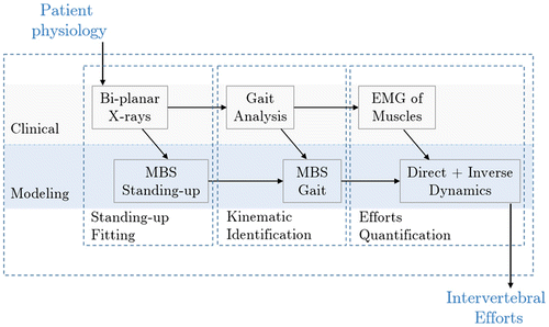
The proposed protocol addresses each of these problems, from both the clinical and the modeling points of view, as shown in Figure . The geometrical identification of the spine, using bi-planar X-rays, the computation of its kinematics from a limited amount of data and the impact of the patient physiology have been addressed in (Abedrabbo Citation2017).
Figure 1. The general protocol is based on both clinical and modeling processes.
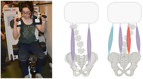
The present contribution focuses on a global analysis of the protocol developed for this study, based on the results of one scoliotic patient, for which a special attention will be paid to the computation of the muscular forces and the intervertebral efforts. The model used includes 6 muscles representing the abdominal and back muscles groups (Figure -right). The muscular forces are estimated through their activation measured by EMGs. The correlation between the forces and activation was established via the David Back protocol (Figure -left).
Figure 2. Muscular forces estimation: EMG calibration (left) and muscular activation (right). High activation in red, middle activation in purple and no activation in blue.
3. Results and discussion
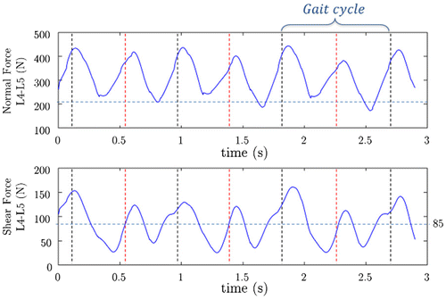
An illustrative result obtained from our biomechanical model of the spine is shown in Figure . It reveals the dynamic impact of the gait, by showing a significantly higher intervertebral – normal and shear – forces than in a standing up position (up to 125% higher).
Figure 3. Dynamic intervertebral forces inside L5-S1: normal force (up) and shear force (down). The horizontal dotted line represents the intervertebral force in the standing up position (Static). The vertical lines refer to the double stance phase of the gait cycle.
4. Conclusion
A preliminary comparison of the results obtained with our approach with the experimental studies carried out by (Damm et al. Citation2016) in which the intervertebral forces are measured using an instrumented vertebral body replacement, shows that our model prediction seems consistent with the reality. For example, the results in (Damm et al. Citation2016) show peak forces during that range from 21 to 83% patient bodyweight for a gait at 4 km/h compared to 50 to 80% of the total bodyweight obtained from our different models for a gait of 6 km/h. We observe a similar order of magnitude for both the maximum force and the dynamic range. However, this comparison cannot be considered as a validation, since the results in (Damm et al. Citation2016) are based on different parameters from those used in our study, namely: non-scoliotic patients, different body mass and a different gait velocity.
Although our biomechanical model is perfectible, the obtained results can already point out the impact of dynamics on the intervertebral efforts during gait for scoliotic patients.
References
- Abedrabbo G. (2017). Quantification of intervertebral efforts using a multibody dynamics approach: application to scoliosis [ PhD thesis]. Louvain-la-Neuve: Université catholique de Louvain.
- Christophy M, Senan NAF, Lotz JC, O’Reilly OM. 2012 Jan. A musculoskeletal model for the lumbar spine. Biomech Model Mechanobiol. 11(1–2):19–34.
- Damm P, Kutzner I, Bergmann G, Rohlmann A, Schmidt H. 2016. Comparison of in vivo measured loads in knee, hip and spinal implants during level walking. J Biomech. 51:128–132.
- Desroches G, Aubin CE, Sucato DJ, Rivard C. 2007 Aug. J and Rivard. C. Simulation of an anterior spine instrumentation in adolescent idiopathic scoliosis using a flexible multi-body model. Med Biol Eng Comput. 45(8):759–768.
- Dubousset J. 2011. Reflections of an orthopaedic surgeon on patient care and research into the condition of scoliosis. J Pediatr Orthop. 31(1):S1–S8.
- Robitaille M, Aubin CE, Labelle H. 2007. Intra and interobserver variability of preoperative planning for surgical instrumentation in adolescent idiopathic scoliosis. Eur Spine J. 16(10):1604–1614.