 ?Mathematical formulae have been encoded as MathML and are displayed in this HTML version using MathJax in order to improve their display. Uncheck the box to turn MathJax off. This feature requires Javascript. Click on a formula to zoom.
1. Introduction
Tibial plateau fracture treatment is carried out in most cases by stable fixation in order to allow early mobilization. Minimally invasive technologies such as tibioplasty or stabilization by locking plate augmentation are used to treat tibial plateau fractures (Vendeuvre et al. Citation2013). This work deals with a numerical approach with a personalized finite element (FE) analysis, to determine the mechanical behaviour of the fractured tibial plateau stabilisation. Mechanical environment, which is an important factor for the fracture healing (Giannoudis et al. Citation2007), is studied from interaction characterization between bone and stabilisation systems. The FE analysis was performed on a 3D patient-specific model of the proximal tibia. The fracture and the stabilization with locking plate were simulated. The goals are estimation of the risks of implant failure, the risks of fracture instability, and the stress studied around the fractured area in accordance with the interactions with mechanical environment.
2. Methods
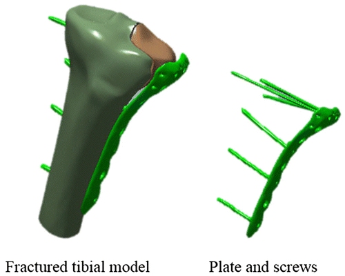
The model was built from a clinical case of a tibial plateau fracture with a lateral split depression on a young female patient (Figure ). This fracture was treated with minimally invasive surgery: bone augmentation and reduction of the depression with the use of a balloon and fixation of a plate with locked screws.
The FE model corresponding to the state after stabilization was created from data provided by computed tomography of the stabilized tibia. Each part was identified and segmented: tibial plateau, separated fragment, PMMA cement, plate and screws. A second FE model was built to simulate an intact tibial plateau by merging the fragment and the plateau and by replacing the cavity corresponding to the cement by cancellous bone material.
The orthotropic elastic material properties of cancellous bone have been incorporated into the FE models from relations between mechanical parameters and local bone density (Rho Citation1996). A titanium alloy material was employed for plate and screws (). The applied loading was chosen to simulate a single leg stance during gait, which corresponds to a joint contact force equal to three times the body weight.
The stress distribution in each component of the fracture fixation was analysed in order to study mechanical effects in terms of rigidity and strength of stabilization of proximal tibial fracture, compared with the intact tibial plateau.
3. Results and discussion
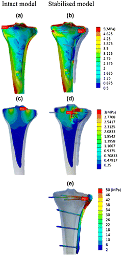
Figure shows a comparison of equivalent stress distributions between intact model and stabilised model in cortical bone (a–b) and cancellous bone (c–d). Figure (e) shows stress distribution in the plate and screws for the stabilised model.
For cortical bone, stress distributions in both cases are similar on the plateau, around 5 MPa. On the lateral wall of the tibia, stress intensity is lower in the stabilised bone (less than 1 MPa) than in the intact one where equivalent stress goes from 3 to 5 MPa. Fractured parts of cortical bone are subjected to small stresses because of the presence of the plate. Stress distribution in plate and screws confirms this observation. Maximum values of equivalent stress in plate and screws, close to fractured bone, increases up to 50 MPa. In the inferior part of the plate and in the screws positioned at the bottom, equivalent stress does not exceed 25 MPa.
Concerning cancellous bone, intact model gives a balanced stress distribution in the tibial plateau. For the stabilised model, stress concentration appears in the fractured area, from each part of the fracture and close to the contact between bone fragments and screws in the separation area.
This biomechanical study investigated the effect of additional stabilisation system after balloon reduction of tibial plateau impression fractures using a computed method. FE approach showed that the plate and screws change stress distribution and leading to a decrease of the stress shielding.
This FE analysis shows the mechanical support offered by plate and screws. The stress concentration is identified in plate with maximum values superior to 50 MPa. Equivalent stress computed in this case for a static loading simulating a single leg stance during gait remains inferior to the elastic limit of the titanium alloy. No damage should appear in the fixations but a fatigue analysis could be performed to verify material durability during the bone healing period. The shape of the plate and screw diameters in inferior part could be optimized because of the low value of stress values.
4. Conclusions
This biomechanical study investigated the effect of implants and fixation systems after balloon reduction of tibial plateau impression fractures using computed method. The finite element model of clinical case was used to analyse stabilization of tibial plateau fracture. The intact tibial plateau case showed the anatomic stabilization used as a reference model. The computed results of stresses or displacements of the fractured models show mechanical effects linked to the implant stability. These results have important implications for implant choice in tibial traumatology and the ability to allow early mobilization of the patient.
References
- Giannoudis PV, Einhorn TA, Marsh D. 2007. Fracture healing: the diamond concept. Injury. 38:S3–S6.10.1016/S0020-1383(08)70003-2
- Rho J-Y. 1996. An ultrasonic method for measuring the elastic properties of human tibial cortical and cancellous bone. Ultrasonics. 34:777–783.10.1016/S0041-624X(96)00078-9
- Vendeuvre T, Babusiaux D, Brèque C, Khiami F, Steiger V, Merienne J-F, Scepi M, Gayet L-E. 2013. Tuberoplasty: minimally invasive osteosynthesis technique for tibial plateau fracture. Orthop Traumatol Surg Res. 99:267–272.10.1016/j.otsr.2013.03.009


