1. Introduction
The measurement of the trunk movements and the estimation of the spine posture is an important field of research in biomechanics, clinical, ergonomic and rehabilitation areas. In fact, regular and prolonged spine deviations from the natural curvature ‘S shape’ cause structural deformity of the spine and back pain which are associated with disability and high costs for health care (Wong and Wong Citation2008).
Quantitative assessment of spine curvature is accessible through the use of medical technology based on two-dimensional images acquired by radiography (X-ray) and three-dimensional (3D) imaging techniques (tomography and magnetic resonance; Vrtovec et al. Citation2009). Optoelectronic systems constitute also a reliable alternative for the assessment of spinal curvature and permit assessing trunk movements and dynamic posture (Gracovetsky et al. Citation1995; Ranavolo et al. Citation2013). The cost and the restricted use to clinical or laboratory environments of the above techniques constitute however their major drawbacks. Other techniques perform spine posture and motion analysis by means of portable potentiometric goniometers and electromagnetic tracking systems. The first ones are limited by their bulkiness which may restricts the patient’s movements and the second ones are limited by their accuracy which relies on transmitter-receivers distance and on the presence of metallic objects.
In recent decades, the miniaturization and the extension of the Micro-Electro-Mechanical-Systems (MEMS) technology has led to lightweight, portable and especially low cost inertial sensors which are reliable alternative to compute acceleration, angular velocity and angles following three axis without the constraints outlined above (Cuesta-Vargas et al. Citation2010). Furthermore, a recent study (Palazzo et al. Citation2016) highlighted the need of technologies to supervise spinal posture changes during a home-based exercise program.
The aim of the current paper is to introduce a new-designed tool to assist patient during home rehabilitation program and to provide visual feedback on spinal movements.
2. Methods
2.1. System design
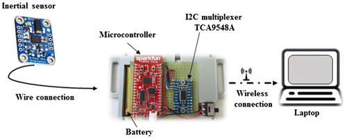
The system (Figure ), composed of hardware and software, was designed for 3D angles measurement which is a prerequisite for spinal movement estimation.
The hardware prototype is composed as follows:
| • | three inertial sensors (Bosh Sensortec BNO055) mounted with a safety pin on a stretch tee-shirt at the level of the seventh cervical vertebrae (C7), the twelfth thoracic vertebrae (T12) and the fifth lumbar vertebrae (L5). Each sensor includes gyroscope, magnetometer and accelerometer; | ||||
| • | a WIFI-compatible microcontroller (SparkFun ESP32 Thing) to firstly gather data from sensors and then transmit them to laptop using wireless connection. | ||||
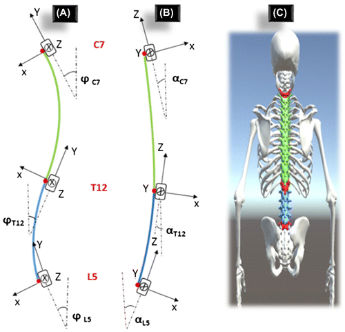
The software was performed to process the sensors data and to provide to users a visual feedback through a graphic interface. This feedback consists on a 3D model of spine developed with Unity 3D, called the Avatar (Figure (C)). Spine is modelled by two rigid segment namely thoracic (green) and lumbar (blue) regions.
2.2. Spine modelling
Based on the inertial data, the 3D postural changes in thoracic (between C7 and T12) and lumbar regions (between T12 and L5) are assessed simultaneously. Angles information is expressed in the form of quaternions that are firstly used to provide a visual feedback to the user by moving the Avatar accordingly. Given that sensors orientations are expressed in terrestrial reference frame, the quaternions are reset at zero in neutral standing position ‘S shape’ prior the trial. At this position, the Cartesian coordinate system (X, Y and Z axis) points the International Society of Biomechanics standard directions: X to anatomical anterior direction, Y to anatomical superior direction and Z to anatomical right side.
Quaternions are also used to estimate spine postural changes. The system detects inclination differences between angles formed by the tangent and vertical lines, and consequently lumbar lordosis, thoracic kyphosis and deviation from the neutral position in the frontal plane can be detected (Figure (A) and (B)). It also detects rotation differences between consecutive sensors, and so the twist of the spine.
3. Results and discussion
Concerning the Avatar animation, the performed trials showed a smooth movement in real time. The lag between user movement and avatar movement was not perceived by the participants, that increases interactivity. Our first trials highlight the usability of the proposed tool for real time monitoring of the Avatar with the inertial sensors. By providing visual feedback during exercises patients will be able to better perform their rehabilitation program (Burghoorn et al. 2015). Furthermore, by visualizing an avatar of their back, patients can also experience less pain (Wand et al. Citation2012). To increase patients’ motivation to be engaged in a home based exercise therapy and to provide assistance, the development of video-game exercise seems to be a real opportunity. Concerning the accuracy of the spine postural changes estimation, a validation against a gold standard system (i.e. optoelectronic system) should be carried out and is in progress in our laboratory.
4. Conclusion
The current paper describes our approach in developing a new system based on wearable inertial sensors to provide visual feedback on spinal posture during home-based exercise program. This preliminary study highlights some parts of its feasibility.
Acknowledgements
This work is performed during the HODOREV project and supported by the Region Île de France, managing authority for the Operational Program 2014-2020, via the European Regional Development Fund (FEDER).
References
- Cuesta-Vargas AI, Galán-Mercant A, Williams JM. 2010. The use of inertial sensors system for human motion analysis. Phys Ther Rev. 15:462–473.10.1179/1743288X11Y.0000000006
- Gracovetsky S, Newman N, Pawlowsky M, Lanzo V, Davey B, Robinson L. 1995. A database for estimating normal spinal motion derived from noninvasive measurements. Spine. 20:1036–1046.10.1097/00007632-199505000-00010
- Palazzo C, Klinger E, Dorner V, Kadri A, Thierry O, Boumenir Y, Martin W, Poiraudeau S, Ville I. 2016. Barriers to home-based exercise program adherence with chronic low back pain: Patient expectations regarding new technologies. Ann Phys Rehabil Med. 59:107–113.10.1016/j.rehab.2016.01.009
- Ranavolo A, Don R, Draicchio F, Bartolo M, Serrao M, Padua L, Cipolla G, Pierelli F, Iavicoli S, Sandrini G. 2013. Modelling the spine as a deformable body: Feasibility of reconstruction using an optoelectronic system. Appl Ergon. 44:192–199.10.1016/j.apergo.2012.07.004
- Vrtovec T, Pernuš F, Likar B. 2009. A review of methods for quantitative evaluation of spinal curvature. Eur Spine J. 18:1–15.
- Wand BM, Tulloch VM, George PJ, Smith AJ, Goucke R, O’Connell NE, Moseley GL. 2012. Seeing it helps: movement-related back pain is reduced by visualization of the back during movement. Clin J Pain. 28:602–608.10.1097/AJP.0b013e31823d480c
- Wong WY, Wong MS. 2008. Smart garment for trunk posture monitoring: a preliminary study. Scoliosis. 3:7.10.1186/1748-7161-3-7


