1. Introduction
Knee osteoarthritis (OA) is a serious health concern, requiring novel therapeutic options. Walking mechanics has long been identified as an important factor in the OA process (Childs et al. Citation2004; Russell et al. Citation2010; Ogrodzka et al. Citation2011). Specially, a larger peak Knee Adduction Moment (KAM) during the first half of stance has been associated with the progression of medial knee OA (Lynn & Costigan Citation2008; Shull et al. Citation2013; Favre et al. Citation2016). Consequently, various gait interventions have been designed to reduce the KAM, including walking with a decreased Foot Progression Angle (FPA). Other gait variables have recently been associated with medial knee OA progression, particularly a larger peak Knee Flexion Moment (KFM) during stance and a larger Knee Flexion Angle (KFA) at heel-strike (van den Noort et al. Citation2013; Simic et al. Citation2013; Favre et al. Citation2016). Currently, there is a paucity of data regarding the effect of reducing the FPA on the KFM and the KFA.
This study aims to test the correlations between the FPA and the KAM, the KFM and the KFA. It is hypothesized that reducing the FPA is beneficial with respect to these three OA-related gait variables.
2. Methods
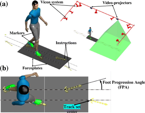
Seven healthy subjects participated in this study after providing informed consent (4 males; 24 ± 5 years old; 21.9 ± 1.5 kg.m−2). Their walking mechanics was determined using a validated procedure based on a camera-based system (Vicon) and floor-mounted forceplates (Kistler) (Figure (a)). Participants were first asked to walk without instructions and these initial trials were used to determine their normal footstep characteristics. Then, footsteps with the same characteristics as during the normal trials, except for the FPA, were displayed on the floor and participants were requested to walk following these footsteps. Nine trials with visual instructions were collected for each participant, corresponding to FPA modifications in the range ± 20° compared to the normal FPA, with a 5° increment (Figure (b)). For each participant, the associations between the FPA and the knee biomechanics (KAM, KFM and KFA) were assessed using Pearson correlations based on data from the 9 trials with FPA variations. A significant level was set a priori to 5%.
3. Results and discussion
Significant relationships were noted between the FPA and the peak KAM for 5 out of 7 subjects (p < 0.03), with R2 comprised between 0.65 and 0.93. Four participants also reported significant correlations between the FPA and the KFA (0.48 < R2 < 0.77). For the KFM, significant relationships were noticed with the FPA for 2 out of 7 subjects (p < 0.05), with inconsistent R2 (0.47 and 0.61, Table ).
Table 1. Linear regressions for the First KFA peak, the first KAM peak and the First KFM peak.
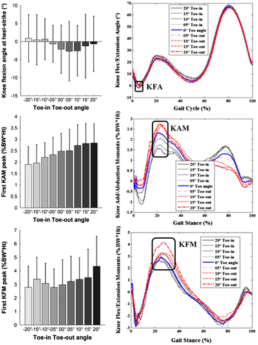
In this study, it is confirmed that the toe-in of the FPA induces the reduction of the KAM (Figure ). However, this toe-in of the FPA is responsible for increasing the KFA (Figure ). In this case, the variation in the toe-in with the KFA should be monitored because increasing the KFA is associated with the OA progression. Still, we notice no or inconsistent FPA- toe-in effect on the KFM (Figure ).
Figure 2. Mean ± standard deviation of the first KFA peak (1st row), first KAM peak (2nd row), and first KFM peak (3rd row) variables divided according to the levels of the instructions of modify gait (foot progression angle).

The results vary among subjects suggesting that individualized modifications should be considered. Consequently, there is no need to be tested on a larger cohort.
4. Conclusions
The KAM, the KFM and the KFA are considered indicators of knee osteoarthritis progression. In this study, a foot progression angle modification (toe-in, normal, toe-out) during gait was altered the characteristics of the KAM. A reduction in the overall magnitude has been found with the toe-in of the FPA during gait modification. An increased KFA has been also found during the toe-in gait, which causes undesirable effects on the OA progression. This finding is novel, indicating that the toe-in of FPA should be controlled during gait modification not to increase KFA and not to vary other parameters in relationship with the OA.
Acknowledgements
This study was partially funded by the Swiss government excellence scholarship for foreign scholar program.
References
- Childs JD, Sparto PJ, Fitzgerald GK, Bizzini M, Irrgang JJ. 2004. Alterations in lower extremity movement and muscle activation patterns in individuals with knee osteoarthritis. Clin Biomech. 19:44–49.10.1016/j.clinbiomech.2003.08.007
- Favre J, Erhart-Hledik JC, Chehab EF, Andriacchi TP. 2016. A general scheme to reduce the knee adduction moment by modifying a combination of gait variables. J Or-thopaedic Res. 34:1547–1556.10.1002/jor.23151
- Lynn SK, Costigan PA. 2008. Effect of foot rotation on knee kinetics and hamstring activation in older adults with and without signs of knee osteoarthritis. Clin Biomech. 23:779–786.10.1016/j.clinbiomech.2008.01.012
- van den Noort JC, Schaffers I, Snijders J, Harlaar J. 2013. The effectiveness of voluntary modifications of gait pattern to reduce the knee adduction moment. Hum Mov Sci. 32:412–424.10.1016/j.humov.2012.02.009
- Ogrodzka K, Niedźwiedzki T, Chwała W. 2011. Evaluation of the kinematic parameters of normal-paced gait in subjects with gonarthrosis and the influence of gonarthrosis on the function of the ankle joint and hip joint. Acta BioEng Biomech. 13:47–54.
- Russell EM, Braun B, Hamill J. 2010. Does stride length influence metabolic cost and biomechanical risk factors for knee osteoarthritis in obese women? Clin Biomech. 25:438–443.10.1016/j.clinbiomech.2010.01.016
- Shull PB, Shultz R, Silder A, Dragoo JL, Besier TF, Cutkosky MR, Delp SL. 2013. Toe-in gait reduces the first peak knee adduction moment in patients with medial compartment knee osteoarthritis. J Biomech. 46:122–128.10.1016/j.jbiomech.2012.10.019
- Simic M, Wrigley TV, Hinman RS, Hunt MA, Bennell KL. 2013. Altering foot progression angle in people with medial knee osteoarthritis: the effects of varying toe-in and toe-out angles are mediated by pain and malalignment. Osteoarthritis Cartilage. 21:1272–1280.10.1016/j.joca.2013.06.001

