
1. Introduction
Lumbar spinal fusion is a surgery to permanently join, or fuse, two or more vertebrae in the human spine eliminating motion between them. It is currently considered as one of the most successful surgical treatment in patients with spinal deformity, degenerative disk diseases, and degenerative spondylolisthesis (Bashkuev and Checa Citation2015). However, pain at the bone, non-fusion, breakage of metal implants, infection may appear and lead to its failure (Chou and Loeser Citation2009). Its success depends critically on the geometrical design of the interbody cages and the material properties (Noailly et al. Citation2014). An appropriate design resulting from the real morphology of the patient can provide adequate primary stability and greatly avoid fixation problems, stress-shielding effects, and load transfer.
The main objective of this work is to introduce a systematic design process of custom-fitted interbody cages for lumbar spinal fusion, based on anatomical reverse engineering (ARE) techniques such as magnetic resonance imaging (MRI) and 3D reconstruction. Finite element analysis (FEA) was used to evaluate and compare the stress distribution for the optimization of the cage design.
2. Methods
2.1. MRI scan data and segmentation
A stack of 2D image slices for the region of interest (ROI) including lumbar spine was obtained using a clinical MRI scanner (GE Signa HDxt 1.5T) with a slice thickness of 0.625 mm, a 512 × 512 pixel resolution (320 × 320 mm2). The software BioImageXD was used to perform the segmentation process and extract the 3D cloud point of the second and third lumbar vertebrae (L2 and L3).
2.2. 3D reconstruction and CAD modelling
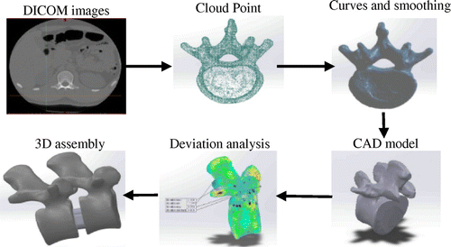
The cloud point was imported in SolidWorks® using ScanTo3D tool which allows noise removing, realignment, extraneous deleting, curves generating and mesh smoothing. Finally, 3D CAD model of the lumbar vertebra was created and exported (Figure ). In order to ensure a good morphological and morphometric reproducibility of the reconstructed model, a deviation analysis was performed to compare between the cloud point model (initial) and the smoothed model (final) in the reconstruction process.
Figure 1. 3D reconstruction steps.
2.3. Design of interbody cage
In order to sketch the outer surface profile as close as possible to the two lumbar vertebrae (L2 and L3), a specific adaptation algorithm was developed using Macro tool, as a result, two profile type of interbody cage were created. For each profile, geometrical parameters were introduced to generate different possible cage dimensions as shown in Figure .
Figure 2. Closest profiles generated (a): first profile type, (b): second profile type.
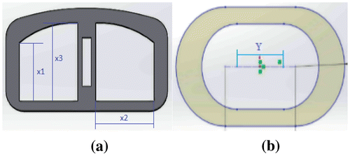
Figure 3. Von Mises stress distribution in first cage.
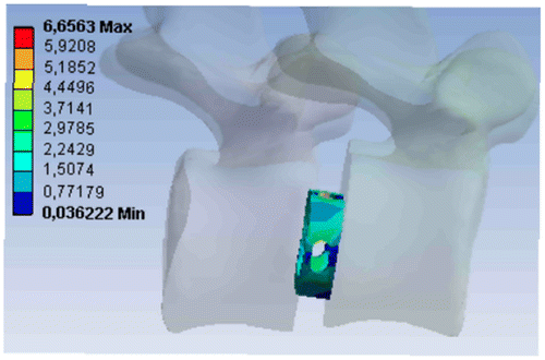
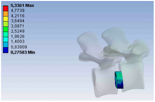
Figure 4. Von Mises stress distribution in second cage.
2.4. Finite element analysis and design optimization
Stress-shielding is considered as one of the main reason leading to the failure of lumbar spinal fusion. Stress distribution in the regions surrounding the interbody cage must be maximum in order to enhance the mechanical stimulus required for bone formation and avoid stress-shielding effects.
For this, a parametric finite element study was performed using ANSYS Workbench software. The 3D model consists of the two lumbar vertebral bodies (bone) and the interbody fusion cage (Titanium alloy Ti6Al4 V). A distributed load corresponding to a 600 N axial compressive force was applied at the top of the upper vertebral body. The average equivalent von-Mises stress within the fusion zone which represents the objective function of optimization was estimated for 14 scenarios representing the variation of the three geometrical parameters of the first design profile (x1, x2 and x3) and 08 scenarios representing the variation of the one geometrical parameter of the second design profile (Y). The geometrical parameters of cages represent optimization variables which affect the objective optimization function.
3. Results and discussion
The average von-Mises stress observed within the fusion zone of all models was widely below the reported ultimate strength for cortical and cancellous bone under compression (139.5 and 35.95 MPa respectively, Cory and Nazarian Citation2010), thereby eliminating overloading risks. The purpose of the FE parametric study is to examine and compare the stress distribution for different designs in order to be able to select the profile and the geometrical parameters leading to minimize stress-shielding effects. Some FE results are presented in Table 1 for the first generated design and Table 2 for the second generated design.
The comparison showed that on the one hand, the stresses in the first interbody cage profile are higher than the second. On the other hand, the stress in the same profile increases with the geometrical parameters increasing the contact area between the cage and two vertebras. Note that a function minimizing the weight has been integrated to control the dimensions.
The increase of the contact surface leads to a good load transfer between the cage, therefore this will minimize stress-shielding risks.
4. Conclusions
On the basis of the anatomical reverse engineering techniques, a systematic process has been carried out for the design of a custom-fitted interbody cage for lumbar spinal fusion. The proposed custom design process starts from the patient morphology which allows the measurement of the vertebra dimensions and extracting the closest possible profile for the fusion cages design. Using the finite element concepts, geometrical parameters compromising stress-shielding effects and the cage weight has been calculated. However, the results must be validated in a future works and other important phenomena must be taken into account such as bone stimulation, wear resistance proprieties and the hostile physiological environment.
Acknowledgements
The authors wish to thank the staff of medical imaging department of “Hôpital Central de l’Armée – Ain Naadja”.
References
- Bashkuev M, Checa S. 2015. Computational analyses of different intervertebral cages for lumbar spinal fusion. J. Biomechan. 48(12):3274–3282.10.1016/j.jbiomech.2015.06.024
- Chou R, Loeser JD. 2009. Interventional therapies, surgery, and interdisciplinary rehabilitation for low back pain. Spine. 34(10):1066–1077.10.1097/BRS.0b013e3181a1390d
- Cory E, Nazarian A. 2010. Compressive axial mechanical properties of rat bone as functions of bone volume fraction, apparent density and micro-ct based mineral density. J Biomechan. 43(5):953–960.10.1016/j.jbiomech.2009.10.047
- Noailly J, Malandrino A, Galbusera F. 2014. Computational modelling of spinal implants. Comput Modell Biomechan Biotribol Musculoskeletal Syst. 447–484.10.1533/9780857096739.4.447