
1. Introduction
Natural breathing is known to induce at each inspiration/expiration, minimal perturbations of body balance that are compensated by movement of trunk and lower limb (Hodges et al. Citation2002). This phenomenon called posturo ventilatory synchronization may be impaired (Manor et al. Citation2012). Its evaluation requires simultaneous assessments of ventilation profile and postural control, using the less disruptive methods. Spirometry is the reference method to assess the ventilatory parameters. It requires however to connect the subject to the spirometer, using a mouthpiece, that is known to modify significantly the respiratory frequency (Gilbert et al. Citation1972) and may influence the postural control. The Optoelectronic Plethysmography (OEP) was validated as an alternative non-disruptive method to assess ventilation as it allows subjects to breathe naturally. Evidently, non-contact measure of ventilation by camera is supposed to induce no perturbation of postural control, and may be more appropriate than spirometer to assess the posturo ventilator synchronization. The objective of this study was to evaluate the postural perturbations due to the use of the spirometer, by comparing it to the OEP method, on postural control.
2. Methods
2.1. Subjects and equipment
Eight healthy subjects participated to the study (4F/4 M; median and [Q1–Q3]: 33y.o. [27–45]; 171 cm [166–175] height; 75 kg [71–80] weight; 26 kg/m² [25–26.7] BMI).
An AMTI® force plate was used to measure the centre of pressure (CoP) displacements. Subjects were equipped with 41 retroreflective markers placed on their trunk. OEP was performed with Vicon® system at 100 Hz synchronized with force plate.
Subjects breathed through spirometer, a low-resistance pneumotachograph (M.E.C. PFT Systems Pocket-Spiro, Medical Electronic Construction, Brussels, Belgium) fixed to the floor by a tripod.
2.2. Protocol
Subjects were placed on the force plate and were asked to keep arms alongside the trunk in a relaxed position. All measurements were done in standing and sitting positions. The subjects were instructed to breathe during at least 30 s. Each subjects performed this exercise without the spirometer (natural breathing (NB)) and breathing through the spirometer (SP).
The following signals were recorded: respiratory profile from OEP, and displacement of the CoP from the force plate.
The protocol was approved by an Ethics Committee (CPP IDF VI Paris),
2.3. Data analysis
Mean respiratory frequency (f) was computed from the breathing profile measured with OEP in both natural and spirometer breathing, using the same duration (between 30 and 50 s, deleting the first cycles). Ventilation profile measured with spirometer was not analysed in this study.
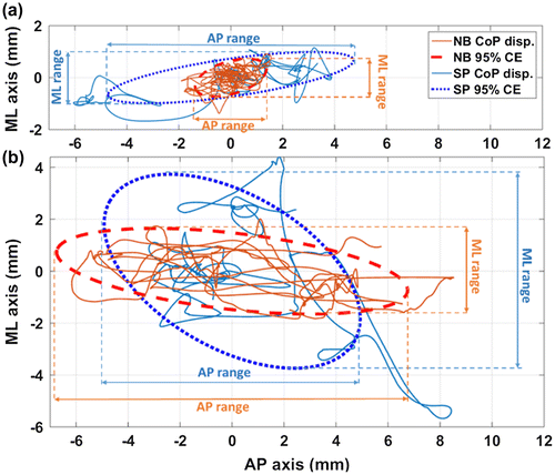
From the force plate data, confidence ellipse (CE) of 95% of the displacement covered by the CoP was computed for each exercise (Chiari et al. Citation2002). Three stabilometric parameters were reported: CE ranges along the antero-posterior (AP) and medio-lateral (ML) axis (Figure ) and the mean CoP velocity (MV). AP and ML range were normalized by the subject height in standing position and by top of skull – stool distance in sitting position,
Figure 1. Centre of pressure displacement and confidence ellipses for natural (NB) and spirometer (SP) breathe in sitting (a) and standing position (b).
NB and SP were compared for all parameters, using a non-parametrical test (Wilcoxon), considering significant difference with a p-value lower than 5%.
3. Results and discussion
CoP displacements and ventilation profiles were analyzed over 32 natural breathing exercises.
In sitting position, differences observed between SP and NB were equal to: Δf = 3.03 min−1 [1.72; 6.89]; ΔAP-range = 1.34 mm [0.80; 1.46]; ΔML-range = 2.45 mm [2.09; 2.57]; ΔMV = 9.66 mm/s [8.64; 9.76].
In standing position, differences were equal to: Δf = 3.40 min−1 [2.61; 4.17]; ΔAP-range = 2.81 mm [1.66; 10.08]; ΔML-range = 2.82 mm [2.38; 4.48]; ΔMV = 3.87 mm/s [1.29; 4.78].
Figure presents the CoP displacement along natural breathing and through the spirometer for one subject, in both sitting and standing positions. During natural breathing, the surface covered by the CoP is smaller in sitting than in standing position. Furthermore, the increase of this surface is more important in sitting position over AP and ML axis, while in standing position a decrease of AP range and an increase of ML range appear.
Table presents results obtained during NB and SP exercises for the four parameters f, AP-range, ML-range and MV in both sitting and standing positions. Results are presented as median [Q1; Q3].
Table 1. Breathing and Stabilometric variables (median [Q1; Q3]) measured in natural breathing (NB) and breathing through a spirometer (SP).
Breathing frequency increases non-significantly when subject breathe through the spirometer. This difference appears similarly in both sitting and standing positions, with an increase upper than 3 min−1. This phenomenon could be explained by the significant breathing modification observed with breathing assessment (Gilbert et al. Citation1972).
The two Stabilometric parameters (ML ranges and MV) significantly increase during spirometer breathing. These increases could be explained by the tripod fixation constraining the postural chain mobility and limiting the counter-perturbation (Bouisset & Zattara Citation1981). Moreover this tripod fixation appears to stabilize AP range in standing position with a significant decrease of it.
4. Conclusions
This study showed the significant perturbation of the body balance during breathing through a spirometer, and validated the use of a non-disruptive device to study human ventilation. As we can observe on Figure, spirometer perturbation differs in sitting and standing position. In standing position SP stabilizes stabilometric perturbation over the AP axis and increases it over ML axis, while in sitting position SP increases perturbation over both AP and ML axis.
From stabilometric results observed in Figure , confidence ellipse angle, appearing to change in standing position, sway path of CoP and frequency analysis could also be studied to analyse spirometer influence on stabilometry.
References
- Bouisset S, Zattara M. 1981. A sequence of postural movements precedes voluntary movement. Neurosci Lett. 22:263–270.10.1016/0304-3940(81)90117-8
- Chiari L, Rocchi L, Cappello A. 2002. Stabilometric parameters are affected by anthropometry and foot placement. Clin Biomech. 17:666–677.10.1016/S0268-0033(02)00107-9
- Gilbert R, Auchincloss JH, Brodsky J, Boden W. 1972. Changes in tidal volume, frequency, and ventilation induced by their measurement. J Appl Physiol. 33.
- Hodges P, Gurfinkel V, Brumagne S, Smith T, Cordo P. 2002. Coexistence of stability and mobility in postural control: evidence from postural compensation for respiration. Exp Brain Res. 144:293–302.10.1007/s00221-002-1040-x
- Manor BD, Hu K, Peng CK, Lipsitz LA, Novak V. 2012. Posturo-respiratory synchronization: Effects of aging and stroke. Gait Posture. 36:254–259.10.1016/j.gaitpost.2012.03.002