
1. Introduction
The skeleton is a common organ affected by metastatic cancers (Coleman Citation1997), such as breast, prostate and lung cancer. Bone metastases are often lytic, i.e. destroying the local bone tissue. Metastatic bones are more likely to fail and therefore have to be monitored by physicians, who have to decide which treatment is the best suited to each case. However, the tools at their disposal do not allow to accurately predict whether a bone will fail or not (Van der Linden et al. Citation2004). Several studies showed that patient-specific finite element models were a promising tool to fulfill this prediction (Derikx et al. Citation2015). In these previous studies, it was assumed that metastases played no mechanical role, and were accordingly modeled as holes in the bone. Nevertheless, practical cases showed that metastases do play a mechanical role in bone strength, because if the previous theory was true, the bone would have failed. Thus, the aim of our core study is to investigate and quantify the real impact of metastases (according to the different types) on bone strength by a mechanical test, and to try to simulate the experimental test through numerical modelling. To reach this final aim, 1-month-old BALB/c nude mice were injected intra-tibialy with different tumor cells in their right limb and by phosphate-buffered saline (PBS) in their contra-lateral limb, as has been advised (Wright et al. Citation2016) to create a sham control. However, the impact of the intra tibial injection of PBS on mechanical properties of the tibia was never quantified. Thus, the current aim of this sub study is to determine the impact of this type of injection on mice tibia assuming that the intra-tibial injection degrades the mechanical properties of the bone.
Table 1. Mean stiffness, max load of each group, statistical significance of tests for each group and parameters inj.: injected; L: left; R: right; n: number of mice.
2. Methods
2.1 Materials
Thirteen one-month-old female BALB/c nude mice (Janvier Laboratories®) were used in this study, after the approval of University Claude Bernard Lyon I Ethical Comity for Animal Experimentation. Mice were divided into 3 groups.
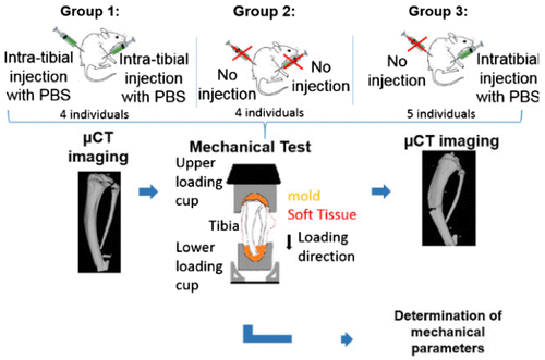
Group 1 (n = 4) was injected intra-tibialy with PBS in both limbs, group 2 (n = 4) was not injected, and group_3 (n = 5) was injected intra-tibialy with PBS in their right limb only (Figure ).
Figure 1. Global process of the study.
At day 30, mice were anesthetized then euthanized by cervico-dislocation, both limbs were excised in bloc and stored in gaze soaked with PBS at -20 °C until mechanical testing.
2.2 Samples preparation
Limbs were thawed at ambient temperature for one hour in gauze soaked with PBS. They were then dissected: the tibia was separated from both the femur and the foot. The skin was removed, but muscles were left in. Next, the tibia was cut at half its length plus two millimeters. Finally, the attachment between tibia and fibula was cut.
After μCT imaging (see next section) the proximal end of the tibia was molded using epoxy paste (Pattex, Ref 1875423) and the distal end was imbedded by 2 mm in the same paste. These features were used for mechanical testing (Figure ).
2.3 μCT imaging
Micro CT imaging (Bruker Skyscan 1176, Kontiche, Belgium; 10 μm nominal resolution) of each sample was performed before and after mechanical testing in order to validate the fracture, as well as assessing its location (Figure ).
2.4 Mechanical test
After the creation of the molds, the samples were placed at 4 °C overnight to be tested the following day. Prior to the test, the samples were left at ambient temperature for 30 min. Tibiae were then placed axially in custom loading fixtures and pre-cycled using a sinusoidal waveform between -0.5 N and -2 N for 30 cycles at 0.5 Hz. The destructive test was conducted immediately after pre-cycling by compressing the tibia at a rate of 0.03 mm/s until failure using an electromagnetic testing machine (Bose Corporation, Eden Prairie, MN: 5500). Load-displacement data were recorded at 60 Hz (WinTest® Digital Control System) and test curves were analyzed to determine stiffness and ultimate force.
2.5 Mechanical parameters
A custom Python program was developed to assess mechanical properties mentioned above. Briefly, stiffness (S) was determined by using the derivative of the experimental curve. It was determined using a linear regression on the longest interval were the derivative function variation was under ± 5 N/mm. Ultimate load (Fmax) was defined as the maximum load.
2.6 Statistical test
All statistical tests were performed using SPSS© software and non-parametric tests were used. In order to assess the side effect (right vs left), a Wilcoxon test for paired samples was performed on all samples of group 1 and 2 separately.
To assess the impact of the injection on the mechanical properties, a Wilcoxon test was performed on group 3, and a Mann-Whitney test was performed on group 4 (=group1 + group2 + group3).
3. Results and discussion
No difference between the right and left tibia of each individual was found (respectively p = 0.068 and 0.715 for the S and 0.068 and 0.715 for the Fmax). Therefore, to increase the statistical power and show the effect of the injection, group 4 was formed with 3 cases: injected bone from both right and left tibia from group 1, non-injected bone from both right and left tibia from group 2 and only right tibia injected from group 3. A significant difference was only found for Fmax in group 3 (p = 0.043). The same result was obtained for group 4 (p = 0.029).
Finally, the relative difference was quantified for both significant results. The ultimate force was found to be 15% greater for the non-injected limb of group 3 and 23% greater for non-injected limb of group 4.
There are several limitations to this study: the first one being that the muscles were not removed and could impact the results. This decision was made in order to respect the protocol of our study, where muscles dissection is prohibited to avoid a discard of the tumor implanted in soft tissue. However, as all the ligaments were cut, this should not have a major impact.
Another limitation is that we assumed from a statistical result on low effectives that there were no differences between each right and left limb. But, as the pooled groups for the Mann-Whitney test were composed of 9 right limbs and 4 left for the injected group and 4 right limbs and 9 left for the non-injected one, this should avoid any unwanted repercussions on the results.
Lastly, the geometry differences could influence the results, but as the paired test on group 3 showed the same results as the impaired one of group 4, we can legitimately assume that this does not affect our results.
4. Conclusions
In light of our results, it can be concluded that this PBS injection has an effect on the ultimate load of the tibiae one-month post injection, but not on its stiffness. This result seems logical as the injection creates a local defect, and therefore should not impact the response of the structure, but only its maximum load. As a consequence, bi lateral injections will have to be considered to perform comparative studies using both limbs.
Acknowledgements
The authors wish to thanks Jean-Paul Roux, Marc Gardegaront for their technical support and Julia Greenfield for english editing. This work is supported by LabEx PRIMES (ANR-11-LABX-0063).
References
- Coleman RE. 1997. Skeletal complications of malignancy. Cancer.
- Derikx et al. 2015. Towards clinical application of biomechanical tools. J of Biomechanics.
- Van der Linden et al. 2004. Comparative analysis of risk factors. J Bone Joint Surg.
- Wright et al. 2016. Murine models of breast cancer bone metastasis. Bonekey.