
1. Introduction
Manual wheelchair (MWC) propulsion is a constraining mode of locomotion for the musculoskeletal system. It results that 30–70% of the MWC users suffer from musculoskeletal disorders, in particular at the shoulder joints (Finley and Rodgers Citation2004). Hence, there is a high interest in investigating shoulder biomechanics during MWC locomotion. However, tracking the scapula motion during MWC locomotion is not obvious because high soft tissue artifact can occur using skin markers. Some studies reported the use of a scapula locator during pseudo-kinematics acquisitions and showed a non-negligible motion of the scapula during MWC propulsion (Koontz et al. Citation2003). However, this methodology cannot be used to track dynamically the scapula during MWC locomotion. For that purpose, others methods were developed, such as multiple-calibration method (de Groot and Brand Citation2001). Nevertheless, this method can be time consuming for the patient. Finally, other studies used a technical cluster composed of at least 3 markers placed on the acromion (Shaheen et al. Citation2011) or on the scapula spine (Prinold and Bull Citation2015). These techniques seem to be efficient for tracking the scapula orientation but failed for the translation (Naaim et al. Citation2017). In addition, validation data did not concern MWC locomotion.
The goal of this study was to evaluate the accuracy of a spinal marker cluster to track the scapula motion during MWC locomotion.
2. Material and methods
Sequential-kinematics acquisitions of 46 scapulae were obtained from 10 subjects who participated to 2 or 3 measurements sessions where both scapulae were tracked. During each session, the subject was equipped with reflective markers on the manubrium, the xyphoid process, the spinous processes of both the 7th cervical and the 8th thoracic vertebrae, and on both acromions. A technical scapula spinal marker cluster (SSMC) equipped with 3 markers was also placed on the spine of each scapula. For right and left sides, four static acquisitions were performed with the subject sat in a MWC, reproducing the initial contact of the hand with the handrim (IC), the contact of the hand at the top of the handrim (TC), the final contact (FC) and an elevation of the arm of 30° in the plane of the scapula (Elev30). During each acquisition, a scapula locator (Shaheen et al. Citation2011) was placed by an experimenter on 3 palpated anatomical landmarks: Angulus Acromialis (AA), Trigonum Scapulae (TS) and Margo Medialis (MM). Locations of all the markers and the scapula locator were acquired simultaneously with a 13-cameras optoelectronic motion capture system (Vicon system, Oxford Metrics Inc., UK).
Anatomical frames of the thorax and the scapula as well as the scapulothoracic rotations (yxz) were calculated according to the ISB recommendations. A segmental optimization based on the SSMC and the acromion was performed taking the TC pose as a reference. This allowed the scapula registration during the others poses (IC, FC and Elev30) and the comparison of the reconstructed positions with those obtained with the scapula locator.
Root mean square distances (RMSD) between palpated and simulated landmarks of the scapula were computed. Differences in orientation of the scapula (palpated vs reconstructed) and in origin location (here placed at AA) were calculated and expressed through the RMSD.
3. Results and discussion
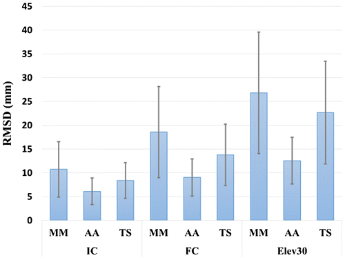
RMSD between reconstructed and palpated markers (Figure ) ranged from 1.5 to 28.5 mm (mean: 8.4 mm; SD: 4.7 mm) for IC; from 1.1 to 48.5 mm (mean 13.8 mm; SD: 8.0 mm) for FC; and from 3.4 to 53.7 mm (mean: 20.7 mm; SD: 11.6 mm) for Elev30. Obviously, the largest errors were found for the furthest markers (TS and MM) from the set composed of the SSMC and the acromion marker.
Figure 1. RMSD between reconstructed and palpated landmarks (MM, AA and TS) for the IC, FC and Elev30 poses.
Across the four static poses, the mean orientation of the scapula ranged from −5.9° ± 7.3° to 0.4° ± 6.6° for up/downward rotation (range: −23.7° to 31.0°); from 27.4° ± 5.9° to 34.8° ± 6.9° for int/external rotation (range: 10.5° to 51.9°); and from −26.6° ± 5.7° and −16.1° ± 5.1° for ant/posterior tilt (range: −37.5° to −3.6°). Hence, in average, ranges of angular motion of the scapula across the 3 investigated poses were 6.3° for up/downward rotation, 7.4° for int/external rotation and 10.5° for tilt.
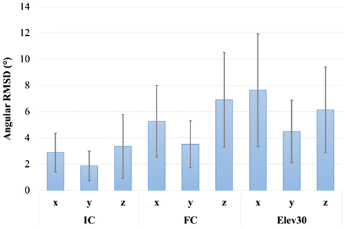
Angular RMSD between palpated and registered scapulae (Figure ) was 5.3° ± 3.6° (range: 0.9° to 16°) for up/downward rotation; 3.3° ± 2.1° (range: 0.4° to 9.8° for int/external rotation; and 5.5° ± 3.5° (range: 0.6° to 15.6°) for tilt. Globally, these results are slightly higher than the existing literature using the acromial marker cluster combined with segmental optimization (Naaim et al. Citation2017).
Figure 2. Angular RMSD between palpated and reconstructed scapulae following up/downward rotation (x), int/external rotation (y) and tilt (z) for the IC, FC and Elev30 poses.
Differences in origin location ranged from 6.1 to 12.6 mm in average for the 3 positions. However, this error could reach 13.6 mm for IC, 17.4 mm for FC and 24.7 mm for Elev30. Nevertheless, this result remains in accordance with the existing literature, where it was noted that the acromial marker cluster combined with segmental optimization often results in large dislocation between the scapula and the clavicle (Naaim et al. Citation2017).
4. Conclusions
This study aimed at evaluating the validity of a scapula spine marker cluster to track the scapula motion during MWC propulsion. The results showed non-negligible uncertainties, even if they remained below the motion of the scapula. This was found both for rotation and location. If the uncertainties reported in the present study are slightly higher than these reported in the literature (Naaim et al. Citation2017), it must be kept in mind that our reference, i.e. scapula locator, is not free of uncertainty (gold standard remains intracortical pins).
Finally, the use of multibody kinematics optimization instead of segmental optimization would allow avoiding dislocation phenomenon but the validity of this method remains to be evaluated.
References
- Finley MA, Rodgers MM. 2004. Prevalence and identification of shoulder pathology in athletic and nonathletic wheelchair users with shoulder pain: A pilot study. J Rehabil Res Dev. 41(3B):395–402.
- de Groot JH, Brand R. 2001. A three-dimensional regression model of the shoulder rhythm. Clin Biomech. 16:735–743.10.1016/S0268-0033(01)00065-1
- Koontz AM, Cooper RA, Boninger ML, Souza AL, Fay BT. 2003. Scapular range of motion in a quasi-wheelchair push. Int J Ind Ergon. 33:237–248.
- Naaim A, Moissenet F, Duprey S, Begon M, Chèze L. 2017. Effect of various upper limb multibody models on soft tissue artefact correction: A case study. J Biomech.
- Prinold JA, Bull AM. 2015. Scapula kinematics of pull-up techniques: Avoiding impingement risk with training changes. J Sci Med Sport. 19:629–635.
- Shaheen AF, Alexander CM, Bull AMJ. 2011. Effects of attachment position and shoulder orientation during calibration on the accuracy of the acromial tracker. J Biomech. 44(7):1410–1413.10.1016/j.jbiomech.2011.01.013