1. Introduction
The swing is the key movement in golf performance. This movement involves a high axial rotation of the spine that can differ significantly between subjects, resulting different amplitudes during the backswing. In addition, the distribution of the rotation along the spine would be variable between subjects. Differences in spine rotation could conduct to low back pain occurrence, which is the m ain injury for golf players and is the first health cause of professional career stop (Lindsay et al. Citation2000).
However, the quantification of the distribution of the axial rotation among the different vertebrae levels during the golf swing is not straightforward. One preliminary study has however attempted to quantify this distribution from biplanar radiographs and 3D reconstruction of the spine (Bourgain et al. Citation2016). However, this method remains to be evaluated in term of reproducibility and to be tested on several subjects.
In this objective, this preliminary study aimed at evaluating a method to quantify the distribution of the axial rotation along the spine based on biplanar-radiographs and 3D-reconstruction of the spine and to evaluate its interest and repeatability.
2. Methods
Three golf players (Table ) were included in this study, all right handed. The protocol was approved by an ethical committee and each volunteer gave his written informed consent prior to the experiment.
Table 1. Population characteristics
Subjects underwent two low-dose biplanar radiographs (EOS® system, EOS imaging, France) allowing the 3D reconstruction of the pelvis and all vertebrae. The first acquisition was performed in a neutral and standard position allowing both the personalization of the bones positions and morphologies (Dubousset et al. Citation2005). The second acquisition was performed with the subject axialy rotatated of about 45° between the shoulder girdle and the pelvis
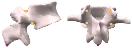
A 3D model of each vertebra was defined from this standard position. Five landmarks (the lower edge of the spinous process and the right and left lower and upper edges of the pedicles insertions) of each vertebra were identified (Figure ) and associated to the vertebra model. These landmarks were chosen as they were visible on both acquisitions and permitted to characterize the axial rotation of the vertebra. Then, these landmarks were identified on the second acquisition to accurately replace the 3D model with a least square method.
Because the process requires manual interventions, the repeatability was evaluated by performing 3 times the landmark identification for every subject.
The 3D models of the vertebrae are regionalized allowing to identify vertebra coordinate system according to ISB recommendation (Wu et al. Citation2002). Finally, the axial rotation of the thoracic spine was calculated from the transformation between the reference frame of the seventh cervical vertebra (C7) and the reference frame of the first lumbar vertebra (L1). In the same way, the lumbar spine axial rotation was computed between the reference frame of L1 and the reference frame of the pelvis. All angles were identified using a ZYX rotation order.
3. Results and discussion
Repeated registrations resulted in standard deviations ranging from 0.4 to 2.7° (S1 and S3 respectively) for the thoracic spine and from 1.0 to 2.3° (S1 and S3 respectively) for the lumbar spine.
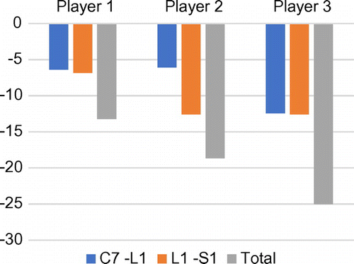
Axial rotations (average of the 3 values) were -6.4°, -6.1° and -12.4°, for S1, S2 and S3 respectively, for the thoracic spine; and -6.9°, -12.6° and -12.6° for the lumbar spine. Hence, axial rotation was homogenously distributed between thoracic and lumbar spine regions for S1 and S3 whereas the lumbar spine represents about 2 thirds of the whole rotation for S2 (Figure ).
Even if not negligible, the uncertainties were lower than the calculated rotation angles. These angles were in accordance with the order of magnitude in the literature (Gregersen & Lucas Citation1967) but the variations obtained on three subjects show the interest of a quantification of this distribution variation in a golfer population. In addition, in our study, the two subjects that report the highest lumbar axial rotation where the most performers but also the two that reported low back pain experienced during the golf practice.
Finally, this preliminary study showed the potential of this method. However, investigations should be performed in order to quantify more precisely the trueness and both inter and intra-operator variability.
Extension of the method on a large golfer population would also be performed in order to inspect the potential relation between lumbar mobility and performance in the one hand, and low back pain in the other hand.
4. Conclusions
The results of this preliminary study show the potential of the method to evaluate the relation between spine mobility and both performance and health. Further investigations should however be performed to quantify more precisely the uncertainties. This will allow to evaluate, on a wider golf player population, the distribution of the axial rotation of the spine, and to determine if some parameters of the spine axial rotation are correlated with both performance and low back pain, in order to enhance injuries prevention.
References
- Bourgain M, Sauret C, Thoreux P, Rouch P, Rouillon O. 2016. Evaluation of the spine axial rotation capacity of golfers and its distribution: a preliminary study.
- Dubousset J, Charpak G, Dorion I, Skalli W, Lavaste F, Deguise J, Kalifa G, Ferey S. 2005. A new 2D and 3D imaging approach to musculoskeletal physiology and pathology with low-dose radiation and the standing position: the EOS system. Bull Acad Natl Med. 189:287–297; discussion 297-300.
- Gregersen GG, Lucas DB. 1967. An in vivo study of the axial rotation of the human thoracolumbar spine. J Bone Joint Surg Am. 49:247–262.
- Lindsay DM, Horton JF, Vandervoort AA. 2000. A review of injury characteristics, aging factors and prevention programmes for the older golfer. Sports Med Auckl NZ. 30:89–103.
- Wu G, Siegler S, Allard P, Kirtley C, Leardini A, Rosenbaum D, Whittle M, D’Lima D, Cristofolini L, Witte H, others. 2002. ISB recommendation on definitions of joint coordinate system of various joints for the reporting of human joint motion—part I: ankle, hip, and spine. J Biomech. 35:543–548.


