
 ?Mathematical formulae have been encoded as MathML and are displayed in this HTML version using MathJax in order to improve their display. Uncheck the box to turn MathJax off. This feature requires Javascript. Click on a formula to zoom.
1. Introduction
Aortic dissection is a sudden and life-threatening delamination event of the aortic wall, with an incidence of 35 cases per 100,000 per year, which requires rapid diagnosis and decision-making. In the majority of cases, dissection initiates with an intimal tear which propagates and allows the blood to flow in the aortic wall creating a false lumen. The mechanical event of dissection can be divided in two different mechanisms: initiation and propagation. Several models of the arterial wall aimed at understanding the underlying phenomena leading to these two (Gasser and Holzapfel Citation2006, 20; Ferrara and Pandolfi Citation2010). However a model closer to the in vivo condition is needed to better understand the initiation and propagation of the aortic dissection. To this aim, a numerical study is proposed involving a model of a tension-inflation test on a complete aorta, first with idealized geometry and then with a real geometry. A tear was present in the medial layer and its propagation was simulated using the extended finite element method (XFEM). A numerical design of experiment (DOE) was performed to quantify the influence of different key parameters on the propagation of the tear.
2. Methods
2.1. The model
2.1.1. Geometry
The geometry was composed of a two-layer perfect cylinder, with 200 mm length and 17 mm diameter (Humphrey and Holzapfel Citation2012), meshed with C3D8H elements in Abaqus. The medial and adventitial thicknesses are listed in . A tear was modeled in the aortic media – disconnecting adjacent elements – to initiate the aortic dissection. The geometry of the tear was part of the DOE.
Table 1. Geometric and material properties of the media and adventitia.
2.1.2. Constitutive material
The aortic wall was modeled with the incompressible Gasser-Holzapfel-Ogden (GHO) hyper-elastic model. The strain energy function ψ̅ is composed of an isotropic neo-Hookean term ψ̅g, representing the ground matrix and the elastin fibers, and anisotropic terms ψ̅fi, accounting for the two symmetrically arranged families of collagen fibers (Holzapfel et al. Citation2000). The model was extended to account for the dispersion of the collagen fiber (Gasser et al. 2005). The strain energy function is defined as:
(1)
(1)
γ and κ are the mean orientation of the fibers and the parameter accounting for their dispersion. µ, k1, k2 are material parameters determined by mechanical testing, here taken from (Weisbecker et al. Citation2012). All parameters are reported in .
2.1.3. Tear propagation
The tear propagation was governed by a linear cohesive traction-separation law defined by three parameters, whose two are independent. Tc is the value of stress at damage initiation. Δc is the maximum separation of the surfaces before complete separation. Finally, the fracture energy Gc is the total energy per unit area dissipated during the separation of the surfaces, defined as:
(2)
(2)
The crack propagation was modeled using XFEM. In this method (Belytschko and Black Citation1999), the propagation of the tear is implemented by incorporating additional enrichment functions to the continuous displacement field to capture the singularity around the crack tip (asymptotic crack tip functions) and the jump in displacement across the crack surfaces (Heaviside jump functions).
2.2. Design of experiment
The simulation model consisted in uniaxial tension followed by inflation. The influence of different parameters on the onset and propagation of the dissection was investigated in three cases: when the tear is in r-θ plane, θ-z plane, and r-z plane. A full factorial experimental design was performed by varying the following parameters:
Axial pre-stretch λ.
Residual stress α
Length and width of the tear.
Radial position (depth) of the tear.
Fracture parameters Tc and Gc.
Thus, 128 simulations were run, 1 more was added to account for the middle points of the experimental designs. The observed output was the critical pressure at which the tear started to propagate.
2.3. Real geometry
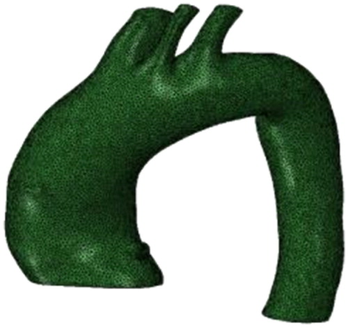
The geometry of the aortic lumen, during diastolic phase, from a patient prior to ascending thoracic aortic aneurysm surgical repair was obtained (Trabelsi et al. Citation2018). The mean thickness of the aortic wall was 1.52 mm. Thus a mesh was created with a constant thickness corresponding to this value (). A tear was introduced in the medial layer, near the most common locations of aortic dissection: along the righ lateral wall and below the ligamentum arteriosum, successively (Roberts Citation1981). The model was then inflated and the critical pressure was assessed.
Figure 1. Mesh of an ascending thoracic aortic aneurysm in the systolic phase.
3. Results and discussion
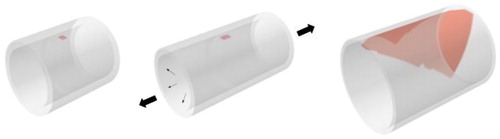
The simulations of tear propagation were run with the perfect geometry and the critical pressure was assessed. The different steps of the simulation are presented in .
Figure 2. Simulation model. (left) initial model with the tear in red. (mid) end of tension step, (right) end of inflation with the propagation of the tear.
Both circumferential and longitudinal propagations were observed in those simulations. Using the full factorial designs, critical pressure was computed as a linear combination of the different parameters and showed that the most influential are the initiation stress Tc and the geometrical parameters of the tear.
The simulation with aneurysm geometry showed a propagation of the tear circumferentially and longitudinally along the aortic wall. The same phenomenon can be seen on CT-scan of patient with aortic dissection.
4. Conclusions
Although this study remains to be validated with experimental data (on-going research), it allowed a better understanding of the mechanisms of aortic dissection propagation. Furthermore, it may lead to improvements in the detection and treatment of this disease.
Additional information
Funding
References
- Belytschko T, Black T. 1999. Elastic crack growth in finite elements with minimal remeshing. Int J Numer Methods Eng. 45(5):601–620.
- Ferrara A, Pandolfi A. 2010. A numerical study of arterial media dissection processes. Int J Fract. 166(1-2):21–33.
- Gasser TC, Holzapfel GA. 2006. Modeling the propagation of arterial dissection. Eur J Mech A Solids. 25(4):617–633.
- Gasser TC, Ogden RW, Holzapfel GA. 2006. Hyperelastic modelling of arterial layers with distributed collagen fibre orientations. J R Soc Interface. 3(6):15–35.
- Holzapfel GA, Gasser TC, Ogden RW. 2000. A new constitutive framework for arterial wall mechanics and a comparative study of material models. J Elast Phys Sci Solids. 61(1/3):1–48.
- Humphrey JD, Holzapfel GA. 2012. Mechanics, mechanobiology, and modeling of human abdominal aorta and aneurysms. J Biomech. 45(5):805–814.
- Roberts WC. 1981. Aortic dissection: anatomy, consequences, and causes. Am Heart J. 101(2):195–214.
- Trabelsi O, Gutierrez M, Farzaneh S, Duprey A, Avril S. 2018. A non-invasive methodology for ATAA rupture risk estimation. J Biomech. 66:119–126.
- Weisbecker H, Pierce DM, Regitnig P, Holzapfel GA. 2012. Layer-specific damage experiments and modeling of human thoracic and abdominal aortas with nonatherosclerotic intimal thickening. J Mech Behav Biomed Mater. 12:93–106.