
 ?Mathematical formulae have been encoded as MathML and are displayed in this HTML version using MathJax in order to improve their display. Uncheck the box to turn MathJax off. This feature requires Javascript. Click on a formula to zoom.
1. Introduction
Distraction Osteogenesis (DO) is a surgical procedure consisting in the progressive lengthening of a bone segment (1 mm/day). The technique requires the implantation of a distractor device, which daily activation is responsible for the lengthening of the bone. Distraction is widespread in the craniofacial area for the treatment of congenital malformations or acquired large bone defects (Adolhs et al. 2014). DO activation requires a transmucosal or transcutaneous rod which may be responsible for multiple adverse events and incomfort. To overcome these issues, we demonstrated recently the feasibility of distant activation with a magnetically activated device for mandibular distraction (Boisson et al. Citation2016). However, to develop new distraction device, one must fully understand the role of the surrounding soft tissues, especially the periosteum, and their participation in the mechanical load opposing the distraction vector.
The periosteum is a bi-layer soft tissue surrounding bone (Evans et al. Citation2013). The outer fibrous layer contains mainly collagen and elastin fibers, whereas the inner layer has a higher cell density. Periosteum is anchored to the bone by Sharpey’s fibers (fibers with a high collagen content) (Evans et al. Citation2013). It also exhibits the properties of a viscoelastic anisotropic material (Popowics et al. Citation2002). It also be demonstrated that, in vivo, the periosteum is pre-stressed on the bone (Bertram et al. Citation1998).
Despite its known role in many bone regeneration aspects, no study, to our knowledge, was conducted on the human mandibular periosteum biomechanics, and none incorporated it in a mandibular DO process (DO may be compared to an uniaxial traction test).
The hypothesis we tested here is that the main load opposing the DO process is provided by the periosteum. In the first part, we measured the torque at the endless screw of the devices during a mandibular DO, to determine the global load of distraction. The second part we measured the stress generated through a uniaxial elongation test to determine the load induced by the periosteum during distraction. Finally, we compared both results.
2. Methods
2.1. Samples harvesting
From January to June 2017, 9 human cadavers (3 males and 6 females) were dissected. Ages at death ranged from 75 to 97 years. Lengths of preservation in cold (at -18 °C) extended from 13 to 460 days. A complete 20 mm horizontal distraction with torque assessment was performed on 7 cadavers. 18 periosteum samples of 40 mm per 10 mm were harvested (2 per left mandibular corpus) for tensile tests ().
Figure 1. A: scheme of the mandible distraction osteogenesis surgical procedure. B: scheme of the harvested samples of periosteum.
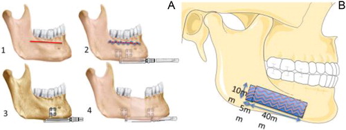
Each periosteum samples were immediately cutted into two pieces:
Samples of 5 mm per 10 mm of each periosteal specimen were harvested, fixed in formol and embedded in paraffin for histological analysis in order to measure the perioste thickness.
Remaining parts of the samples were immediately stored in saline solution to be submitted to tensile test.
2.2. Determination of the load of the cadaveric distraction osteogenesis: 
The torque-based load was obtained following a two-stage procedure: all the distractors were calibrated prior to the cadaveric experiments in order to determine their specific torque load relationship, and then the torque was measured for each millimeter at the activation rod during standard 20 mm right horizontal distractions.
2.3. Determination of the load induced by the periosteum during a DO:
Tensile test protocol was determined in order to simulate the periosteum condition during DO. Tensile tests were performed with a uniaxial elongation machine. Displacement was defined by the distance between the two grips. Load was recorded using a static load cell with a 100 N capacity. Traction rate was set at 0.25 mm/s, which corresponds to the distraction speed in vivo. Strain homogeneity was verified using digital correlation image on 5 samples.
From the geometrical parameter, the load and the displacement, we were able to determine the periosteum stress-strain curves.
To determine the load () induced by the periosteum during a DO, we have evaluated the periosteum stress and strain on the bone. According to Bertram et al., this corresponds to the transition point between the toe and steep regions of the stress-strain curve. Then from characteristic lengths of the DO procedure performed on cadaver, we size the typical periosteum strain on the sample corresponding to a 1 mm distraction. Then
where
are the stress from the lingual and vestibular part respectively, while
are the corresponding periosteum section.
3. Results and discussion
We showed an increasing load through the 20 mm distractions in this study, with a median per millimetre ranging from 18.0 to 50.6 N ().
Figure 2. The grey points correspond to all Lt for all cadavers plotted against the length of the distraction during a 20 mm distraction. The black squares (□) are their medians.
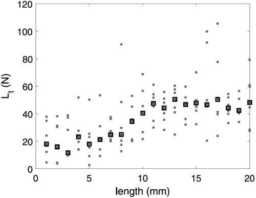
Using the stress-strain data of the tensile tests, we evaluated the median load applied on the distractor during a distraction to be N.
The median accounted for more than
of the median
for
mm. No statistical difference was found between
and
(
). This result confirmed that the periosteum is the primary tissue opposing distraction, which had been suggested previously without biomechanical proof.
4. Discussion
Periosteum pretension value used in this article is based on literature (Bertram et al. Citation1998), and need to be experimentally assessed. The present cadaver model does not include in-vivo specific features like the bony callus and periosteum remodelling during DO.
5. Conclusions
We showed that the periosteum is directly stretched by the distraction activation and that the periosteum is the principal contributor to the stress involved in a distraction at the beginning of activation.
Additional information
Funding
References
- Adolphs N, Ernst N, Menneking H, Hoffmeister B. 2014. Signicance of distraction osteogenesis of the craniomaxillofacial skeleton a clinical review after 10 years of experience with the technique. J. Craniomaxillofac. Surg. 42(6):966–975.
- Bertram JE, Polevoy Y, Cullinane DM. 1998. Mechanics of avian fibrous periosteum: tensile and adhesion properties during growth. Bone. 22(6):669–675.
- Boisson J, Strozyk H, Diner P, Picard A, Kadlub N. 2016. Feasibility of magnetic activation of a maxillofacial distraction osteogenesis, design of a new device. J. Craniomaxillofac. Surg. 44(6):684–688.
- Debelmas A, Picard A, Kadlub N, Boisson J. 2018. Contribution of the periosteum to mandibular distraction. PLoS One. 13(6):e0199116.
- Evans SF, Chang H, Knothe Tate ML. 2013. Elucidating mult scale periosteal mechanobiology: A key to unlocking the smart properties and regenerative capacity of the periosteum? Tissue Eng Part B: Rev. 19:147–159.
- Popowics TE, Zhu Z, Herring SW. 2002. Mechanical properties of the periosteum in the pig, sus scrofa. Arch Oral Biol. 47(10):733–741.