
1. Introduction
Dental implants are widely used in the clinic. However, there remain risks of failure, which depend on the implant primary and secondary stability. Implant stability is an important factor for the implant success and is determined by the biomechanical quality of bone tissue at the bone-implant interface. Radiofrequency analyses (RFA) and quantitative ultrasound (QUS) methods have been suggested to assess implant stability. The first purpose of this study was to compare the results obtained using these two techniques applied to the same dental implants inserted in bone mimicking phantoms. Then, the second aim of this study is to compare the results obtained using these two techniques for dental implants inserted in sheep iliac crest at different healing time.
2. Methods
2.1. In vitro study
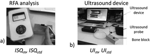
In the in vitro study, the reproducibility of the two techniques was determined for each implant using the device shown in . Different values of trabecular bone density #10, #20, #30 PCF (PCF corresponds to the notation used by the phantom manufacturer to rank the phantom quality) and cortical thickness were considered to assess the effect of bone quality on the ultrasonic indicator (UI) and on the Implant Stability Quotient (ISQ) values. The effect of the implant insertion depth and of the final drill diameter (1 or 2 mm) was also investigated.
Figure 1. Description of the experimental protocol. Measurement realized using a), the resonance frequency analysis device and b) the quantitative ultrasound device.
2.2. In vivo study
In the in vivo study, eighty-one identical implants were inserted in the iliac crests of 11 sheep. The QUS and RFA measurements were realized after different healing times (0, 5, 7 and 15 weeks).
3. Results and discussion
3.1. In vitro study
ISQ values increase and UI values decrease when i) the bone density increase, ii) cortical thickness increase, and iii) the implant is screwed in the phantom. The UI values are significantly different for all final drill diameters except for 2.8 mm and 2.9 mm, while the ISQ values are similar for all final drill diameters lower than 3.2 mm and higher than 3.3 mm. The error realized on the estimation of the trabecular density (respectively cortical thickness and insertion depth) with the QUS device is around 4 (respectively 8 and 4) times lower compared to that made with the RFA technique (Vayron et al. 2018a, 2018b).
3.2. In vivo study
The results obtained with the QUS (respectively RFA) method were significantly different when comparing two consecutive healing time for 97% (respectively 18%) of the implants. The error made on the estimation of the healing time when analyzing the results obtained with the QUS technique was around 10 times lower than that made when using the RFA technique. The results corresponding to the dependence of the ISQ versus healing time were significantly different when comparing two directions of RFA measurement (Vayron et al. 2018a, Citation2018b).
3.3. Physical interpretation
The results obtained in vitro and in vivo may be explained by the dependence of the transmission coefficient on the biomechanical properties of the bone-implant interface. When implant stability increases, it leads to a more important quality and quantity of bone in contact with the implant, which explains the higher transmission coefficient at the bone-implant interface, which allows in turn the results obtained herein.
4. Conclusions
The results show that ultrasound technique provides a better estimation of different parameters related to the implant stability compared to the RFA technique.
This study paves the way towards the development of a medical device thus providing a decision support system to dental surgeons. However, the clinical applicability of the QUS method should be investigated in the future, together with a clinical comparison.
Additional information
Funding
References
- Vayron R, Nguyen V, Lecuelle B, Albini Lomami H, Meningaud J, Bosc R, Haiat G. 2018a. Comparison of resonance frequency analysis and of quantitative ultrasound to assess dental implant osseointegration. Sensors (Basel, Switzerland). 18(5):1397.
- Vayron R, Nguyen VH, Lecuelle B, Haiat G. 2018b. Evaluation of dental implant stability in bone phantoms: Comparison between a quantitative ultrasound technique and resonance frequency analysis. Clin Implant Dent Relat Res. 20(4):470–478.